
Binasal Hemianopia occurs when the lesion is present in: a) Right optic nerve b) Optic Chiasma c) Temporal retina d) Left geniculocalcarine tract
- The nasal visual field is processed by the temporal retina of each eye.
- Fibers from the temporal retina are the uncrossed fibers - they travel on the same side through the optic chiasma.
- These uncrossed (lateral) fibers run along the outer edges of the optic chiasma.
visual field defects optic chiasma binasal hemianopia diagram
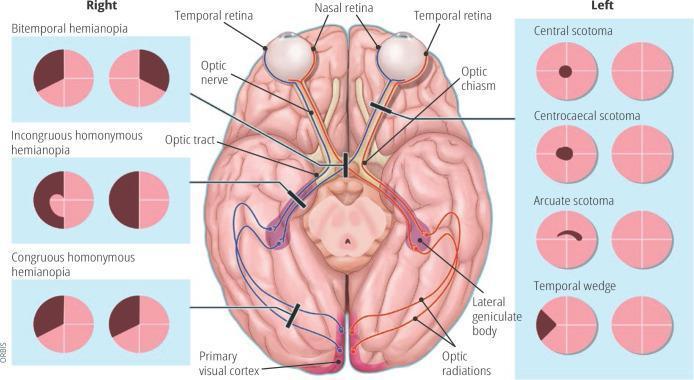
Anatomical diagram and educational infographic detailing the human visual pathway and associated visual field defects. The central illustration shows a superior (dorsal) view of the brain, highlighting the optic nerve, optic chiasm (decussation point), optic tract, lateral geniculate body, optic radiations, and the primary visual cortex in the occipital lobes. Pathways are color-coded (red and blue) to demonstrate the transition from nasal and temporal retinal fibers to the contralateral and ipsilateral hemispheres. Black lesion bars indicate specific sites of damage along this pathway. Flanking the central diagram are clinical representations of visual field loss patterns. The 'Right' panel displays bilateral defects including bitemporal hemianopia, incongruous homonymous hemianopia, and congruous homonymous hemianopia. The 'Left' panel displays unilateral or localized defects including central scotoma, centrocaecal scotoma, arcuate scotoma, and temporal wedge. This diagram serves as a neuro-ophthalmological guide for localizing lesions within the central nervous system based on perimetry findings.
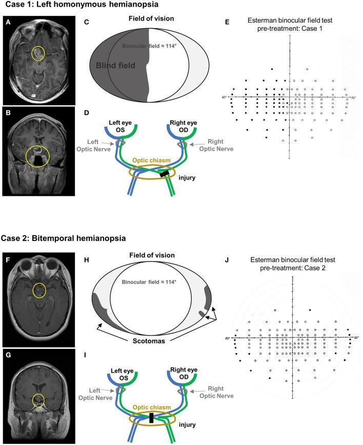
This composite educational image presents two clinical cases correlating neuroimaging findings with visual field defects. Case 1 (A-E) depicts left homonymous hemianopia. T1-weighted contrast-enhanced MRI in axial (A) and coronal (B) planes shows a heterogeneously enhancing mass in the suprasellar region (yellow circles), involving the optic chiasm and third ventricle floor. Schematic (C) and anatomical diagram (D) illustrate the corresponding lesion in the right optic tract and the resulting left hemifield blindness. The Esterman binocular field test (E) confirms significant unseen points (black dots) in the left visual field. Case 2 (F-J) depicts bitemporal hemianopia. MRI scans (F, G) reveal a residual suprasellar mass (yellow circles) inseparable from the hypothalamus and optic chiasm. The schematic (H) and optic pathway diagram (I) show a midline chiasmal injury, leading to peripheral scotomas. The Esterman test (J) demonstrates peripheral blind spots consistent with bitemporal vision loss. This comparison illustrates how specific anatomical injuries along the optic pathway correlate with distinct patterns of binocular visual impairment.
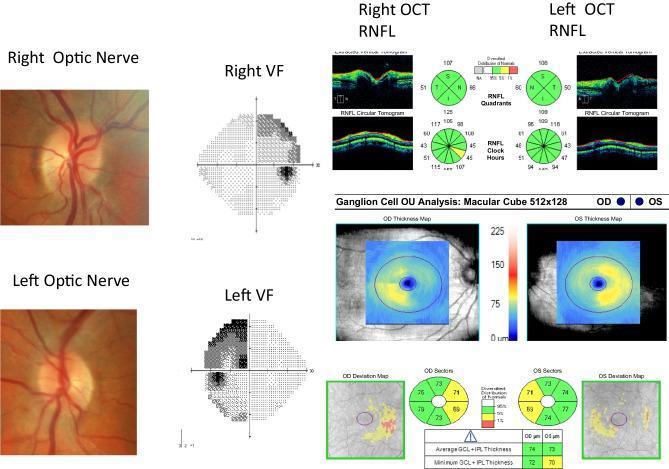
A multi-modal neuro-ophthalmic assessment of a patient with chiasmal compression. The composite includes fundus photographs, Humphrey visual field (VF) tests, and Optical Coherence Tomography (OCT) of the Retinal Nerve Fiber Layer (RNFL) and Ganglion Cell Layer (GCL). The fundus photographs of the right and left optic nerves show a clinically normal appearance with healthy disc margins and vasculature. Automated perimetry (VF) reveals a classic bitemporal hemianopia, characterized by significant sensitivity loss in the temporal hemifields of both eyes. The OCT RNFL thickness maps and circular tomograms appear largely within normal limits (coded green in quadrant/clock-hour maps), indicating minimal retrograde axonal loss at the disc level. However, the Ganglion Cell Analysis (GCL+IPL) thickness and deviation maps demonstrate clear 'binasal' macular thinning that respects the vertical midline, corresponding to the temporal visual field defects. This clinical scenario illustrates that macular GCL thinning can precede detectable RNFL atrophy in chronic compressive chiasmopathy, such as that caused by a pituitary adenoma.
Answer: (b) Optic Chiasma
Understanding the Visual Pathway and Field Defects
| Retinal Fiber | Origin | Crosses in Chiasma? | Carries Signal From |
|---|---|---|---|
| Nasal retinal fibers | Medial (nasal) half of retina | YES - cross to opposite side | Temporal visual field |
| Temporal retinal fibers | Lateral (temporal) half of retina | NO - stay ipsilateral | Nasal visual field |
What is Binasal Hemianopia?
- Loss of both nasal visual fields (medial fields of each eye)
- The nasal visual field is subserved by the temporal retina of each eye
- Temporal retinal fibers travel as uncrossed fibers along the lateral edges of the optic chiasma
Why the Other Options Are Wrong
| Option | Lesion Site | Visual Field Defect Produced |
|---|---|---|
| (a) Right optic nerve | Pre-chiasmal | Monocular blindness (right eye only) |
| (b) Optic chiasma | ✅ CORRECT (lateral edges) | Binasal hemianopia |
| (c) Temporal retina | Retinal level | Affects nasal visual field of that eye (monocular) |
| (d) Left geniculocalcarine tract | Post-chiasmal | Right homonymous hemianopia |
Visual Pathway Lesion Summary (Classic Exam Map)
Optic nerve lesion         → Monocular blindness (ipsilateral eye)
Optic chiasma (center)     → Bitemporal hemianopia (pituitary tumour - commonest)
Optic chiasma (lateral)    → Binasal hemianopia (rare - bilateral carotid aneurysms)
Optic tract lesion         → Contralateral homonymous hemianopia
Temporal lobe radiation    → Superior quadrantanopia ("pie in the sky")
Parietal lobe radiation    → Inferior quadrantanopia ("pie on the floor")
Occipital cortex           → Congruous homonymous hemianopia (macular sparing)
Are you sure ?
binasal hemianopia lesion site optic chiasma lateral edges temporal retinal fibers
The Answer is (b) Optic Chiasma - Confirmed
Why I'm sure:
"Lateral chiasmal lesions may produce binasal hemianopia."
"Temporal retinal fibers of each eye serve the nasal visual fields, and each eye's temporal retinal fibers pass closest to one another at the optic chiasm... lesions involving the optic chiasm might also impinge upon the temporal retinal fibers bilaterally as these fibers course temporally through the optic chiasm."
"A lesion compressing the chiasm laterally is more likely to cause a nasal visual field defect due to compression of nerve fibres supplying the temporal retina."
The Core Anatomy (No Ambiguity)
| Field lost | Retina involved | Fiber type | Location in chiasma |
|---|---|---|---|
| Temporal field | Nasal retina | Crossed fibers | Center of chiasma |
| Nasal field | Temporal retina | Uncrossed fibers | Lateral edges of chiasma |
- Bitemporal hemianopia = central chiasmal lesion (e.g. pituitary adenoma pressing from below)
- Binasal hemianopia = lateral chiasmal lesion (e.g. bilateral carotid aneurysms, sclerosis pressing from the sides)