
Hypertrophied obstructive cardiomyopathy
Reading File
Hypertrophic obstructive cardiomyopathy (HOCM) is a genetic disorder of the heart muscle characterized by asymmetric left ventricular hypertrophy - most often involving the interventricular septum - that causes dynamic left ventricular outflow tract (LVOT) obstruction. It is one of the most common causes of sudden cardiac death in young athletes.
Searching sources...
Finding Sources
Searching PubMed
"hypertrophic cardiomyopathy" AND "management"
Reading File
Reading File
Reading File
Reading File
Reading File
Finding Sources
Finding Sources
Reading File
Reading File
Reading File
Loading Image
Loading Image
Reading File
I now have comprehensive material from multiple authoritative sources. Here is a complete overview of Hypertrophic Obstructive Cardiomyopathy (HOCM):
Hypertrophic Obstructive Cardiomyopathy (HOCM)
Definition
Hypertrophic cardiomyopathy (HCM) is defined as unexplained left ventricular (LV) hypertrophy in the absence of abnormal loading conditions (valve disease, hypertension, congenital defects) that would suffice to explain the degree of hypertrophy. When this hypertrophy causes dynamic obstruction of the LV outflow tract (LVOT), the condition is specifically termed hypertrophic obstructive cardiomyopathy (HOCM). Prevalence is 0.2-0.5% worldwide, affecting all racial groups.
- Goldman-Cecil Medicine, p. 494
- Robbins & Kumar Basic Pathology, p. 373
Genetics and Pathogenesis
HCM is usually autosomal dominant with variable penetrance and expression. Sarcomeric contractile protein gene mutations account for 50-60% of cases, with over 1,400 distinct mutations identified. These are uniformly gain-of-function mutations that enhance myofilament activity, leading to:
- Myocyte hypercontractility
- Increased energy consumption
- Net negative energy balance
The most commonly mutated genes:
| Gene | Protein | Frequency |
|---|---|---|
| MYH7 | Beta-myosin heavy chain | Most frequent |
| MYBPC3 | Myosin-binding protein C | Second most |
| TNNT2 | Troponin T | Third most |
Together, mutations in these three genes account for 70-80% of all HCM cases.
Note: the same genes (e.g., beta-myosin) can be mutated in dilated cardiomyopathy (DCM), but those are loss-of-function mutations, in contrast to the gain-of-function mutations in HCM.
- Robbins & Kumar Basic Pathology, p. 373-374
Pathology / Morphology
Gross:
- Massive myocardial hypertrophy without ventricular dilation
- 90% of cases: Asymmetric septal hypertrophy - disproportionate thickening of the interventricular septum relative to the LV free wall
- 10% of cases: Concentric hypertrophy
- The ventricular cavity is compressed into a characteristic "banana-shaped" configuration
- The anterior mitral leaflet contacts the septum during systole, producing a fibrous endocardial plaque in the LVOT
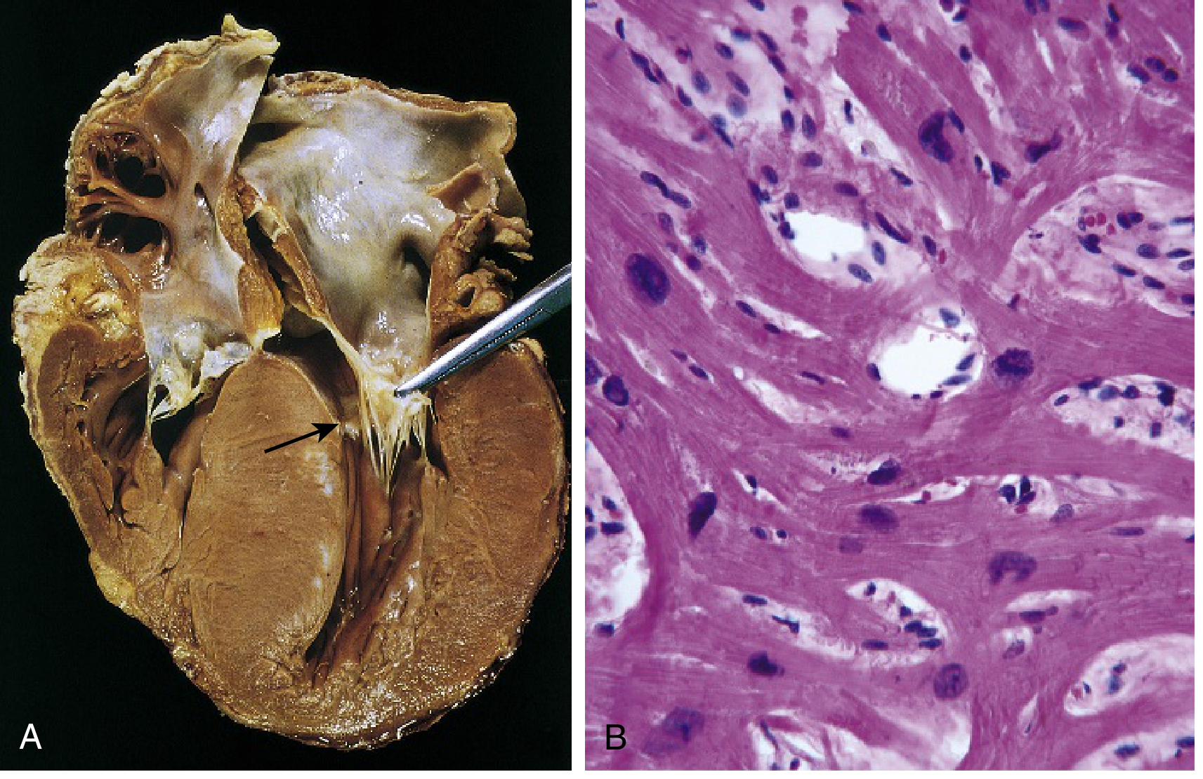
FIG. 9.27 - Hypertrophic cardiomyopathy: (A) Septal muscle bulging into LVOT forming "banana-shaped" lumen, with fibrous endocardial plaque (arrow). (B) Histology showing myocyte disarray, extreme hypertrophy, branching, and interstitial fibrosis.
Histology (pathognomonic triad):
- Marked myocyte hypertrophy
- Haphazard myocyte and myofiber disarray (whorled pattern)
- Interstitial fibrosis
- Robbins & Kumar Basic Pathology, p. 373-374
Mechanism of LVOT Obstruction
The obstruction in HOCM is dynamic (not fixed), caused by two interacting mechanisms:
- Asymmetric septal hypertrophy - the bulging septum narrows the LVOT
- Systolic Anterior Motion (SAM) of the mitral valve - ventricular septal hypertrophy creates a Venturi effect pulling the anterior mitral leaflet forward into the outflow tract during systole
This produces:
- A dynamic gradient across the LVOT that worsens with reduced preload, reduced afterload, or increased contractility
- Functional mitral regurgitation from incomplete coaptation of the mitral leaflets
Conditions that worsen the gradient (increase obstruction):
- Standing, Valsalva maneuver, dehydration, tachycardia, amyl nitrite inhalation, exercise
Conditions that reduce the gradient (decrease obstruction):
- Squatting, handgrip exercise, leg raising, increased preload/afterload, beta-blockers
Clinical Features
Most patients are asymptomatic at initial diagnosis, often discovered incidentally on family screening, routine ECG, or murmur. Symptoms can develop at any age.
Classic symptom triad:
- Exertional dyspnea (most common - diastolic dysfunction + LVOT obstruction)
- Angina/chest pain (in 20-30%; due to microvascular ischemia even without CAD)
- Syncope (in ~20%; often exertional, serious warning sign)
Murmur: Harsh systolic ejection murmur, loudest at the left sternal edge, radiates to the right upper sternal edge and apex. Accompanied by functional mitral regurgitation murmur. The murmur is dynamic - it increases with Valsalva/standing and decreases with squatting/handgrip (distinguishing it from fixed obstruction like aortic stenosis).
Major complications:
- Atrial fibrillation with mural thrombus and embolic stroke
- Ventricular fibrillation causing sudden cardiac death (SCD)
- Infectious endocarditis of the mitral valve
- Progressive congestive heart failure (<5% of patients)
HCM is responsible for ~1/3 of sudden cardiac deaths in athletes under age 35.
- Harrison's Principles of Internal Medicine 22E, p. 2051
- Goldman-Cecil Medicine, p. 494-495
- Robbins & Kumar Basic Pathology, p. 374
Diagnosis
Initial evaluation should include:
- 12-lead ECG - typically abnormal in 90%+ of symptomatic patients; shows LV hypertrophy, deep Q waves (especially II, III, aVF, V5-V6), ST changes
- 2D Echocardiography (gold standard) - demonstrates asymmetric septal hypertrophy, SAM of the mitral valve, LVOT gradient, diastolic dysfunction
- Cardiac MRI - provides superior tissue characterization; late gadolinium enhancement (LGE) represents fibrosis and is an important risk factor for SCD
- Holter monitoring / exercise stress testing - detects NSVT, assesses exercise-induced gradient
- Genetic testing - next-generation sequencing panels; pathogenic variants found in <50% of probands
- Family history (3-4 generation) - all first-degree relatives should be screened
Differential diagnosis to exclude:
- Aortic stenosis
- Hypertensive heart disease
- Cardiac amyloidosis (Fabry disease, Danon disease, Pompe disease)
- Noonan syndrome, LEOPARD syndrome
Risk Stratification for Sudden Cardiac Death
Major risk factors:
| Risk Factor | Notes |
|---|---|
| Prior cardiac arrest or sustained VT | Strongest predictor |
| Unexplained syncope | Especially exertional |
| Family history of SCD | In first-degree relatives |
| LV wall thickness >30 mm | Present in <10% |
| LV systolic dysfunction (EF <50%) | Present in <10% |
| LV apical aneurysm | Especially with apical HCM |
| LVOT gradient, LA diameter, age | Used in ESC risk calculator |
| Late gadolinium enhancement on MRI | Modifying risk factor |
| Nonsustained VT on Holter | >3 beats at rate >120 |
The ESC HCM Risk Calculator (doc2do.com/hcm/webHCM.html) provides a 5-year estimated SCD risk:
-
Risk ≥6%: ICD generally recommended
-
Risk 4-6%: ICD can be considered
-
Risk <4%: ICD not routinely advised
-
Harrison's Principles of Internal Medicine 22E, p. 2051
Management
Treatment Algorithm
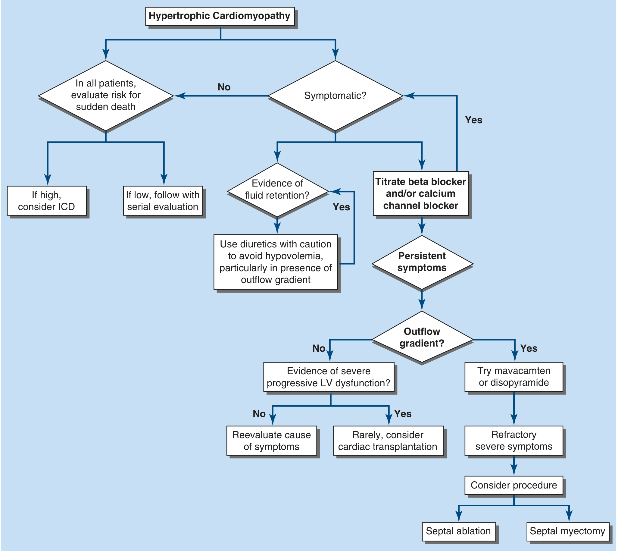
FIGURE 267-5 (Harrison's 22E) - HCM treatment algorithm: all patients evaluated for SCD risk; symptomatic patients treated with beta-blockers/CCBs, then mavacamten or disopyramide for outflow gradient, and septal reduction for refractory cases.
Medical Therapy
Step 1 - First-line agents (reduce heart rate, increase diastolic filling, reduce contractility):
- Beta-blockers (first choice): metoprolol (target 150 mg/day) or propranolol (target 120-480 mg/day); titrate to resting HR 50-70 bpm. Effective in 60-70% with LVOT obstruction.
- Non-dihydropyridine calcium channel blockers: verapamil (120 mg/day, up to >480 mg/day) or diltiazem (180 mg/day, up to >360 mg/day) - alternatives, especially for refractory chest pain
Step 2 - Add-on for persistent LVOT obstruction:
- Disopyramide: negative inotropic antiarrhythmic agent; dose 400-600 mg/day; effective in up to 2/3 of patients. Side effects include anticholinergic effects (dry eyes/mouth, urinary retention). Should always be combined with a beta-blocker or verapamil to prevent excessive AV nodal conduction from the anticholinergic effect.
- Mavacamten (FDA-approved): small-molecule cardiac myosin inhibitor - directly reduces myosin cross-bridge formation, reducing hypercontractility and LVOT gradient. High efficacy in symptomatic obstructive HCM, including in patients with persistent symptoms on beta-blockers.
- Aficamten (under investigation): another cardiac myosin inhibitor
Diuretics: Used cautiously for fluid overload symptoms; avoid hypovolemia, especially in the presence of outflow gradient.
Drugs to AVOID in HOCM:
- Vasodilators (nitrates, ACE inhibitors, hydralazine) - reduce preload/afterload and worsen obstruction
- Digoxin/cardiac glycosides - increase contractility and worsen obstruction
- Dihydropyridine CCBs
Atrial Fibrillation Management
- Beta-blockers or CCBs (verapamil, diltiazem) for rate control
- Disopyramide + beta-blocker or verapamil/diltiazem for rhythm control in HCM-setting AF
- Amiodarone as alternative
- Anticoagulation for all HCM patients with AF (regardless of CHA₂DS₂-VASc score)
- Cardiac glycosides should be avoided (worsen obstruction)
Invasive / Interventional Therapy (for medically refractory cases, ~5%)
Both procedures reduce the gradient but neither has been shown to improve survival - only symptoms.
| Procedure | Mechanism | Notes |
|---|---|---|
| Surgical septal myectomy | Excision of proximal septal myocardium | Gold standard; 60+ year track record; very low perioperative mortality at expert centers; durable long-term relief |
| Alcohol septal ablation | Controlled infarction of proximal septum via injection of alcohol into septal perforators | Percutaneous; reserved for patients wishing to avoid surgery or with prohibitive comorbidities; requires suitable coronary anatomy |
- Both carry risk of complete heart block requiring permanent pacing
- Mitral valve repair/replacement usually unnecessary after myectomy (MR resolves with septal relief)
- Dual-chamber pacing alone is not effective as primary therapy for LVOT obstruction
Cardiac transplantation: Rarely considered for patients with refractory HF and severe progressive LV dysfunction.
ICD Implantation
- Primary prevention: based on risk stratification (ESC risk calculator ≥6%)
- Secondary prevention: all patients with prior cardiac arrest or sustained VT (regardless of other factors)
- Long-term device-related complications must be weighed, especially in young patients
Genetic Counseling and Family Screening
- Offered to all newly diagnosed patients
- First-degree relatives screened with ECG and echo every 12-18 months from age 12 until adulthood (18-21 years)
- Adults with normal screening evaluated every few years (late-onset LVH possible)
Lifestyle Advice
-
Competitive sports and vigorous exercise were historically restricted; however, recent studies have failed to show a clear link between exertion and ventricular arrhythmias in HCM, leading to more individualized, shared decision-making regarding exercise
-
Avoid dehydration and excessive alcohol consumption
-
Harrison's Principles of Internal Medicine 22E, p. 2051
-
Goldman-Cecil Medicine, p. 495-496
Recent Evidence (2024-2026)
A systematic review on obstructive HCM management (Butzner et al., 2024) analyzed evidence gaps in HOCM therapy, highlighting the expanding role of cardiac myosin inhibitors. A 2025 meta-analysis (Lee et al.) compared outcomes in apical vs. nonapical HCM subtypes.
The most notable recent advance is mavacamten (FDA-approved), which represents the first disease-targeted medical therapy for obstructive HCM by directly inhibiting cardiac myosin. It has been shown to reduce LVOT gradients and improve symptoms in patients who remain symptomatic on traditional beta-blockers/CCBs.
Summary Table
| Feature | HOCM |
|---|---|
| Inheritance | Autosomal dominant |
| Key genes | MYH7, MYBPC3, TNNT2 |
| Mutation type | Gain-of-function (sarcomeric proteins) |
| LV morphology | Asymmetric septal hypertrophy (90%) |
| Obstruction mechanism | SAM of mitral valve + septal bulge |
| Murmur | Systolic ejection; increases with Valsalva/standing |
| Histology | Myocyte disarray + hypertrophy + interstitial fibrosis |
| First-line medical Rx | Beta-blockers, verapamil/diltiazem |
| Add-on for gradient | Disopyramide, mavacamten |
| Invasive Rx | Surgical myectomy or alcohol septal ablation |
| SCD prevention | ICD (risk-stratified) |
| SCD statistic | ~1/3 of SCD in athletes <35 years |
This is a shared conversation. Sign in to Orris to start your own chat.