
G6PD Deficiency
Reading File
Now let me search the medical library and PubMed simultaneously for G6PD deficiency.
Finding Sources
Searching PubMed
"G6PD deficiency"[MeSH Terms] AND management
Excellent - I found G6PD sections in Harrison's, Lippincott Biochemistry, and more. Let me read all key sections in parallel.
Reading File
Reading File
Finding Sources
Excellent - rich data from multiple sources. Let me now read the Robbins pathology and Lippincott sections in parallel, and also fetch the metabolic pathway diagram.
Reading File
Reading File
Reading File
Loading Image
I now have thorough information from multiple authoritative sources. Let me also grab the Heinz body image and read the Goldman-Cecil clinical manifestations section.
Loading Image
Reading File
I now have comprehensive data from Harrison's, Robbins, Goldman-Cecil, Lippincott Biochemistry, and Tietz Lab Medicine. Let me compile the complete answer.
G6PD Deficiency (Glucose-6-Phosphate Dehydrogenase Deficiency)
Overview
G6PD deficiency is the most common enzymopathy in humans, affecting an estimated 400+ million people worldwide. It is an X-linked disorder that leaves red blood cells (RBCs) unable to defend against oxidant stress, triggering episodic or chronic hemolytic anemia.
- Lippincott Illustrated Reviews: Biochemistry, 8th ed, p. 445
- Tietz Textbook of Laboratory Medicine, 7th ed, p. 3054
Biochemical Mechanism
The enzyme G6PD catalyzes the first and rate-limiting step of the pentose phosphate pathway (PPP): conversion of glucose-6-phosphate to 6-phosphogluconolactone, generating NADPH. In RBCs, the PPP is the only source of NADPH.
NADPH is required to:
- Reduce oxidized glutathione (G-S-S-G) back to reduced glutathione (G-SH) via glutathione reductase
- G-SH then detoxifies H₂O₂ via glutathione peroxidase
When G6PD is deficient → less NADPH → depleted G-SH → H₂O₂ accumulates → oxidizes hemoglobin → Heinz bodies form → RBC membrane rigidity and hemolysis.
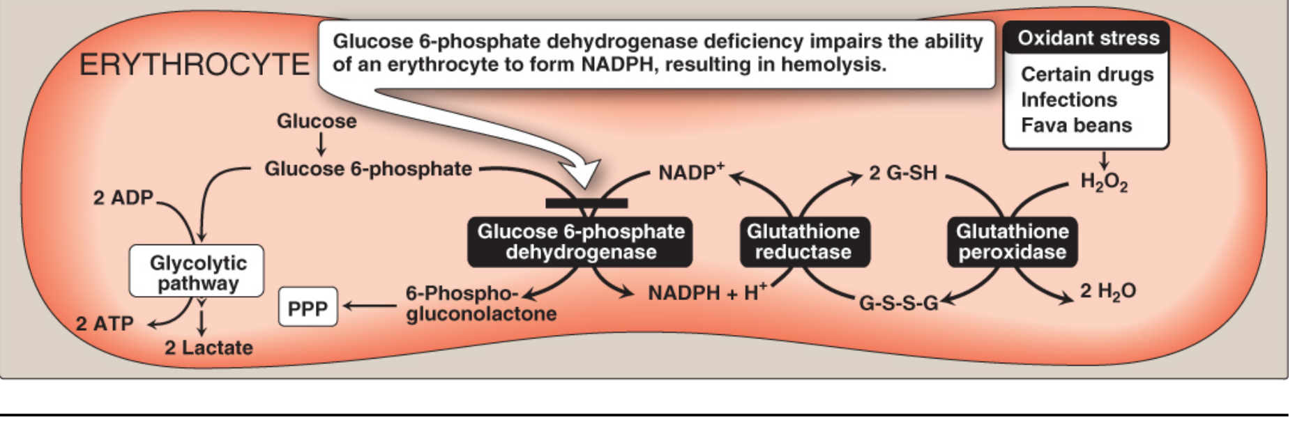
Key point: Other tissues can generate NADPH via the malic enzyme pathway, but RBCs cannot - making them uniquely vulnerable. Additionally, mature RBCs have no nucleus or ribosomes and cannot synthesize new G6PD enzyme, leaving them entirely dependent on the activity of the enzyme already present.
Epidemiology & Genetics
| Feature | Details |
|---|---|
| Inheritance | X-linked recessive |
| Prevalence | ~400-500 million affected worldwide |
| Sex | Primarily males (hemizygous); females usually carriers but can be affected with unfavorable lyonization |
| Gene locus | Xq28, 18 kb, 13 exons |
| Variants | >400 described; >200 different mutations (mostly missense point mutations) |
High-prevalence regions overlap with malaria-endemic zones (sub-Saharan Africa, Mediterranean, Middle East, South and Southeast Asia). This is not coincidental - G6PD-deficient RBCs lysed by oxidant stress induced by Plasmodium falciparum actually protect the host from severe malaria, providing a selective evolutionary advantage.
- Goldman-Cecil Medicine, International Edition
- Tietz Textbook of Laboratory Medicine, 7th ed
WHO Classification of Variants
| Class | Enzyme Activity | Example Variant | Distribution | Clinical Features |
|---|---|---|---|---|
| Class I | <10% (severe) | G6PD Guadalajara | Sporadic | Chronic nonspherocytic hemolytic anemia (CNSHA) + NNJ + acute exacerbations |
| Class II | <10% (severe) | G6PD Mediterranean (Ser188Phe) | Middle East, South/Central Asia | Acute hemolysis; favism common |
| Class III | 10-60% (moderate) | G6PD A- (Val68Met + Asn126Asp) | Africa (10% of African-Americans) | Acute hemolysis after oxidant stress; self-limited |
| Class IV | 60-150% (normal) | G6PD A, G6PD B | Worldwide | No clinical manifestations |
| Class V | Increased activity | G6PD Hektoen | Rare | None |
Tietz Textbook of Laboratory Medicine, 7th ed, Table 78.2
G6PD A- (African variant): Half-life only 13 days vs. ~60 days for normal enzyme. Only older RBCs are G6PD-deficient; younger cells generated during the hemolytic episode have near-normal activity. This self-limiting nature means hemolysis abates even if the offending drug is continued.
G6PD Mediterranean: Much lower residual activity (<5%). Young and old RBCs are deficient; hemolysis is therefore more severe and prolonged. Associated with favism.
Precipitating Factors ("Triggers")
1. Oxidant Drugs
Commonly remembered as categories starting with "A":
- Antibiotics: Sulfonamides (sulfamethoxazole), dapsone, nitrofurantoin, nalidixic acid
- Antimalarials: Primaquine, chloroquine (high doses), pamaquine
- Analgesics: Aspirin (high doses), phenacetin
- Antipyretics: (various)
- Other: Methylene blue, rasburicase, vitamin K derivatives
2. Favism
Fava beans (broad beans) contain oxidant compounds (vicine, convicine). Particularly associated with the G6PD Mediterranean variant. All patients with favism have G6PD deficiency, but not all G6PD-deficient individuals develop favism.
3. Infection
The most common precipitant in practice. The inflammatory response generates free radicals in macrophages (as part of the host defense), which diffuse into RBCs and cause oxidative damage.
- Lippincott Illustrated Reviews: Biochemistry, 8th ed, p. 447-448
- Robbins & Kumar Basic Pathology, p. 389
Pathological Features
Heinz Bodies
Oxidized hemoglobin denatures and precipitates, forming intracellular inclusions called Heinz bodies, attached to the RBC membrane. These are visible on supravital staining (crystal violet or brilliant cresyl blue), not on routine Giemsa stain.
Bite Cells (Degmacytes)
Splenic macrophages attempt to "pluck out" Heinz bodies from RBCs. This creates characteristic bite cells (notched cells) visible on peripheral blood smear. These cells have reduced deformability and are subsequently trapped and destroyed in the spleen.

Clinical Manifestations
G6PD deficiency presents in 5 clinical syndromes:
| Syndrome | Features |
|---|---|
| Acute Hemolytic Anemia (AHA) | Onset 2-3 days after oxidant exposure; pallor, jaundice, dark urine (hemoglobinuria), fatigue |
| Favism | Severe hemolytic crisis after fava bean ingestion; more severe than drug-induced |
| Neonatal Jaundice (NNJ) | Appears 1-4 days after birth; unconjugated hyperbilirubinemia; can be severe; not always accompanied by significant anemia |
| Chronic Nonspherocytic Hemolytic Anemia (CNSHA) | Class I variants; ongoing hemolysis without clear precipitant; lifelong |
| Infection-induced hemolysis | Most common trigger in clinical practice |
- Harrison's Principles of Internal Medicine, 22E, p. (block 11)
- Tietz Textbook of Laboratory Medicine, 7th ed
Diagnosis
- Peripheral blood smear: Bite cells, blister cells, Heinz bodies (on supravital stain)
- G6PD enzyme assay (fluorescent spot test or spectrophotometric): Quantitative measurement of enzyme activity. Important caveat: Test should ideally be done 2-3 months after a hemolytic episode - during acute hemolysis, older deficient cells have been destroyed and the remaining younger cells have near-normal activity, giving a false-normal result (especially with G6PD A-)
- DNA mutation analysis: For definitive variant identification
- Reticulocytosis, elevated unconjugated bilirubin, elevated LDH, decreased haptoglobin: Markers of hemolysis
Management
Acute Hemolytic Anemia
- Identify and remove the trigger (stop offending drug, treat infection)
- Mild-moderate cases: Supportive care only; hemolysis is self-limiting (especially G6PD A-)
- Severe anemia: Blood transfusion (especially in children - can be a medical emergency)
- Acute renal failure (hemoglobinuria): Hemodialysis may be needed, but recovery is the rule without pre-existing kidney disease
- Ensure adequate hydration
Neonatal Jaundice
- Managed as for any neonatal jaundice: phototherapy or exchange transfusion depending on bilirubin levels
Chronic Nonspherocytic Hemolytic Anemia (Class I)
- Regular folic acid supplementation
- Regular hematologic surveillance
- Transfusions during exacerbations
- In severe cases with significant transfusion requirement: Splenectomy (evidence is limited but beneficial in practice, unlike hereditary spherocytosis where it is well-established)
- Iron chelation if regular transfusions required
Prevention
-
Screen before prescribing oxidant drugs (primaquine, dapsone, rasburicase)
-
Patient education: avoid fava beans, inform all healthcare providers
-
Neonatal screening programs in high-prevalence populations
-
Harrison's Principles of Internal Medicine, 22E
-
Miller's Anesthesia, 10e
Key Points Summary
| Feature | Value |
|---|---|
| Inheritance | X-linked recessive (Xq28) |
| Mechanism | ↓ NADPH → ↓ GSH → oxidative damage → Heinz bodies → bite cells → hemolysis |
| Most common worldwide variant | G6PD A- (Africa), G6PD Mediterranean (Middle East/Mediterranean) |
| Key triggers | Infections > drugs (sulfonamides, primaquine, dapsone) > fava beans |
| Blood smear | Bite cells + Heinz bodies (supravital stain) |
| Enzyme testing pitfall | False normal during/after acute episode (destroyed older cells) |
| Evolutionary advantage | Protection against P. falciparum malaria |
| Self-limiting feature | G6PD A- hemolysis stops as young cells with normal activity replace old ones |
Sources: Harrison's Principles of Internal Medicine 22E (2025) | Robbins & Kumar Basic Pathology | Goldman-Cecil Medicine | Lippincott Illustrated Reviews: Biochemistry, 8th ed | Tietz Textbook of Laboratory Medicine, 7th ed | Miller's Anesthesia, 10e
This is a shared conversation. Sign in to Orris to start your own chat.