

history og hemmorhagic bulla formation which converted into large erosive plaque 20 days ago
hemorrhagic bulla bullous pemphigoid erosive plaque lower leg
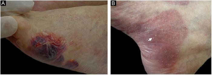
Clinical photograph of dermatological manifestations of Bullous Pemphigoid (BP) on the lower extremities, divided into two panels. Image A displays the acute phase, showing a large, tense hemorrhagic bulla with a characteristic dark purple-red hue on the foot. The bulla is situated atop an ill-defined erythematous plaque, indicating underlying cutaneous inflammation. Image B demonstrates a later stage of disease progression on the skin. It depicts large, confluent erythematous and violaceous plaques with evidence of cicatricial (scarring) changes from prior blistering. Multiple small, white, firm papules known as milia are visible; one milium is specifically indicated by a white arrow. The presence of milia over healed or scarring areas is a recognized clinical feature in certain subsets of Bullous Pemphigoid. This comparison illustrates the transition from active hemorrhagic vesicobullous lesions to chronic cicatricial stages with secondary milium formation.
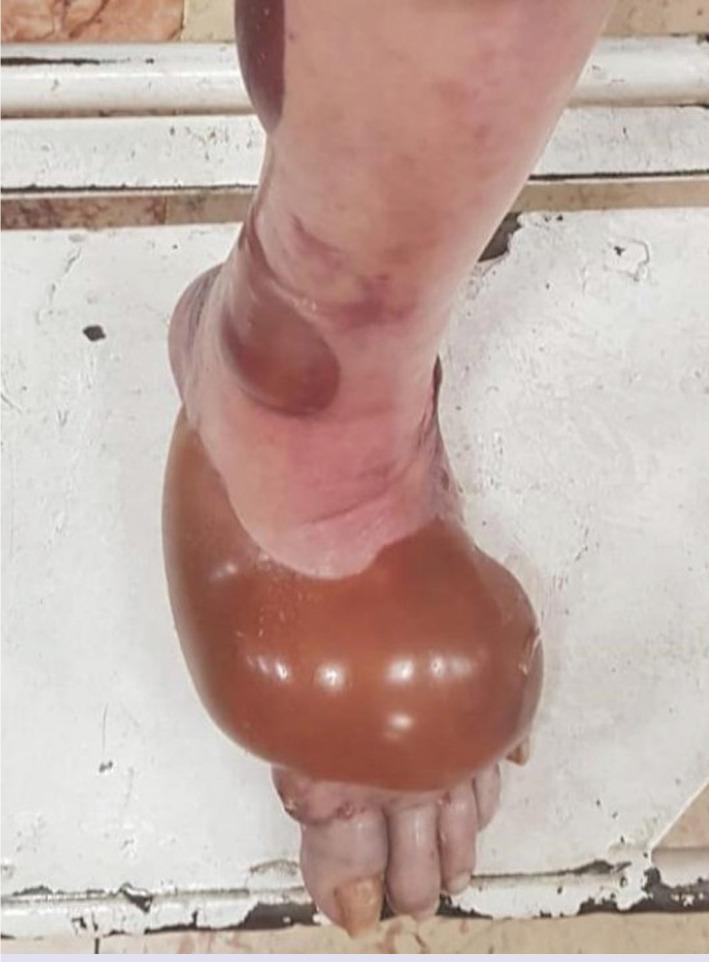
This clinical photograph shows the lower leg and dorsal aspect of the left foot of a 71-year-old patient, demonstrating significant bullous lesions. The primary feature is a large, tense, spherical bulla covering a major portion of the dorsal foot. This lesion is filled with translucent amber or serosanguinous fluid. Secondary, smaller, more irregular bullae with a purplish or hemorrhagic appearance are visible on the lateral and posterior aspect of the distal leg. The surrounding skin exhibits mild erythema and localized edema, particularly at the margins of the largest bulla. The morphology is characteristic of a bullous drug eruption, specifically following methotrexate administration in this clinical context. Educational focus includes the recognition of tense bullae in geriatric patients and the differential diagnosis of drug-induced dermatologic toxicity versus autoimmune blistering diseases like bullous pemphigoid.
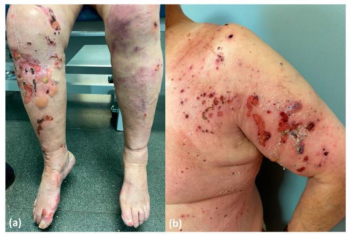
This clinical photograph displays two distinct phenotypes of Bullous Pemphigoid (BP) associated with DPP4 inhibitors (DPP4i). Image (a) represents a non-inflammatory, drug-induced phenotype localized to the lower extremities. The left leg shows large, tense subepidermal blisters (bullae), areas of denuded skin with exposed dermis, and serous weeping, while the right leg exhibits scattered erythematous macules and smaller vesicles. Image (b) demonstrates an inflammatory, drug-triggered phenotype on the right shoulder and upper arm. This presentation is characterized by a polymorphic eruption including widespread erythematous papules, urticarial plaques, and multiple hemorrhagic erosions with significant crusting and exudate. The surrounding skin shows marked inflammation (erythema), typical of the classic BP presentation involving NC16A autoantibodies. These images serve as a comparison of morphological variations in drug-associated autoimmune blistering diseases, highlighting the difference between a primarily bullous, non-inflammatory presentation and an erosive, highly inflammatory cutaneous response.
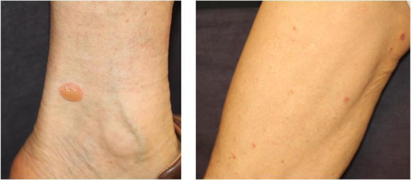
Two clinical photographs demonstrate a bullous pemphigoid eruption on the lower extremities. The left image shows a solitary, tense, fluid-filled bulla located near the medial malleolus of the ankle. The lesion is approximately 1-1.5 cm in diameter, circular, with a translucent, yellowish serous content and a smooth, intact roof. The right image shows multiple, smaller, erythematous papules and early bullae distributed across the leg. In both views, the surrounding skin appears largely non-erythematous and lacks significant peri-lesional inflammation. These findings are characteristic of an autoimmune bullous disease, often presenting as an immune-related adverse event (irAE) following immune checkpoint inhibitor therapy. The clinical significance of these tense bullae lies in their subepidermal origin, which distinguishes them from the flaccid bullae seen in pemphigus vulgaris. This material is suitable for dermatology and oncology educational indexing.
bullous pemphigoid hemorrhagic bulla diagnosis management
Clinical Analysis: Hemorrhagic Bulla → Large Erosive Plaque (20 days)
Image Findings
- Proximal lesion: Large geographic erosive plaque with bright-red/beefy dermal bed, thick black necrotic eschar superiorly, peeling epidermal margins, and surrounding hemosiderin-stained skin
- Distal lesion (near ankle): Smaller elongated erosion with necrotic flaps still partially attached
Differential Diagnosis (Ranked by Likelihood)
1. Bullous Pemphigoid (Hemorrhagic Variant) - Most Likely
- Hemorrhagic bullae as primary lesions
- Conversion to large denuded erosions with epidermal peeling at margins
- Lower leg distribution
- Focal dark necrosis (ischemia within bulla contents)
- Skin biopsy (perilesional skin): H&E shows subepidermal blister with eosinophil-rich infiltrate; no acantholysis
- Direct Immunofluorescence (DIF): Linear IgG ± C3 at the dermoepidermal junction (basement membrane zone) - the gold standard
- Indirect IF / ELISA: Anti-BP180 (BPAG2) antibodies, often >200 U/mL in active disease; anti-BP230
2. Epidermolysis Bullosa Acquisita (EBA) - Important Mimic
3. Pemphigus Vulgaris
4. Calciphylaxis / Ischemic Necrosis
5. Drug-Induced Bullous Eruption
Recommended Workup
| Investigation | Purpose |
|---|---|
| Skin punch biopsy (perilesional) - H&E | Blister level, eosinophils, acantholysis |
| Direct Immunofluorescence (DIF) - perilesional | IgG/C3 pattern (linear vs fishnet vs granular) |
| Indirect IF / ELISA for anti-BP180, anti-BP230 | Quantify BP antibodies |
| Salt-split skin DIF | Distinguish BP (epidermal) from EBA (dermal) |
| CBC, BMP, renal function, Ca×P product | Rule out calciphylaxis |
| Medication review | Identify culprit drugs |
| Wound swab culture | Rule out secondary infection (given necrotic eschar) |
| Nikolsky sign assessment | Positive in PV, negative in BP |
Management (Presumptive BP, Pending Biopsy)
- Wound care: Non-adherent dressings; gentle debridement of necrotic eschar if viable wound bed beneath; avoid traumatizing peri-wound skin
- Topical superpotent corticosteroids (clobetasol 0.05% cream): First-line for localized BP; proven as effective as systemic steroids in moderate BP with fewer systemic side effects
- Systemic corticosteroids (prednisolone 0.5 mg/kg/day): If widespread or rapidly progressive; taper once blisters controlled
- Steroid-sparing agents: Doxycycline 200 mg/day ± nicotinamide (for mild-moderate BP); azathioprine or mycophenolate mofetil for maintenance
- Treat superinfection if wound culture positive
- Correct any contributing drug if drug-induced BP suspected
Key Clinical Pearls
- The progression from hemorrhagic bulla → large erosive plaque over 20 days is classic for BP or EBA, not PV (PV erosions form quickly from fragile flaccid blisters)
- Black eschar in BP represents ischemic necrosis within a hemorrhagic bulla content, not primary infarction - but calciphylaxis must always be excluded
- DIF is mandatory - cannot reliably distinguish BP from EBA, PG, or linear IgA disease on clinical grounds alone
- Untreated BP carries significant morbidity in elderly patients; mortality is associated with both disease severity and treatment toxicity
there is history of eploratory laparotomy for bladder rupture and history of cvp line in this pt followed by this
purpura fulminans DIC skin necrosis hemorrhagic bulla lower limb post surgical
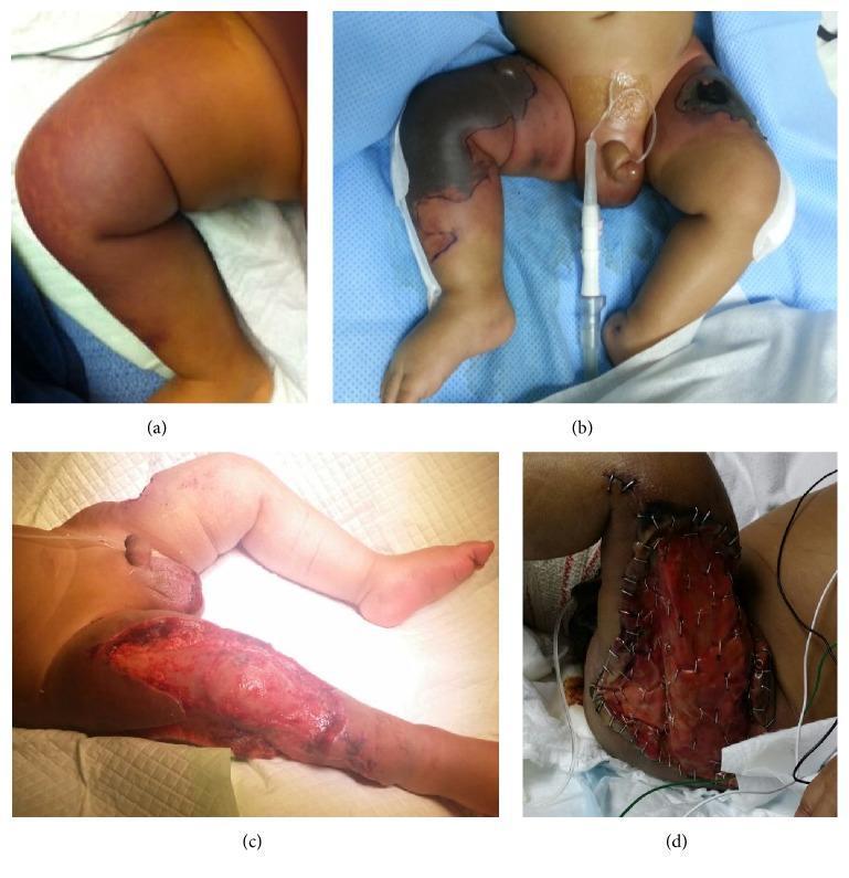
A series of four clinical photographs (a-d) documenting the rapid progression and surgical management of purpura fulminans in an infant. Panel (a) displays early reddish-purple discoloration and induration on the thigh. Panel (b) shows the evolution into extensive hemorrhagic necrosis, characterized by sharply demarcated, blackened eschar on the lower extremities and groin. Panel (c) illustrates the post-operative state following emergency fasciotomy and surgical debridement for compartment syndrome, revealing a deep, open wound with exposed subcutaneous tissues and muscle. Panel (d) demonstrates the application of an artificial dermal regeneration template (Integra) over the debrided area, secured with surgical staples to prepare the wound bed for future grafting. This sequence serves as a clinical teaching tool for recognizing life-threatening thrombotic skin necrosis associated with protein C deficiency and DIC, as well as the multidisciplinary surgical approach involving tissue debridement and dermal substitutes.
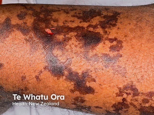
Clinical photography of an affected extremity demonstrates incipient skin necrosis in the setting of purpura fulminans. The image shows mottled, retiform purpuric patches with dark brown to black centralized necrosis against a background of erythematous to coppery skin. Lesions are irregular in shape, coalescing in some areas, with peripheral hemorrhagic crust formation emerging at the lesion margins. The distribution is patchy across the distal leg, consistent with cutaneous microvascular occlusion and ischemic injury. This appearance reflects progression from palpable purpura to early necrosis associated with disseminated intravascular coagulation (DIC) and severe sepsis in PF. Key visual cues include confluent retiform patterning, non-blanching discoloration, and evolving tissue necrosis that risks desquamation and potential gangrene if untreated. The photograph, obtained under standard ambient lighting without dermatoscopic magnification, provides rapid bedside documentation of skin viability and guides urgent clinical decisions such as escalation of antimicrobial therapy, hemodynamic support, and surgical consultation for potential debridement or amputation planning if progression occurs. The image is useful for education on PF cutaneous manifestations, differentiation from leukocytoclastic vasculitis, and radiologic or laboratory correlation in suspected septic DIC. It supports differential diagnoses of vascular occlusive pityriasis vs infection-related necrosis and underscores the urgency of prompt intervention. Urgent multidisciplinary care is essential.
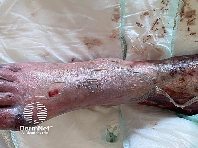
Imaging modality: Clinical photography of the distal lower extremity shows acral skin necrosis with extensive epidermal detachment, consistent with purpura fulminans–associated ischemic skin injury. The lesion spans the dorsal and plantar aspects of the foot and may extend toward the ankle. The skin appears initially erythematous with progressive darkening and thinning, culminating in necrotic eschar formation and superficial slough; there is evident edema and compromised capillary refill. Surrounding regions show purpuric markings and hemorrhagic crusts, indicating microvascular injury. The appearance suggests acute vasculopathy with intravascular coagulation, typical of disseminated intravascular coagulation in severe sepsis or meningococcemia. Clinically this pattern denotes critical illness with high risk of further tissue loss, secondary infection, and systemic shock if not rapidly managed. This image is valuable for dermatology and critical care education, illustrating acral necrosis as a cutaneous marker of systemic coagulopathy. Potential diagnostic considerations include purpura fulminans, necrotizing fasciitis, calciphylaxis, and extensive cutaneous DIC. Management implications center on aggressive hemodynamic support, broad-spectrum antimicrobial therapy, correction of coagulopathy, and consideration of surgical debridement in salvageable tissue. The photograph complements clinical examination and laboratory data in recognizing life-threatening coagulopathy in septic patients. Clinical correlation with laboratory coagulation tests is essential for therapeutic adjustment and prognosis.
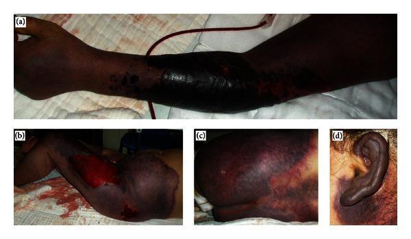
This composite clinical photograph series illustrates extensive, sharply demarcated purpura fulminans across multiple anatomical regions in a patient. (a) The left arm demonstrates a progression from discrete petechial and purpuric spots distally to large, confluent, dark purple and black patches proximally, suggestive of full-thickness skin necrosis and deep tissue ischemia. (b) The left lower limb shows massive, deep violaceous discoloration with a large central ulcerated area exposing subcutaneous tissue, indicating advanced hemorrhagic necrosis. (c) The buttocks present a mottled, retiform pattern of purpuric patches with varying intensities of erythema and cyanosis. (d) The left ear displays diffuse, dark purplish discoloration involving the entire pinna, a classic finding in severe systemic coagulopathies. Collectively, these images illustrate the typical visual manifestations of purpura fulminans, characterized by rapidly progressive, painful, cutaneous hemorrhage and necrosis associated with disseminated intravascular coagulation (DIC). The presentation is critical for educational indexing in dermatology and critical care, focusing on the recognition of life-threatening dermatological emergencies.
Revised Clinical Analysis - Critical Context: Post-Laparotomy + CVP Line
- Exploratory laparotomy for bladder rupture - major abdominal surgery, likely prolonged operative time, supine positioning, urinary extravasation/sepsis risk
- CVP line - indicating critical illness, likely ICU admission, vasopressor use possible
#1 - Surgical/Positional Pressure-Induced Ischemic Bullae (Most Common in This Scenario)
- Tense hemorrhagic bullae at pressure points, appearing within 24-72 hours post-op
- Conversion to large erosive plaques/ulcers over days-weeks
- Distribution exactly matching the anterior and lateral lower leg as seen in the image - consistent with a calf resting on an operating table edge or leg support
- Lower leg bilateral/patchy distribution
- Onset post-operatively
- No systemic coagulopathy needed
- Hemorrhagic nature from dermal capillary rupture under pressure
#2 - Purpura Fulminans / DIC from Post-Surgical Sepsis (Life-threatening - Must Exclude)
#3 - CVP Line-Associated Complications
- Heparin-Induced Thrombocytopenia (HIT): If heparin was used for line flushing/DVT prophylaxis, HIT produces paradoxical venous/arterial thrombosis → skin necrosis appearing 5-14 days after heparin exposure. This timeline (20 days ago onset, matching post-op CVP line period) fits precisely.
- Warfarin/anticoagulant-induced skin necrosis: If anticoagulation was started post-op for DVT prophylaxis
- Septic emboli from infected CVP line (catheter-related bloodstream infection, CRBSI): Septic emboli to skin produce hemorrhagic pustules/bullae/infarcts
Revised Differential Diagnosis Table
| Diagnosis | Supporting Features | Priority |
|---|---|---|
| Pressure/positional ischemic bullae | Post-laparotomy, lower leg distribution, onset within days of surgery | High - Most common |
| Purpura Fulminans / Sepsis-DIC | Bladder rupture → urosepsis, black eschar, large necrotic areas | High - Most dangerous |
| HIT (Heparin-Induced Thrombocytopenia) | CVP line → heparin exposure, 5-14 day onset, thrombotic skin infarcts | High - Often missed |
| Warfarin-induced skin necrosis | If anticoagulation started post-op, protein C depletion, fat-rich areas | Moderate |
| Septic emboli (CRBSI) | CVP line infection, hemorrhagic bullae + systemic sepsis | Moderate |
| Calciphylaxis | If renal failure present (bladder rupture → AKI) | Moderate |
| Autoimmune BP (prior analysis) | Less likely now - surgery/sepsis context dominates | Lower |
Urgent Investigations
For DIC / Purpura Fulminans:
- CBC - thrombocytopenia, fragmented RBCs (schistocytes)
- PT, aPTT - prolonged
- D-dimer - markedly elevated
- Fibrinogen - low (<100 mg/dL in severe DIC)
- Peripheral blood smear - microangiopathic hemolytic anemia
- Blood cultures × 2 (peripheral + from CVP line)
- Protein C and S levels
For HIT:
- Platelet count trend (characteristic fall of >50% from baseline 5-14 days after heparin)
- HIT ELISA (anti-PF4/heparin antibodies)
- 4T score calculation (Thrombocytopenia, Timing, Thrombosis, oTher causes)
- Serotonin release assay (gold standard for HIT confirmation)
For Pressure Injury:
- Clinical diagnosis - wound mapping correlating lesion sites to operative positioning
- No specific labs needed; however rule out DIC/HIT first
Other:
- Wound/skin biopsy with culture (rule out secondary infection, assess necrosis depth)
- Doppler USS lower limbs - rule out DVT (especially if unilateral involvement)
- Urine/wound cultures - identify sepsis source
- LFTs, renal function - organ failure assessment
Management Based on Suspected Etiology
If DIC/Purpura Fulminans confirmed (Sabiston Textbook of Surgery):
- Treat the source: antibiotics targeting gram-negative organisms (piperacillin-tazobactam + vancomycin empirically); source control of any residual septic focus
- FFP for PT/aPTT >2× normal with active bleeding
- Cryoprecipitate for fibrinogen <100 mg/dL
- Platelet transfusion if <50,000/mm³ with bleeding
- LMWH (low-dose) if platelet count >30,000 and no active bleeding - prevents further microvascular thrombosis
- Protein C concentrate or recombinant activated protein C (drotrecogin alfa) in severe cases - Tintinalli's EM, p. 1670
If HIT confirmed:
- STOP all heparin immediately - including flushes, LMWH, heparin-coated catheters
- Switch to argatroban (direct thrombin inhibitor) or fondaparinux
- Do NOT give warfarin until platelet count recovers
- Hematology consult
If Pressure Injury:
- Offload - specialized pressure-redistributing mattress, regular repositioning
- Wound care: Non-adherent dressings (e.g., Mepitel), moist wound healing
- Debridement of necrotic eschar once wound bed assessed
- Nutritional support (critical in post-op patients)
- Plastic surgery/wound care team referral
Bottom Line
Find recent literature on HIT and sepsis-induced skin necrosis