


Differential diagnosis of lower eye lid swelling
lower eyelid swelling infraorbital ecchymosis periorbital edema
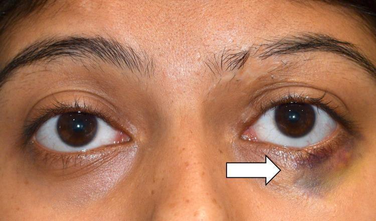
This clinical photograph shows a close-up, frontal view of a patient's periorbital region, demonstrating unilateral ecchymosis of the left lower eyelid. A white arrow points to a localized area of significant skin discoloration characterized by deep purple and blue hues, transitioning into yellowish-green tones at the periphery, indicative of blood extravasation in different stages of breakdown. There is associated mild edema and a palpable nodular mass noted in the clinical history, though only the superficial bruising and slight swelling of the infraorbital soft tissue are visually evident. The right eye and periorbital area appear normal, providing a clear comparison of the pathology. This image is used to illustrate clinical findings associated with orbital trauma or an occult foreign body, emphasizing the importance of detailed physical examination and imaging in cases of localized periorbital bruising.
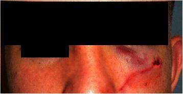
This clinical photograph displays a frontal view of a patient's midface, focusing on the left periorbital and suborbital regions. The image demonstrates significant moderate edema (swelling) and extensive ecchymosis (bruising) localized to the left suborbital area and the lower eyelid, characterized by deep purple and reddish discoloration. A linear incision site is visible at the lateral aspect of the infraorbital rim, secured with medical skin sutures, indicating recent surgical intervention. The surrounding facial skin shows generalized erythema, potentially reflecting inflammatory changes or trauma-induced irritation. The contralateral (right) side of the face appears unremarkable, providing a baseline for assessing the unilateral swelling and facial asymmetry. Clinically, this presentation is consistent with post-traumatic or post-surgical changes following a maxillofacial injury or orbital floor exploration. The visual features highlight key signs of soft tissue trauma and the immediate postoperative state of the orbital frame.
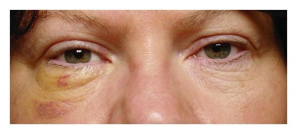
This clinical photograph shows a close-up, frontal view of a 49-year-old female's mid-face, specifically the periorbital region, six days post-orbital surgery. The primary clinical focus is on the postoperative appearance of the left eye compared to the normal right eye. The left infraorbital and periorbital area exhibits significant ecchymosis and edema. A prominent yellowish-green hue (indicative of resolving bilirubin) is visible across the lower eyelid, extending toward the lateral canthus. Within this yellow region, there is a localized reddish-purple area. Inferior to this, on the malar eminence, a larger purplish-red ecchymotic patch suggests deeper tissue involvement or recent hematoma resolution. The left lower eyelid also demonstrates increased skin wrinkling and mild drooping (ectropion or pseudoptosis due to swelling) compared to the contralateral side. The right eye appears healthy with normal skin texture and no discoloration. This image serves as an educational tool for monitoring normal postoperative healing stages, tissue trauma recovery, and the typical color progression of bruising following ophthalmologic or orbital tumor surgery.
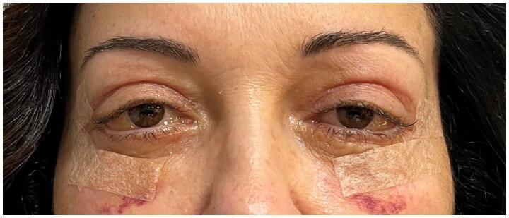
This clinical photograph shows a close-up frontal view of a patient's periorbital region, three weeks following bilateral blepharoplasty. The most prominent finding is severe bilateral conjunctival chemosis, characterized by translucent, bulbous swelling and elevation of the bulbar conjunctiva, making it appear thickened and puffy around the corneas. Visible erythema and vascular congestion are present in the ocular tissue. The periocular area shows typical postoperative changes, including moderate edema of the upper and lower eyelids and mild infraorbital ecchymosis (bruising) on the cheeks. Surgical adhesive tapes are applied to the lower eyelids/cheeks, and fine sutures are visible at the lateral canthi. The lower lids appear slightly retracted, likely due to the ongoing edema and surgical healing process. This image serves as an educational example of persistent postoperative chemosis, a known complication of eyelid surgery that requires clinical monitoring and potential medical or surgical intervention.
differential diagnosis lower eyelid swelling periorbital ecchymosis unilateral causes ophthalmology
Image Analysis - Key Clinical Findings
- Smooth, convex, crescent-shaped infraorbital puffiness spanning the full width of the lower lid
- Overlying hyperpigmentation (dusky brownish discoloration)
- No erythema, discharge, or surface change
- Skin intact, sclera quiet
- Left eye (right side of photo): Moderate lower lid swelling with prominent dark purple-reddish-brown discoloration (ecchymosis) extending into the upper cheek - narrowing of the palpebral fissure
- Right eye (left side of photo): Appears normal/unaffected
Differential Diagnosis of Lower Eyelid Swelling
GROUP 1 - Most Likely in This Case
| Diagnosis | Key Features | How It Fits |
|---|---|---|
| Periorbital ecchymosis ("black eye") | Blunt trauma causing blood vessel disruption; swelling + purple-brown bruising; self-resolving over 2-4 weeks | Strong match - unilateral, ecchymotic discoloration, swelling |
| Orbital fat herniation | Age-related prolapse of orbital fat through the orbital septum; bilateral but asymmetric; painless, chronic | Matches the contralateral eye's chronic puffiness with hyperpigmentation |
| Periorbital/preseptal cellulitis | Bacterial infection anterior to the orbital septum; redness, swelling, warmth, tenderness; no proptosis or restricted EOM | Less likely given the dark bruise color rather than erythema |
GROUP 2 - Inflammatory / Allergic
- Allergic contact dermatitis - reaction to cosmetics, eye drops, or topical agents; bilateral more common; associated with itching
- Angioedema - rapid-onset, non-pitting, painless swelling; often bilateral; can be triggered by ACE inhibitors, NSAIDs, allergens
- Herpes zoster ophthalmicus - vesicular eruption following a dermatome; unilateral; look for Hutchinson's sign
- Insect bite / sting - sudden onset, localized, usually resolves within 24-48 hours
- Orbital inflammatory pseudotumor (Idiopathic Orbital Inflammation) - painful proptosis, restricted EOM, periorbital edema; often unilateral
GROUP 3 - Structural / Glandular
- Chalazion / Meibomian gland cyst - focal, firm nodule in the lower lid; non-tender; may cause diffuse lid swelling if large
- Hordeolum (stye) - acute infection of a lid gland; tender, localized abscess with surrounding edema
- Dacryocystitis - infection of the lacrimal sac at the medial canthus; localized swelling at inner corner
- Dermoid cyst / Epidermoid cyst - smooth, slow-growing subcutaneous mass; often near orbital rim
GROUP 4 - Systemic Causes (usually bilateral)
- Nephrotic syndrome / renal failure - bilateral periorbital puffiness, worse in morning; proteinuria
- Hypothyroidism (myxedema) - non-pitting edema, periorbital puffiness, facial puffiness; bilateral
- Superior vena cava (SVC) syndrome - bilateral facial/periorbital edema with venous engorgement
- Hypoalbuminemia (liver disease, malnutrition) - diffuse dependent edema including periorbital
- Cushing's syndrome - bilateral facial fullness ("moon face") with periorbital fat deposition
GROUP 5 - Vascular / Traumatic
- Subperiosteal hematoma - post-trauma blood collection under the periorbeum; can track down to lower lid
- Ruptured dermoid - sudden inflammation after rupture of a pre-existing cyst
- Orbital floor fracture ("blowout fracture") - periorbital ecchymosis + enophthalmos + diplopia on upward gaze after facial trauma
- Carotid-cavernous fistula - pulsatile proptosis, chemosis, arterialized conjunctival vessels (rare)
GROUP 6 - Neoplastic (rule out)
- Eyelid carcinoma (BCC, SCC) - indurated, ulcerative lid lesion; irregular margins; loss of lashes
- Lymphoma - painless, salmon-pink, "fish-flesh" colored conjunctival or lid mass
- Rhabdomyosarcoma (in children) - rapid-onset proptosis and lid swelling
- Metastatic disease - hard, fixed orbital mass; breast/lung/prostate common primaries
- Neuroblastoma (children) - bilateral periorbital ecchymosis ("raccoon eyes") - a red flag sign
Most Likely Diagnosis for This Patient
- Periorbital ecchymosis from blunt trauma - the purple-brown discoloration and unilateral swelling fit this perfectly
- Preseptal cellulitis - if there is warmth/tenderness and a preceding skin break or sinusitis
- Age-related orbital fat herniation (bilateral, asymmetric) - for the chronic puffiness component
Red Flags Requiring Urgent Ophthalmology Review
- Proptosis (eye pushed forward)
- Restricted or painful eye movements (suggests orbital cellulitis or orbital floor fracture)
- Vision changes - blurring, diplopia, or decreased acuity
- Fever + systemic toxicity (orbital cellulitis / cavernous sinus thrombosis)
- Rapid progression of swelling
- No history of trauma in a child with ecchymosis (consider neuroblastoma or non-accidental injury)