
Endocrine pancreas
Reading File
Now let me search the medical library for comprehensive content on the endocrine pancreas.
Finding Sources
Finding Sources
Excellent sources found. Let me read the key sections in parallel - physiology, histology, and pathology.
Reading File
Reading File
Reading File
Good content so far. Now let me read the physiology detail on insulin and glucagon, and fetch the histology image. Also read the Medical Physiology book for deeper content.
Reading File
Reading File
Loading Image
Loading Image
Excellent content. Now let me get more on insulin actions, glucagon, and read the immunofluorescence image plus more details on insulin mechanism.
Reading File
Loading Image
I now have comprehensive content. Let me read a bit more on insulin actions and somatostatin, plus fetch one more key image (glucagon nutrient flow diagram).
Reading File
Loading Image
I now have thorough content from multiple authoritative textbooks. Let me compile the full comprehensive answer.
The Endocrine Pancreas
Overview
The endocrine pancreas is a diffuse organ scattered within the exocrine gland whose primary function is regulating blood glucose, fatty acid, and amino acid metabolism. It secretes four major hormones: insulin, glucagon, somatostatin, and pancreatic polypeptide (PP).
1. Islets of Langerhans - Structure & Histology
The endocrine cells are organized into clusters called the islets of Langerhans, which make up only 1-2% of total pancreatic mass. There are approximately 1 million islets, each containing about 2,500 cells, most numerous in the tail of the pancreas.
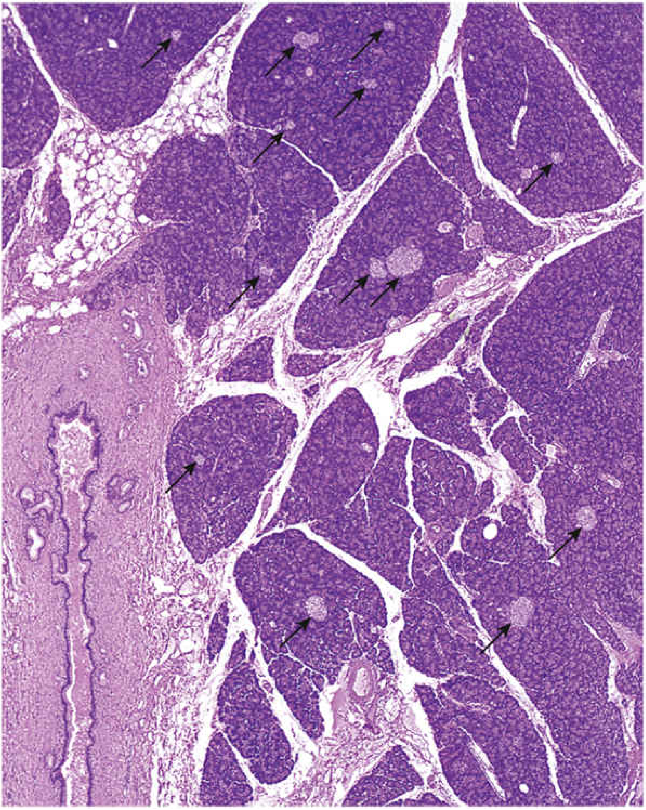
H&E-stained pancreas: islets appear as round, pale-staining profiles (arrows) scattered among more intensely staining exocrine acini. - Histology: A Text and Atlas
Each islet consists of polygonal cells arranged in short, irregular cords, profusely invested with fenestrated capillaries. The cells develop between weeks 9-12 of gestation.
Cell Types
| Cell Type | % of Islet | Hormone | Mallory-Azan Stain | TEM Granules |
|---|---|---|---|---|
| B (β) | 60-70% | Insulin | Brownish orange | ~300 nm; dense, crystalline angular core |
| A (α) | 15-20% | Glucagon | Red | ~250 nm; dense eccentric core, light halo |
| D (δ) | 5-10% | Somatostatin | Blue | ~325 nm; homogeneous matrix |
| PP | ~5% | Pancreatic polypeptide | - | Small, irregular granules |
Spatial arrangement: β cells occupy the central core; α cells line the outer rim; δ cells are interposed between α and β cells, allowing paracrine communication.
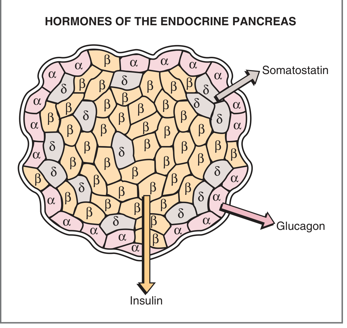
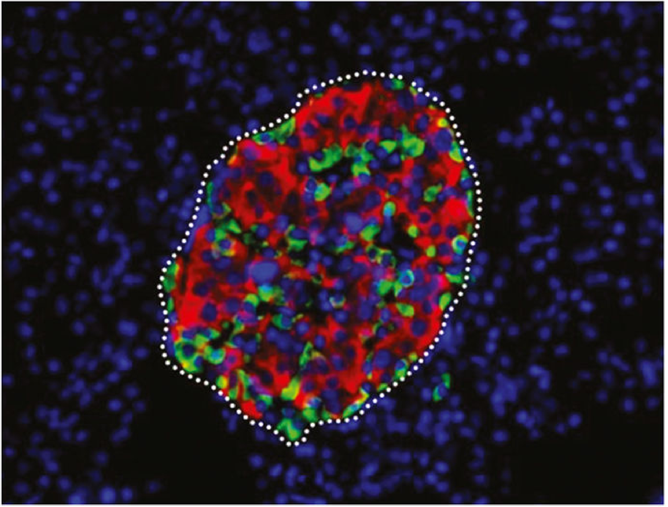
Intra-islet Communication (Paracrine Mechanisms)
There are three communication pathways between islet cells:
- Gap junctions - connect α-to-α, β-to-β, and α-to-β cells for rapid ionic and molecular transfer
- Blood supply - arteries enter the islet core and flow outward; venous blood from β cells (carrying insulin) bathes α and δ cells before leaving the islet
- Innervation - adrenergic, cholinergic, and peptidergic neurons; δ cells have neuron-like processes extending onto β cells
2. Insulin
Synthesis
Insulin is encoded on chromosome 11. The biosynthetic sequence is:
Preproinsulin → (signal peptide cleaved) → Proinsulin → (C-peptide cleaved) → Insulin + C-peptide
- Insulin = two chains: A chain (21 amino acids) + B chain (30 amino acids), linked by disulfide bridges
- Proinsulin folds in the ER; packaged in Golgi secretory granules; proteases cleave C-peptide during packaging
- Insulin and C-peptide are co-secreted in equimolar amounts - C-peptide measurement reflects endogenous β-cell function (useful in insulin-treated patients)
- Metabolized in liver and kidney by disulfide-cleaving enzymes
Regulation of Insulin Secretion
Mechanism (glucose-stimulated):
- Glucose enters β cell via GLUT2 transporter
- Phosphorylated to glucose-6-phosphate by glucokinase (rate-limiting step)
- Oxidative metabolism generates ATP
- ↑ ATP/ADP ratio closes ATP-sensitive K⁺ channels (K_ATP)
- Membrane depolarization opens voltage-gated Ca²⁺ channels
- ↑ Intracellular Ca²⁺ triggers exocytosis of insulin granules
| Stimulatory Factors | Inhibitory Factors |
|---|---|
| ↑ Blood glucose | ↓ Blood glucose / fasting |
| ↑ Amino acids | Somatostatin |
| ↑ Fatty acids & ketoacids | α-Adrenergic agonists |
| Glucagon | Diazoxide |
| Cortisol | Exercise |
| GIP (incretin) | - |
| Vagal stimulation / acetylcholine | - |
| Sulfonylurea drugs (close K_ATP) | - |
| Obesity | - |
Actions of Insulin ("Hormone of the Fed State")
Insulin acts on liver, muscle, and adipose tissue via its tyrosine kinase receptor:
Carbohydrate metabolism:
- ↑ GLUT4 insertion in muscle and adipose → ↑ glucose uptake → ↓ blood glucose
- ↑ Glycogen synthesis (liver and muscle)
- ↓ Glycogenolysis
- ↓ Gluconeogenesis (via ↑ fructose-2,6-bisphosphate → ↑ phosphofructokinase)
Fat metabolism:
- ↑ Fat deposition; ↓ lipolysis in adipose
- ↓ Ketoacid (β-hydroxybutyrate, acetoacetate) formation in liver
Protein metabolism (anabolic):
- ↑ Amino acid uptake into cells; ↑ protein synthesis; ↓ protein degradation
Other:
- ↑ Na⁺-K⁺ ATPase activity → ↑ K⁺ uptake into cells → ↓ serum K⁺
- Growth-promoting effects
3. Glucagon
Synthesis & Secretion
Glucagon is a 29-amino acid peptide secreted by α cells. It is the hormone of starvation/fasting, counter-regulatory to insulin.
| Stimulatory | Inhibitory |
|---|---|
| Fasting / ↓ blood glucose | Insulin |
| ↑ Amino acids (arginine, alanine) | Somatostatin |
| CCK (fat/protein ingestion) | ↑ Fatty acids & ketoacids |
| β-Adrenergic agonists | - |
| Acetylcholine | - |
Key point: Insulin modulates glucagon release - in type 1 DM (no insulin), the glucagon response to hypoglycemia is exaggerated. Amino acids stimulate glucagon (but this effect is blunted by simultaneous glucose - opposite to insulin, which is stimulated by both).
Mechanism of Action
Glucagon binds its receptor → Gs protein → adenylyl cyclase → ↑ cAMP → PKA → phosphorylation of enzymes
Actions of Glucagon (primarily on liver)
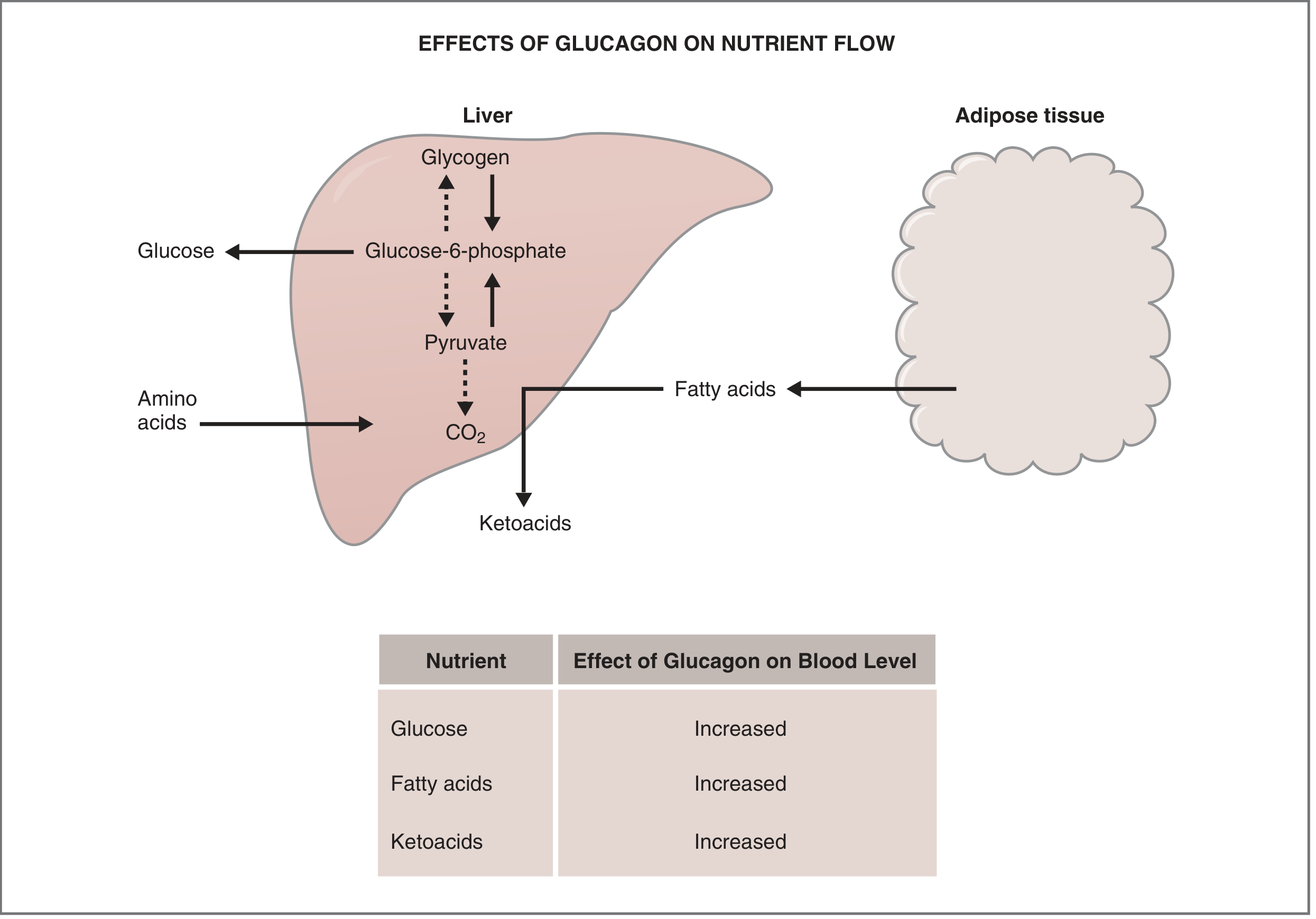
| Action | Effect on Blood Level |
|---|---|
| ↑ Glycogenolysis (liver) | ↑ Glucose |
| ↑ Gluconeogenesis | ↑ Glucose |
| ↑ Lipolysis (adipose) | ↑ Fatty acids |
| ↑ Ketoacid formation | ↑ Ketoacids |
4. Somatostatin (δ cells)
- Suppresses both insulin and glucagon release (paracrine inhibition)
- Has "neuronal" cytology - δ cells send dendrite-like processes onto β cells
- Inhibited by: low blood glucose, certain nutrients
- Also inhibits GI motility and other GI secretions
5. Pancreatic Polypeptide (PP cells, ~5%)
- Stimulates gastric and intestinal enzyme secretion
- Inhibits intestinal motility
- Released after protein meals and fasting
6. Clinical Relevance
Diabetes Mellitus (most important endocrine pancreas disease)
A group of metabolic disorders characterized by hyperglycemia due to defects in insulin secretion, insulin action, or both.
ADA/WHO Diagnostic Criteria (any one, confirmed on separate day):
- Fasting plasma glucose ≥ 126 mg/dL
- Random plasma glucose ≥ 200 mg/dL (with symptoms)
- 2-hour glucose ≥ 200 mg/dL on 75 g OGTT
- HbA1c ≥ 6.5%
Prediabetes: FPG 100-125 mg/dL or HbA1c 5.7-6.4%
| Type 1 DM | Type 2 DM | |
|---|---|---|
| Mechanism | Autoimmune β-cell destruction | Insulin resistance + relative β-cell failure |
| Insulin | Absent | Relative deficiency |
| Prevalence | Minority | Vast majority (>90%) |
| Key feature | Prone to DKA | Prone to hyperosmolar state |
Chronic hyperglycemia causes damage to kidneys (leading cause of ESRD), eyes (leading cause of adult-onset blindness), nerves, and blood vessels. In the US, diabetes affects >37 million people (~11% of population).
Sources:
- Costanzo Physiology, 7th Edition, pp. 444-452
- Histology: A Text and Atlas with Correlated Cell and Molecular Biology, pp. 1718-1722
- Robbins & Kumar Basic Pathology (Robbins Pathology), pp. 742-743
This is a shared conversation. Sign in to Orris to start your own chat.