
Ronchi
What aspect of Ronchi are you asking about?
Explain
What are you asking about?
rhonchi lung auscultation breath sounds diagram
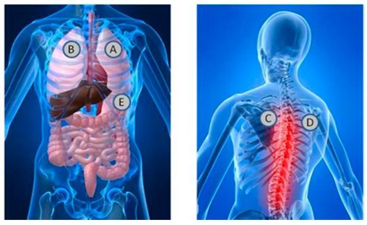
This medical anatomical diagram illustrates the standard stethoscope placement locations for auscultation of heart and lung sounds. The image consists of two panels: an anterior (front) view and a posterior (back) view of a semi-transparent human torso. In the anterior view, labels A and B are positioned over the apical regions of the right and left lungs, respectively, while label E is located at the lower left costal margin, corresponding to the cardiac apex and mitral valve area. Visible internal structures include the lungs, heart, liver, and intestinal tract. In the posterior view, labels C and D are placed in the interscapular region at the level of the upper thoracic spine, typically used for assessing posterior bronchial and vesicular breath sounds. This view highlights the vertebral column, ribs, and scapulae, with a red glow emphasizing the spinal region. This illustration serves as an educational guide for clinical physical examination techniques, demonstrating the relationship between surface landmarks and underlying thoracic and abdominal viscera.
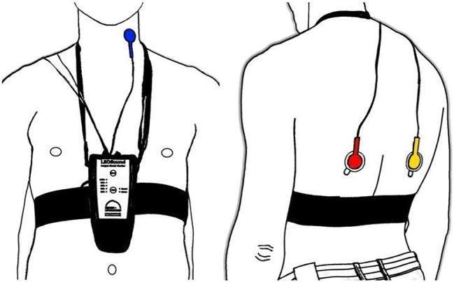
An anatomical diagram illustrating the placement of the LEOSound lung-sound monitor and its bio-acoustical sensors on a patient. The illustration includes two views: an anterior (front) view and a posterior (back) view. In the anterior view, a recording device is secured to the mid-thorax by a black chest belt and a neck strap. A blue tracheal microphone is positioned on the neck near the trachea. In the posterior view, two additional microphones are attached using adhesive pads to the inferior region of the left (red microphone) and right (yellow microphone) scapula. The system is designed for mobile, long-term recording and automated analysis of respiratory sounds such as coughing and wheezing. The diagrams emphasize the standardized anatomical landmarks required for optimal auscultation and data collection in clinical or home settings.
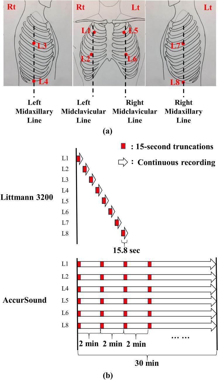
This educational graphic illustrates standardized auscultation locations and lung sound recording protocols. Panel (a) presents anatomical diagrams of the thorax showing eight specific auscultation points (L1–L8). Right-side locations include the second intercostal space (ICS) on the midclavicular line (L1), the fifth ICS on the midclavicular line (L2), the fourth ICS on the midaxillary line (L3), and the tenth ICS on the midaxillary line (L4). Left-side locations (L5–L8) mirror these positions. Panel (b) compares two digital recording methodologies. The Littmann 3200 protocol demonstrates a sequential approach, capturing 15.8-second recordings from L1 through L8 one by one. In contrast, the AccurSound (HF-Type-1) protocol depicts a simultaneous multi-channel approach, recording from six locations (L1, L2, L4, L5, L6, L8) concurrently for a continuous 30-minute duration. The diagram highlights how these recordings are truncated into 15-second segments for analysis. This visual is designed to explain standardized pulmonary physical examination techniques and data acquisition for digital stethoscopes.
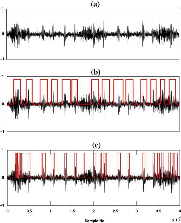
A comparison chart consisting of three time-domain waveforms (a, b, and c) illustrating heart sound (HS) localization within real recorded high-flow lung sound (LS) data. Subfigure (a) presents the raw acoustic signal, showing amplitude fluctuations with periodic bursts corresponding to mechanical cardiac events obscured by respiratory noise. Subfigures (b) and (c) demonstrate the performance of two different computational methods for identifying cardiac segments. The black waveforms represent the sound signal, while the overlaid red rectangular pulses indicate the identified temporal boundaries of heart sound components. Subfigure (b) shows the output of the HHA method, characterized by wider, continuous red blocks that capture the duration of heart sound complexes. Subfigure (c) displays the result of the SSA method, showing fragmented, narrower red pulses that often miss segments or produce discontinuous localization. This clinical signal processing diagram is used in cardiology and pulmonology research to evaluate algorithms for separating cardiac and respiratory sounds in auscultation data.
Rhonchi (Ronchi)
What They Are
- Murray & Nadel's Textbook of Respiratory Medicine, p. 3711
How They Are Produced
- Rupture of fluid films within the airways
- Vibration of the airway walls - caused by secretions or partial obstruction of medium-sized bronchi
- Fishman's Pulmonary Diseases and Disorders; Murray & Nadel's, p. 3711
Key Clinical Feature: Clearance with Cough
Classification of Breath Sounds (Where Rhonchi Fit)
| Sound | Pitch | Duration | Character |
|---|---|---|---|
| Rhonchi | Low (<200 Hz) | Continuous (>250 ms) | Gurgling/snoring quality |
| Wheezes | High (>400 Hz) | Continuous | Musical/whistling |
| Stridor | High | Continuous, inspiratory | Focal, heard over neck |
| Crackles (Rales) | Varied | Discontinuous | Popping/crackling |
Causes and Clinical Associations
- Viral or bacterial bronchitis
- Aspiration (with feeding difficulties)
- Excessive secretions (indicating need for suctioning in ventilated patients)
- Bronchiectasis - pooling of secretions in dilated bronchi
- COPD - chronic airflow obstruction with secretions
- Chronic bronchitis
- Harrison's Principles of Internal Medicine 22E, p. 1590-1592
- Mulholland and Greenfield's Surgery, p. 3816
Rhonchi vs. Wheezes - Key Distinction
| Feature | Rhonchi | Wheezes |
|---|---|---|
| Pitch | Low (<200 Hz) | High (>400 Hz) |
| Mechanism | Secretions / fluid films in medium airways | Airway wall flutter from narrowing |
| Clears with cough? | Yes | No |
| Typical cause | Bronchitis, COPD, bronchiectasis | Asthma, CHF, airway obstruction |
Clinical Significance
- Rhonchi in the context of COPD/chronic bronchitis indicate increased mucus burden and may signal an acute exacerbation.
- Their presence in a patient with worsening dyspnea and decreased oxygenation is an indication for airway suctioning in ventilated patients.
- In a patient with right atrial abnormality on ECG, rhonchi alongside prolonged expiration and hyper-resonance point toward underlying chronic lung disease (COPD, occupational lung disease).
Summary
What are crackles and how do they differ from rhonchi?
Crackles - Definition and Mechanism
- Murray & Nadel's Textbook of Respiratory Medicine, p. 3697
Fine vs. Coarse Crackles
| Feature | Fine Crackles | Coarse Crackles |
|---|---|---|
| Pitch | Higher | Lower |
| Duration | Shorter | Longer |
| Intensity | Softer | Louder |
| Timing | Late inspiration | Early/throughout inspiration |
| Transmitted to mouth? | No | Yes |
| Clear with coughing? | Yes (if postural) | Rarely disappear entirely |
| Causes | Heart failure, interstitial fibrosis (IPF), pulmonary edema | Bronchitis, COPD, secretions |
- Murray & Nadel's, p. 3698-3699; Fishman's Pulmonary Diseases, p. 3485
Causes of Crackles
| Condition | Type of Crackle |
|---|---|
| Heart failure (pulmonary edema) | Fine, bilateral basal |
| Interstitial lung disease / IPF | Fine, late inspiratory ("Velcro") |
| Pneumonia | Fine or coarse (alveolar filling) |
| Bronchitis / COPD | Coarse (secretions in airways) |
| Atelectasis (bedridden) | Fine, clear with deep breaths |
| Emphysema | Altered elastic recoil crackles |
- Goldman-Cecil Medicine, p. 301; Fishman's, p. 3485; Harrison's 22E, p. 1594
Crackles vs. Rhonchi - Side-by-Side Comparison
| Feature | Crackles | Rhonchi |
|---|---|---|
| Sound quality | Non-musical, explosive pops | Musical, low-pitched, continuous |
| Duration | Discontinuous (<250 ms each) | Continuous (>250 ms) |
| Frequency | Varied (fine = high; coarse = low) | Low (<200 Hz) |
| Mechanism | Explosive opening of collapsed small airways | Secretions/fluid films vibrating in medium-sized airways |
| Phase | Predominantly inspiratory | Both inspiratory and expiratory |
| Clears with cough? | Fine: yes (transiently); Coarse: rarely | Yes - a hallmark feature |
| Typical diseases | Heart failure, ILD, pneumonia (fine); Bronchitis, COPD (coarse) | Bronchitis, COPD, bronchiectasis |
| Airways involved | Small airways and alveoli | Medium-sized bronchi |
- Crackles = brief pops (think of individual bubbles bursting)
- Rhonchi = sustained, low rumbling sound (think of air moving through a mucus-coated tube)
- Goldman-Cecil Medicine, p. 301; Murray & Nadel's, p. 3689
Quick Reference: All Adventitious Sounds
| Sound | Type | Pitch | Cause | Clears with cough? |
|---|---|---|---|---|
| Fine crackles | Discontinuous | High | ILD, heart failure, edema | Sometimes |
| Coarse crackles | Discontinuous | Low | Bronchitis, COPD, secretions | Rarely |
| Rhonchi | Continuous | Low (<200 Hz) | Secretions in medium airways | Yes |
| Wheezes | Continuous | High (>400 Hz) | Airway wall flutter (asthma, CHF) | No |
| Stridor | Continuous | High | Upper airway obstruction | No |
| Pleural rub | Grating | - | Pleural inflammation | No |