
Hemangioma of left oral mucosa
Reading File
Now let me search the medical library for hemangioma of the oral mucosa.
Finding Sources
Finding Sources
Finding Sources
Finding Sources
Let me read the most relevant sections - Cummings Otolaryngology (oral/head and neck) and check for oral cavity-specific content on hemangioma.
Reading File
Reading File
Finding Sources
I have excellent content from Cummings Otolaryngology. Let me now search for dermatology/pathology content on hemangioma types, and also fetch the image from the block19 content.
Finding Sources
Finding Sources
Finding Sources
Searching PubMed
hemangioma oral mucosa treatment
Let me read the Cummings salivary gland hemangioma section and also fetch any relevant images.
Reading File
Searching PubMed
oral hemangioma vascular malformation management
Now let me fetch the relevant histology image and also search for a clinical image of oral hemangioma.
Loading Image
Searching the Web
hemangioma oral mucosa clinical features treatment 2024
I now have comprehensive information to provide a thorough answer. Let me compile everything.
Hemangioma of the Left Oral Mucosa
Definition & Overview
A hemangioma of the oral mucosa is a benign vascular neoplasm characterized by abnormal proliferation of blood vessels lined by endothelial cells. It is the most common benign vascular tumor of the head and neck region. Oral cavity hemangiomas account for approximately 14% of all hemangiomas of the head and neck.
- Cummings Otolaryngology Head and Neck Surgery, Ch. 90
Classification
Hemangiomas are classified under the ISSVA (International Society for the Study of Vascular Anomalies) framework, distinguishing them from true vascular malformations:
| Type | Features |
|---|---|
| Infantile hemangioma | Absent/small at birth, rapid proliferative phase (age 1-6 months), then involutes over 1-12 years |
| Congenital hemangioma | Fully formed at birth; includes RICH (rapidly involuting), NICH (non-involuting), and PICH (partially involuting) |
| Capillary hemangioma | Small, thin-walled capillaries; common superficial type |
| Cavernous hemangioma | Large, dilated vascular channels; deeper, more common in adults |
Epidemiology & Site Predilection
- Infantile hemangiomas occur in up to 10% of all births; female predominance
- The lip is the most frequent site of hemangioma in the oral cavity, followed by tongue, buccal mucosa, and floor of mouth
- Usually present at birth or become evident in the first few weeks/months of life; can occasionally become evident later
Associated Syndromes
Oral/head-neck hemangiomas may be associated with:
- Sturge-Weber-Dimitri syndrome (facial port-wine stain, leptomeningeal angiomatosis, glaucoma)
- Von Hippel-Lindau syndrome (hemangioblastomas + visceral lesions)
- Multiple cutaneous lesions are not uncommon alongside parotid/salivary gland hemangiomas
Clinical Features
| Feature | Description |
|---|---|
| Appearance | Soft, painless mass; red or blue in color (blanches with pressure/diascopy) |
| Size | Typically < 2 cm; can become extensive, involving the tongue or oropharynx |
| Consistency | Soft, compressible |
| Symptoms | Usually asymptomatic; large lesions can cause functional problems (speech, swallowing, aesthetics) |
| Behavior | Tend to spontaneously regress over years; involution may be incomplete; fibrosis can develop |
Histopathology
Microscopically, hemangiomas comprise capillaries lined by proliferative endothelial cells with uniform vessel size. Mitoses are frequent but are not indicators of malignant behavior.
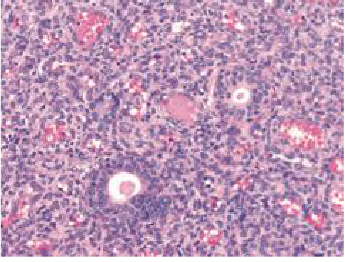
Histology: Juvenile lobular hemangioma showing dense vascular proliferation with proliferative endothelial cells - Cummings Otolaryngology
Diagnosis
- Clinical diagnosis in most straightforward cases (blanching on pressure is pathognomonic)
- MRI is the imaging modality of choice to determine extent - important before treatment, as extensive spread may not be evident clinically
- Biopsy if diagnosis is in doubt (risk of bleeding; use with caution)
- Distinguish from vascular malformations (low-flow vs. high-flow), pyogenic granuloma, and lymphangioma
Treatment
The treatment approach depends on lesion behavior (proliferating vs. involuting) and functional impact:
Conservative / Observation
- The majority will involute spontaneously over time; watchful waiting is appropriate for small, asymptomatic lesions
- Indicated for non-problematic infantile hemangiomas in proliferative or involuting phases
Medical Therapy
| Agent | Notes |
|---|---|
| Propranolol (beta-blocker) | First-line for infantile hemangiomas; dose 2-3 mg/kg/day in 3 divided doses; mechanism - inhibits proangiogenic factor secretion and endothelial growth; rapid and excellent results |
| Corticosteroids (local and systemic) | Older mainstay; both local injection and systemic use described |
| Interferon | Used for refractory cases; systemic side effects limit use |
| Vincristine | Used in refractory/large lesions |
Surgical / Invasive Therapy
| Modality | Notes |
|---|---|
| Conservative surgical excision | For lesions limiting oral form and function; recurrence/persistence is not unusual |
| Sclerotherapy (intralesional sclerosing agents) | Effective for localized lesions; agents include sodium tetradecyl sulfate, bleomycin |
| Laser therapy | CO2, Nd:YAG, KTP lasers used; good hemostasis; useful for mucosal lesions |
| Embolization | For high-flow or extensive lesions; often adjunctive |
| Radiation | Rarely used; adjunctive in refractory cases |
For adults with cavernous hemangiomas (the common adult type in salivary glands), surgical excision is the primary treatment.
Key Points for Left Oral Mucosa Hemangioma
- Most are benign with favorable natural history - spontaneous involution is common
- Lip is the most common site, but buccal mucosa, tongue, and floor of mouth are also frequently involved
- Get MRI to define extent before any intervention
- For infantile/pediatric: propranolol is now first-line medical therapy
- Surgical excision is reserved for lesions that are functionally limiting or fail to involute
- Recurrence after excision is known - counsel the patient
- Always differentiate from vascular malformation (malformations do not involute, require different management)
Sources:
- Cummings Otolaryngology Head and Neck Surgery, Ch. 90 (Oral Cavity Tumors) and Ch. 84 (Salivary Gland Tumors)
- Medical management of vascular anomalies - J Oral Pathol Med 2022 (PMID 35668029)
- StatPearls: Oral Hemangiomas - Lyssy et al., 2023

12013.jpg
Diagnosisi dentigerous cyst or okc
analyse_media
Reading File
Finding Sources
Finding Sources
Finding Sources
Finding Sources
Reading File
Finding Sources
Finding Sources
Reading File
Reading File
Reading File
Loading Image
Now I have all the information needed. The textbook OKC/KCOT image above matches very closely with the submitted OPG. Let me compile the full analysis.
OPG Analysis: Dentigerous Cyst vs. OKC/KCOT
Radiographic Findings on Your OPG
| Feature | Observation |
|---|---|
| Location | Left posterior mandible - angle and ascending ramus region |
| Associated tooth | Impacted left mandibular third molar (#38), displaced inferiorly |
| Shape | Unilocular radiolucency, with a scalloped superior border |
| Borders | Well-defined, well-corticated, radiopaque rim |
| Internal structure | Homogeneous radiolucency - no septations or calcifications |
| Size/extension | Moderately large; extends significantly superiorly into the ascending ramus |
| Growth direction | Anteroposterior - runs along the medullary canal into the ramus with minimal obvious cortical ballooning |
| Cortical expansion | Minimal to mild despite the lesion's size - no gross buccal/lingual ballooning visible |
| Adjacent teeth | #37 shows slight displacement; no aggressive root resorption |
| Contralateral side | Right third molar has a normal, thin follicular space |
Differential Diagnosis: Key Distinguishing Features
| Radiographic Feature | Dentigerous Cyst | OKC / KCOT | Your OPG |
|---|---|---|---|
| Attachment to tooth | Strictly at the CEJ | May extend apical to CEJ; tooth "suspended" in a larger lumen | Crown enclosed, but lesion extends well beyond CEJ into ramus |
| Shape of borders | Round, smooth, oval | Often scalloped | Scalloped - favors OKC |
| Growth direction | Concentric hydraulic expansion | Anteroposteriorly along medullary canal | AP extension into ramus - favors OKC |
| Cortical expansion | Noticeable, concentric ballooning | Minimal expansion relative to size | Minimal expansion for the lesion size - favors OKC |
| Size | Usually moderate | Can become very large with minimal symptoms | Large extension into ramus - favors OKC |
| Internal pattern | Unilocular (usually) | Unilocular or multilocular | Unilocular (compatible with both) |
| Inferior border | Usually convex downward | May show bowing of inferior border | Possible inferior bowing (textbook sign of KCOT) |
Textbook Reference: OKC/KCOT Radiograph
This is the textbook OPG of a KCOT from Cummings Otolaryngology, showing the classic pattern of bowing of the inferior border and scalloping - note the similarity to your case:
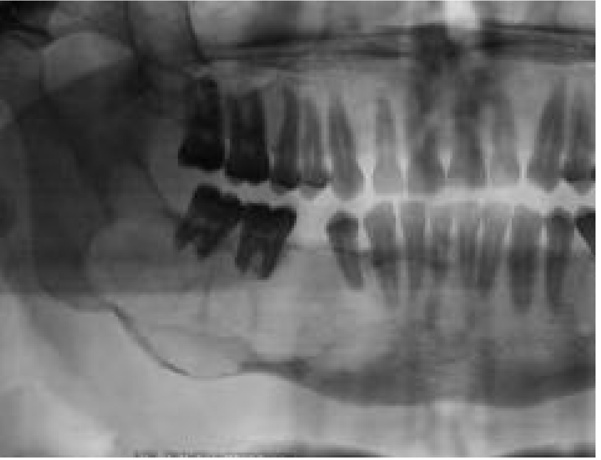
Radiological Verdict
The features on this OPG are more consistent with OKC/KCOT than a simple dentigerous cyst.
The three most discriminating findings pointing toward OKC/KCOT are:
- Scalloped borders - not typical of a dentigerous cyst
- Anteroposterior extension into the ramus with minimal cortical expansion (OKC "creeps" along medullary bone)
- Disproportionately large lesion for a pericoronal cyst in a young patient
That said, both lesions are pericoronal and can mimic each other. Unicystic ameloblastoma is also in the differential.
What to Do Next
| Step | Rationale |
|---|---|
| CBCT (Cone Beam CT) | Assess buccolingual cortical integrity; perforation would strongly favor OKC/KCOT or ameloblastoma |
| Biopsy / Histopathology | Definitive diagnosis - OKC shows parakeratinized stratified squamous epithelium, uniform 4-10 cells thick, palisaded basal cells with "tombstone" appearance; dentigerous cyst has non-keratinized lining |
| Screen for Gorlin syndrome | Multiple OKC/KCOTs + rib anomalies + calcified falx cerebri = Basal Cell Nevus Syndrome |
Why It Matters Clinically
| Dentigerous Cyst | OKC / KCOT | |
|---|---|---|
| Treatment | Enucleation ± marsupialization | Enucleation + peripheral osteotomy + Carnoy's solution chemical curettage |
| Recurrence | Low | Up to 62.5% with simple enucleation; modern rates <10% with thorough curettage |
| Malignant potential | Rare | Rare but documented; aggressive local behavior |
| Follow-up | Standard | Long-term radiographic surveillance required; recurrences can appear 40 years later |
Bottom line: Histopathology is mandatory before finalizing the diagnosis. Do not manage this as a simple dentigerous cyst without tissue confirmation, as the treatment protocols differ significantly.
- Cummings Otolaryngology Head and Neck Surgery, Ch. 88
- Scott-Brown's Otorhinolaryngology Head & Neck Surgery, Ch. on Jaw Cysts
Localized aggressive periodontists
Finding Sources
Finding Sources
Reading File
Finding Sources
Finding Sources
Finding Sources
Searching PubMed
localized aggressive periodontitis treatment management
Finding Sources
Searching PubMed
aggressive periodontitis 2018 classification molar incisor
Reading File
Searching the Web
localized aggressive periodontitis 2018 classification Grade C Stage III treatment guidelines
I now have all the information needed for a comprehensive answer. Let me compile everything.
Localized Aggressive Periodontitis (LAP)
Current Classification - Important Update
The term "Localized Aggressive Periodontitis" (LAP) was retired by the 2018 AAP/EFP World Workshop Classification. It is now classified as: "Localized Stage III, Grade C Periodontitis - Molar/Incisor Pattern"
This change reflects that the old "chronic vs. aggressive" distinction was unreliable (you couldn't determine rate of progression on a first visit). The new system uses Stage (severity) and Grade (risk of progression) instead.
Definition (Classic Concept)
LAP is a rapidly progressive, highly destructive form of periodontitis occurring predominantly in otherwise systemically healthy adolescents and young adults, characterized by severe alveolar bone loss localized to the first molars and incisors, with minimal involvement of other teeth.
Epidemiology
| Feature | Details |
|---|---|
| Age | Adolescents (puberty) to young adults (~10-30 years) |
| Sex | Female > male predominance (some studies) |
| Race | Higher prevalence in individuals of African descent |
| Prevalence | ~0.1-0.2% of the general population; higher in some African populations (~2%) |
| Systemic health | Patient is otherwise healthy |
Etiology & Pathogenesis
LAP is driven by a combination of microbial and host factors:
Key Microorganism
Aggregatibacter actinomycetemcomitans (Aa) - formerly called Actinobacillus actinomycetemcomitans
- Small, capnophilic (CO₂-requiring) Gram-negative rod
- Highly virulent strains produce a leukotoxin that destroys neutrophils and monocytes
- JP2 clone of Aa (prevalent in African-descent populations) produces 10-20x more leukotoxin - strongly associated with LAP
- Other organisms: Porphyromonas gingivalis, Tannerella forsythia, Treponema denticola
Host Defects
- Impaired neutrophil chemotaxis - neutrophils fail to migrate normally to the periodontal tissues
- Reduced neutrophil phagocytic capacity
- Elevated prostaglandin E2 and IL-1β production - drives bone resorption
- Strong genetic predisposition - familial clustering is common
Classic Clinical Features
| Feature | Description |
|---|---|
| Pattern | Bone loss localized to permanent first molars and incisors - often symmetrical ("arc-shaped" bone loss) |
| Amount of plaque | Disproportionately little plaque relative to the severity of destruction |
| Gingival appearance | May look nearly normal clinically despite deep pockets |
| Pocket depth | Deep periodontal pockets (≥6 mm) at affected sites |
| Bone loss | Rapid and severe - can lose 3-4 mm of attachment per year (vs. ~0.1 mm/year in chronic periodontitis) |
| Mobility | Affected teeth may show pathological mobility |
| Systemic | No systemic disease |
| Family history | Positive family history in many cases |
Radiographic Features
- Arc-shaped (angular) bone loss around first molars - vertical bone defects on mesial and distal of first molars
- "Mirror image" pattern - bilateral and often symmetric
- Bone loss extending from the cervical third to the middle/apical third of roots
- Incisors show crestal bone loss
- Normal bone levels around other teeth (distinguishes from generalized disease)
Diagnosis (2018 Classification Framework)
| Criterion | Finding in LAP / Grade C |
|---|---|
| Extent | Localized (<30% of teeth) OR Molar/incisor pattern |
| Stage | Stage III (CAL ≥5 mm, RBL to middle third of root, probing depths ≥6 mm) |
| Grade C | Bone loss/age ratio >1.0; or evidence of rapid progression; or JP2 Aa clone; or associated risk factors |
| Primary evidence of Grade C | % bone loss ÷ age >1.0 (e.g., 30% bone loss at age 25 = 1.2 → Grade C) |
Treatment
Treatment is staged following the EFP/AAP Step-wise approach:
Step 1 - Risk Factor Control / Patient Education
- Oral hygiene instruction
- Smoking cessation
- Dietary counseling
- Systemic disease control if applicable
Step 2 - Cause-Related / Non-Surgical Therapy
- Scaling and root planing (SRP) - full-mouth within 24-48 hours ("full-mouth disinfection")
- Systemic antibiotics - critical in LAP because Aa invades tissue and SRP alone cannot eradicate it:
- Amoxicillin 500 mg + Metronidazole 500 mg, TID × 7-14 days - most evidence-based combination
- Doxycycline 100 mg OD × 14-21 days (alternative, especially if penicillin-allergic)
- Antibiotics should be given adjunctively with SRP, not alone
Step 3 - Surgical Therapy (if needed after reassessment ~8-12 weeks)
- Flap surgery for residual pockets ≥6 mm
- Osseous surgery for intrabony defects
- Regenerative procedures - guided tissue regeneration (GTR), bone grafts for angular defects at first molars (LAP tends to respond well to regeneration due to young age and good healing potential)
Step 4 - Supportive Periodontal Therapy (Maintenance)
- 3-monthly recall for at least 1 year
- Monitor for disease recurrence and tooth loss
- Long-term maintenance is essential - recurrence rate is high
Systemic Associations to Screen For
When LAP is diagnosed, especially in young patients, rule out:
- Papillon-Lefèvre syndrome (palmar-plantar hyperkeratosis + severe periodontitis; cathepsin C mutation)
- Chediak-Higashi syndrome (neutrophil granule defect)
- Leukocyte adhesion deficiency (LAD)
- Cyclic neutropenia
- Hypophosphatasia (premature loss of deciduous teeth)
- Down syndrome (trisomy 21)
Prognosis
- Good with early, aggressive treatment - young age means excellent regenerative capacity
- Poor without treatment - can progress to generalized disease
- First molars are most frequently lost if untreated
- Antibiotic adjunctive therapy significantly improves outcomes compared to SRP alone
Sources:
- Sherris & Ryan's Medical Microbiology, 8th ed. - Ch. 41
- Harrison's Principles of Internal Medicine, 22nd ed. (2025) - Ch. on Oral Diseases
- 2018 AAP/EFP Staging and Grading of Periodontitis - Tonetti et al., 2018 (PMID 29926952)
- Grade C Periodontitis in Young Adults - Oral Dis 2026 - Hamdoun et al. (PMID 41841411)

12014.jpg
This is LAP with respect to 37/47
analyse_media
Excellent radiographic analysis. Here is the full interpretation:
OPG Analysis - Bishnu Psd Neupane, 36Y, Male
Stated Diagnosis: LAP involving 37 and 47
Radiographic Findings
Tooth 47 (Lower Right Second Molar)
- Severe vertical (angular) bone loss - predominantly on the mesial aspect
- Bone loss extends to the apical third / near root apex (>50% root length loss)
- Radiographic furcation involvement (bifurcation radiolucency) - Class II/III
- Distal aspect also shows vertical loss reaching middle third of root
- Appears to have a deep restoration or carious lesion in the crown
- Prognosis: Poor to Hopeless without intervention; high risk of tooth loss
Tooth 37 (Lower Left Second Molar)
- Vertical (angular) bone loss on the mesial aspect
- Extends to the middle third of root (~33-50% root length)
- Possible early furcation involvement
- Distal bone level relatively preserved
- Prognosis: Guarded to Poor - Stage III, Grade C
Rest of Dentition
| Region | Finding |
|---|---|
| Lower/upper incisors | Normal bone levels - no bone loss |
| First molars (16, 26, 36, 46) | Largely intact; mild distal bone loss on 36/46 contiguous with the 37/47 defects only |
| Premolars, canines | Normal bone levels throughout |
| Anterior segments | Alveolar crests high, intact, sharp lamina dura visible |
Critical Diagnostic Discussion
While the clinician has labeled this as LAP, the radiographic pattern raises an important clinical question:
The bone loss is confined to second molars (37/47), NOT the classic first molar/incisor pattern of LAP.
| Feature | Classic LAP | This Case |
|---|---|---|
| Primary sites | First molars + incisors (bilateral) | Second molars only (37/47) |
| Incisors | Always involved | Not involved |
| First molars (36/46) | Severely affected | Relatively spared |
| Age | Adolescent onset | 36 years old |
| Pattern | Arc-shaped / vertical around 6s and 1s | Vertical, mesial 7s |
| Symmetry | Yes - bilateral | Yes - bilateral |
Alternative Diagnoses to Consider
1. Retrograde Endo-Periodontal (Endo-Perio) Lesion - Tooth 47
- Tooth 47 shows a deep restoration/carious lesion suggesting possible pulpal necrosis
- A primary endodontic infection can drain through the furcation or lateral canals, creating a radiolucency mimicking periodontal bone loss
- The furcation involvement on 47 may be endodontic in origin
- Do: Pulp vitality test on 47 - if non-vital, treat endodontically first before periodontal surgery
2. History of Mesioangularly Impacted Third Molars (38/48)
- The third molars are absent on this OPG
- Mesioangularly impacted wisdom teeth are the most common cause of isolated deep mesial vertical defects on second molars
- If 38/48 were extracted late, they often leave a non-healing deep periodontal pocket and vertical bone defect on the mesial of the adjacent second molar
- Ask the patient: When were wisdom teeth extracted? Were there pericoronal infections?
3. Localized Grade C Periodontitis - Second Molar Pattern
- Under the 2018 AAP/EFP classification, this can still be classified as "Localized Stage III-IV, Grade C Periodontitis" - the molar-incisor pattern is the classic form, but localized Grade C affecting second molars in isolation is recognized
- Bone loss/age ratio: if bone loss ~50% at age 36 = >1.0 → Grade C confirmed
Recommended Next Steps
| Investigation | Purpose |
|---|---|
| Pulp vitality test on 47 | Rule out primary endodontic origin |
| Periapical X-rays of 37 and 47 | Better resolution for furcation defect, periapical status, bone defect morphology |
| Probing chart | Record full 6-point pocket depths; BOP; furcation classification |
| CBCT | Assess exact 3D morphology of bone defect; buccolingual width; decide if regeneration is feasible |
| History of 38/48 | Was there a mesioangularly impacted wisdom tooth? History of pericoronitis? |
Treatment Planning
If confirmed as Grade C Periodontitis / LAP involving 37/47:
| Phase | Treatment |
|---|---|
| Systemic | Rule out diabetes (HbA1c), full blood count for neutrophil defects |
| Cause-related | Full-mouth SRP + Amoxicillin 500 mg + Metronidazole 500 mg TID × 7-14 days |
| Tooth 47 | If non-vital → RCT first, then re-evaluate periodontally. If prognosis hopeless → consider extraction + socket preservation for future implant |
| Tooth 37 | SRP, reassess at 3 months; if residual pocket ≥6 mm → periodontal flap + regenerative surgery (bone graft + GTR membrane) |
| Maintenance | 3-monthly recall, microbiological testing for Aa |
Bottom line: This is a bilateral, symmetric, highly localized pattern of severe periodontal destruction - consistent behaviorally with Grade C (formerly aggressive) periodontitis. However, a primary endodontic origin on 47 and a wisdom tooth extraction sequela must be actively excluded before finalizing the diagnosis and treatment plan, as these would change management significantly.
Maxillary sinusitis inhalation treatment
Finding Sources
Finding Sources
Finding Sources
Finding Sources
Reading File
Reading File
Reading File
Finding Sources
Reading File
Finding Sources
Finding Sources
Searching the Web
maxillary sinusitis steam inhalation treatment evidence saline nasal corticosteroid 2024
I now have comprehensive information. Let me compile the full answer covering inhalation and all other treatment modalities for maxillary sinusitis.
Maxillary Sinusitis: Inhalation Therapy & Full Treatment
What "Inhalation Treatment" Means in Sinusitis
There are two distinct inhalation-based treatments commonly used:
- Steam inhalation - breathing hot water vapour
- Intranasal drug delivery - nasal sprays, drops, and irrigations applied directly to the sinus mucosa
Steam Inhalation - Evidence Assessment
Steam inhalation is NOT evidence-based for sinusitis.
| Study / Source | Finding |
|---|---|
| University of Southampton RCT (Little et al.) | Steam inhalation was NOT effective for chronic sinus congestion; no benefit over control |
| NICE CKS Guidelines | "There is no evidence for using steam inhalation" in acute sinusitis |
| EPOS 2020 | Acknowledges poor evidence; not recommended as a primary treatment |
Why people still use it: Steam may temporarily relieve nasal congestion by warming and humidifying inspired air, which can feel soothing - but it does not improve mucociliary clearance, reduce inflammation, or treat the underlying infection.
Safety note: Steam inhalation carries a risk of facial scalding burns, especially in children and elderly. Not recommended in paediatric patients.
Intranasal Drug Delivery - The Effective "Inhalation" Treatments
These ARE evidence-based and form the core of maxillary sinusitis management:
1. Saline Nasal Irrigation (Most Important Non-Drug Treatment)
| Type | Description |
|---|---|
| Isotonic saline (0.9%) | Gentle; good for daily maintenance |
| Hypertonic saline (2-3%) | Better mucociliary clearance; more effective |
| High-volume (neti pot / syringe) | 240 mL delivered with gravity or pressure; proven effective |
Mechanism: Mechanically flushes debris, allergens, and thick mucus from nasal passages and sinus ostia; improves mucociliary function; reduces mucosal edema.
Evidence: NIHR Systematic Review - high-volume saline irrigation is one of the two first-line treatments recommended. The SNIFS II RCT 2024 confirmed it is feasible and acceptable in primary care for acute sinusitis.
How to use: Tilt head 45° to one side over a sink; instill 240 mL into the upper nostril; allow drainage from the lower nostril. Repeat twice daily.
2. Intranasal Corticosteroids (INCS) - First-Line Medical Treatment
| Drug | Dose |
|---|---|
| Mometasone furoate | 2 sprays each nostril OD (200 mcg/day) |
| Fluticasone propionate | 2 sprays each nostril OD (200 mcg/day) |
| Budesonide | 2 sprays each nostril OD |
| Beclomethasone | 2 sprays each nostril BD |
| Triamcinolone | 2 sprays each nostril OD |
Evidence:
- Reduce mucosal edema → open sinus ostia → restore drainage
- Recommended by NICE, EPOS 2020, and NIHR for both acute and chronic sinusitis
- High-dose INCS (e.g., mometasone 400 mcg/day) for 14 days in acute sinusitis
- Ongoing use for chronic sinusitis and nasal polyps
- Minimal systemic absorption at standard doses
Correct technique (critical for maxillary sinus):
- Blow nose first
- Tilt head forward slightly
- Direct spray laterally (away from nasal septum) toward the lateral wall / inferior turbinate
- Breathe in gently through the nose
3. Topical Nasal Decongestants (Short-term only)
| Drug | Dose |
|---|---|
| Xylometazoline 0.1% | 2-3 drops/sprays each nostril TDS, max 3 days |
| Oxymetazoline 0.05% | 2-3 sprays each nostril BD, max 3 days |
Mechanism: Alpha-1 adrenergic agonist → vasoconstriction → rapid reduction of mucosal edema → opens sinus ostia → restores drainage
Warning: Use for ≤3 days only - longer use causes rhinitis medicamentosa (rebound congestion, dependency). Reduces mucosal edema to aid drainage but does not treat infection.
Full Treatment Algorithm for Maxillary Sinusitis
Acute Bacterial Rhinosinusitis (ABRS)
Diagnose bacterial cause if:
- Symptoms NOT improving after 10 days, OR
- Symptoms worsen after initial 5-7 days (double sickening), OR
- Severe symptoms from the start (fever >39°C, unilateral facial pain/swelling, purulent discharge)
| Step | Treatment |
|---|---|
| Mucociliary restoration | Saline nasal irrigation BD + topical decongestant ≤3 days |
| Anti-inflammatory | Intranasal corticosteroid (mometasone 400 mcg/day × 14 days) |
| Mucolytic | Guaifenesin 600-1200 mg BD (thins mucus) |
| Analgesic | Paracetamol / Ibuprofen for facial pain and fever |
Antibiotics (if bacterial strongly suspected):
| Severity | First-line | Penicillin allergy |
|---|---|---|
| Mild (no recent ABx) | Amoxicillin 500 mg TDS × 7-10 days | Doxycycline 100 mg OD × 7-10 days |
| Mild (recent ABx) | Amoxicillin-clavulanate 625 mg TDS × 7-10 days | Levofloxacin 500 mg OD × 5-7 days |
| Moderate-Severe | Amoxicillin-clavulanate 1 g BD × 10-14 days | Moxifloxacin 400 mg OD × 10 days |
Note: 8 out of 10 patients improve without antibiotics within 2 weeks (Cochrane Collaboration). Reserve antibiotics for confirmed or probable bacterial cases.
Chronic Rhinosinusitis (CRS) - >12 weeks
| Treatment | Details |
|---|---|
| Saline irrigation | High-volume hypertonic saline, daily - first-line |
| INCS | Long-term use (months); foundation of CRS treatment |
| With nasal polyps | Oral corticosteroids (short course 2-3 weeks for exacerbations); dupilumab (IL-4/IL-13 biologic) for refractory CRS with polyps |
| Without polyps | Low-dose macrolide (clarithromycin 250 mg OD × 3 months) - immunomodulatory effect |
| Antifungal | For allergic fungal sinusitis |
| Surgery (FESS) | Functional Endoscopic Sinus Surgery if 12+ weeks of medical treatment fails |
Odontogenic Maxillary Sinusitis (special consideration)
If sinusitis is unilateral and associated with an upper molar or premolar:
- Rule out periapical abscess, periodontal disease, or failed root canal involving the maxillary posterior teeth as the primary cause
- Dental treatment (extraction, RCT, or periodontal treatment) must precede or accompany sinus treatment
- Often requires FESS if dental origin causes sinus polyps/biofilm
Key Take-Home Points:
- Steam inhalation is NOT evidence-based and carries burn risk - do not recommend
- Saline nasal irrigation (high-volume, hypertonic) is the most effective non-drug inhalation treatment
- Intranasal corticosteroids are the first-line medical "inhalation" treatment - both acute and chronic
- Topical decongestants give fast relief but must be limited to ≤3 days
- Most uncomplicated acute sinusitis resolves without antibiotics
- Always consider dental cause for unilateral maxillary sinusitis
- Textbook of Family Medicine, 9e - Sinusitis and Rhinosinusitis Chapter
- Tintinalli's Emergency Medicine - Ch. 244
- NICE CKS: Acute Sinusitis
This is a shared conversation. Sign in to Orris to start your own chat.