
Hemorrhoidectomy surgery producer hemorrhoidectomy surgerynd instrument
hemorrhoidectomy surgical procedure instruments
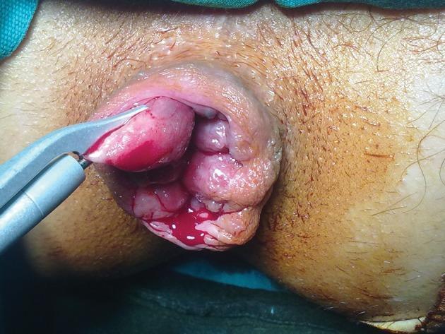
This clinical photograph captures an intraoperative view of a surgical procedure for grade IV internal hemorrhoids. The patient is in the lithotomy position, revealing significant protrusion of prolapsed hemorrhoidal tissue from the anal canal. The tissue is characterized by multiple erythematous, edematous, and lobulated masses with visible vascular congestion and minor active bleeding. A harmonic scalpel (ultrasonic energy device) is shown in use, with its metallic, curved blades grasping and coagulating a portion of the hemorrhoidal pile for excision. This visual demonstrates the Milligan-Morgan open hemorrhoidectomy technique, highlighting the use of advanced energy devices to achieve simultaneous dissection and hemostasis. The surrounding perianal skin and surgical draping are visible, providing clinical context for proctological surgery within a general or colorectal surgery specialty. The educational focus is on surgical management of severe hemorrhoidal disease and the application of energy-based surgical instruments in anorectum procedures.
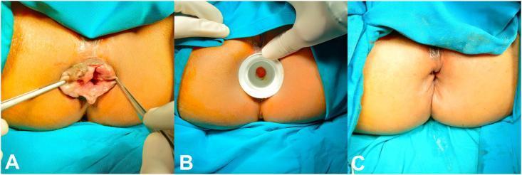
This composite of three clinical photographs documents a surgical hemorrhoidectomy procedure for Grade III or IV hemorrhoids. Panel A shows the preoperative state with significant prolapsed, irregular, reddish-brown hemorrhoidal piles protruding from the anal canal; surgical forceps are used to retract and demonstrate the extent of the prolapse. Panel B illustrates the intraoperative phase, showing the use of a circular white anal dilator to manage internal anal sphincter hypertonicity and provide surgical access to the hemorrhoidal columns. Panel C displays the immediate postoperative result, demonstrating a restored anal anatomy with no visible protruding tissue. The surgical site shows radial suture lines where the hemorrhoidal masses were excised and the mucosa was closed with simple interrupted sutures. This sequence highlights the surgical transition from symptomatic prolapse through intraoperative dilation to postoperative anatomical correction, serving as an educational resource for colorectal surgery and proctology.
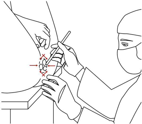
This medical illustration depicts a patient in the lithotomy position during a surgical hemorrhoidectomy. The diagram shows the patient’s hips flexed and legs elevated and abducted to provide optimal exposure of the perineum and anorectal region. A surgeon, wearing sterile gloves, a surgical mask, and a cap, is shown performing the procedure. One hand stabilizes the surgical site while the other holds a slender surgical instrument, likely for tissue dissection or local anesthetic infiltration. Red arrows and symbols overlaying the anal verge indicate the multi-quadrant nature of the hemorrhoidal lesions and the surgical approach for excision. This image serves as an educational resource for demonstrating perioperative patient positioning and the clinical setup for anorectal surgeries, highlighting the importance of anatomical access and sterile technique in a colorectal surgical context.
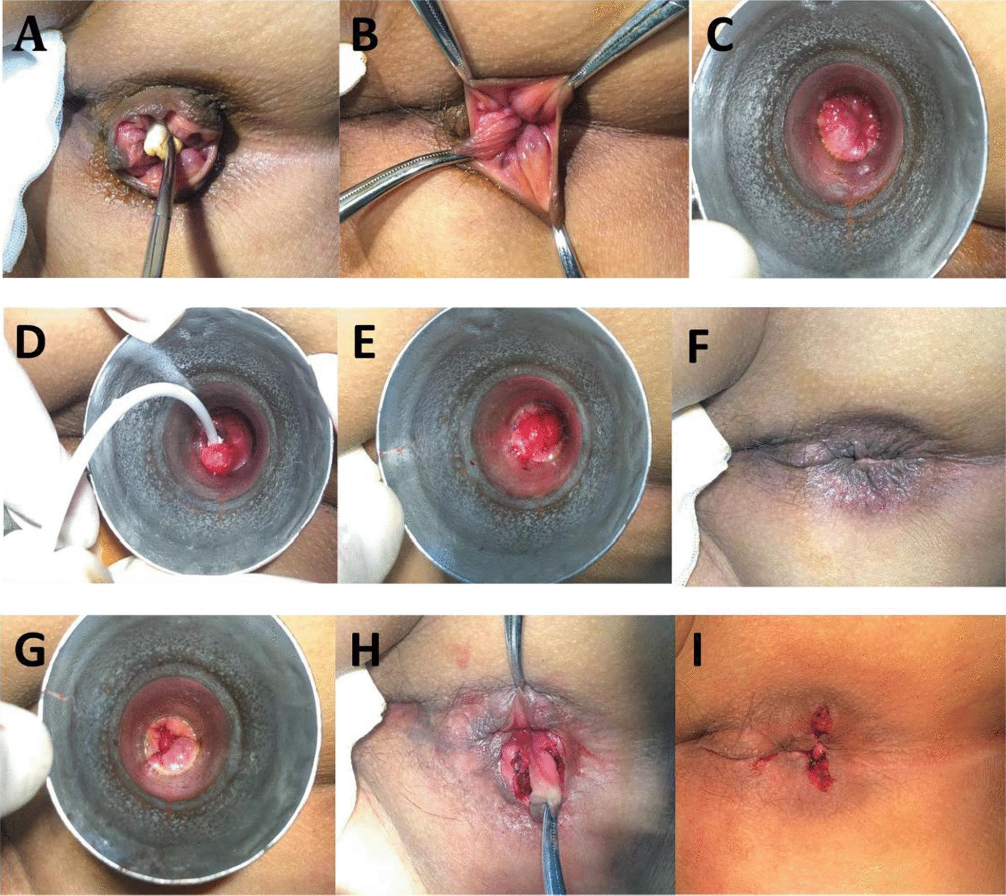
This nine-panel clinical photographic series (A-I) documents a combined surgical procedure for hemorrhoids: the Ruiyun procedure for hemorrhoids (RPH) and a simplified Milligan-Morgan hemorrhoidectomy (sMMH) with dentate line-sparing. Panel A displays circumferential prolapsed hemorrhoids with associated skin tags. Panels B and C illustrate the pre-operative evaluation of the anal canal using forceps and a horn-shaped anoscope to identify internal hemorrhoids and loose mucosa above the dentate line. Panels D and E demonstrate the RPH technique, showing the suctioning of internal hemorrhoidal tissue and subsequent ligation with an elastic suture ring to restrict blood flow. Panel F shows the immediate improvement in mucosal prolapse following ligation. Panels G through I depict the sMMH phase targeting external components below the dentate line: Panel G shows the local anatomy via anoscope, Panel H captures a radial fusiform incision and stripping of the subcutaneous vascular cluster forming an inverted 'V' shape, and Panel I shows the final surgical site after clamping, ligation, and excision of the hemorrhoidal tissue.
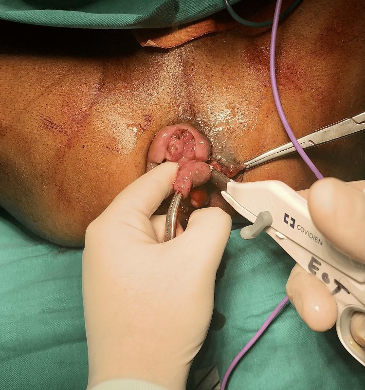
This clinical photograph captures an intraoperative view of a hemorrhoidectomy being performed in a colorectal surgical setting. The surgical field focuses on the perianal region, where a prolapsed, edematous hemorrhoidal mass is visible. The surgeon is employing a bipolar electrosurgical vessel-sealing device (LigaSure, Covidien) to excise the hemorrhoidal tissue while simultaneously achieving hemostasis through thermal fusion of the vascular pedicle. Artery forceps are used to provide traction and stabilize the tissue, allowing for precise dissection at the mucocutaneous junction. The photograph demonstrates the application of energy-based surgical instruments in proctology for reducing blood loss and operative time compared to traditional diathermy or excisional techniques. Key anatomical features include the external anal sphincter area and the diseased vascular cushions of the anal canal. The image is intended for medical education regarding modern surgical management of Grade III or IV hemorrhoids.
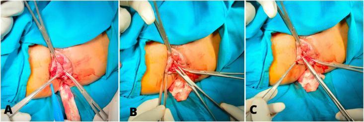
This clinical photograph set illustrates three sequential stages of a surgical hemorrhoidectomy. Image A shows the initial surgical phase where a triangle incision is being made above the dentate line on the protruding hemorrhoidal tissue, with surgical clamps and forceps used to provide traction and exposure. Image B depicts the intraoperative stage of hemorrhoidal excision, showing the removal of the piles while maintaining hemostasis with surgical instruments. Image C demonstrates the final stage of wound closure, where simple interrupted sutures are placed radially to close the excision site. The radial orientation of the sutures is designed to prevent mucosal prolapse and minimize the risk of anal stenosis. The images provide a detailed view of the surgical field, including the use of multifilament absorbable sutures and surgical drapes, highlighting a specific technique for treating circular hemorrhoids at the 3, 7, and 11 o'clock positions.
Hemorrhoidectomy: Surgical Procedure and Instruments
What is Hemorrhoidectomy?
Patient Positioning
- Lithotomy position - preferred for open (Milligan-Morgan) hemorrhoidectomy; patient on back, legs elevated in stirrups
- Prone jack-knife position - preferred for closed (Ferguson) hemorrhoidectomy; patient face-down, buttocks strapped apart with 40° hip flexion
Anesthesia
- General anesthesia (most common)
- Spinal or caudal/regional anesthesia
- Always supplement with local infiltration of bupivacaine + adrenaline 1:200,000 (10 mL per hemorrhoid into submucosal and subcutaneous planes) - reduces bleeding and facilitates dissection from internal sphincter
Types of Hemorrhoidectomy
1. Open Hemorrhoidectomy (Milligan-Morgan)
- Insert proctoscope to identify the 3 primary hemorrhoid sites (typically at 3, 7, and 11 o'clock positions)
- Infiltrate weak adrenaline solution (1:200,000 saline) around each hemorrhoid and in the lower intersphincteric space
- Apply tissue forceps to each pile and adjacent skin
- Make a V-shaped incision in perianal skin with scissors
- Deepen the incision to reveal lower fibers of internal anal sphincter
- Sweep the sphincter away from hemorrhoid with tissue forceps
- Excise hemorrhoidal tissue within the anal canal, leaving the apex (arterial/venous pedicle) intact
- Enclose pedicle in an arterial clip and transfix with non-absorbable suture
- Secure hemostasis from the hemorrhoid bed with cautery; then ligate the pedicle (leave ligature long for easy identification)
- Preserve skin bridges between V-shaped excision segments - critical to prevent stenosis
- Confirm complete hemostasis with anal speculum; apply gauze dressings
- Operate in order: 7 o'clock → 3 o'clock → 11 o'clock to avoid obscuring the field with bleeding
2. Closed Hemorrhoidectomy (Ferguson)
- Insert Fansler proctoscope to identify hemorrhoid sites
- Insert soft gauze swab into anal canal, withdraw to bring hemorrhoids into view
- Infiltrate bupivacaine + adrenaline 1:200,000 into submucosal and subcutaneous planes
- Make a V-shaped incision at the anoderm
- Elevate hemorrhoid off external and internal sphincter; carry dissection cephalad using short curved scissors
- Identify and display the circular internal sphincter fibers at the base of the wound
- Dissect residual hemorrhoidal tissue from under the cut mucosa edges to create two mucosal flaps
- Clamp vascular pedicle at apex; suture ligate pedicle (2-0 Vicryl on tapered needle)
- Close the mucosal defect with a locking running absorbable suture (cutting needle) from apex down to skin
- Close skin with simple running sutures, leaving the last few mm open to drain
3. Energy-Device Hemorrhoidectomy
- Electrothermal bipolar device with intelligence sensors; audible signal when vessel obliteration complete
- Applied across hemorrhoid and skin tag; hemorrhoidal tissue excised along the coagulation line
- Reduces lateral thermal damage; suitable for 2nd degree hemorrhoids
- Significantly increases cost
- Uses high-frequency ultrasound current to cut and coagulate by destroying protein-hydrogen bonds
- Achieves coagulation at low temperature - reduces lateral thermal injury
- Less postoperative pain, reduced bleeding risk
- Effective as a closed hemorrhoidectomy without skin excision
- Limited intra-anal procedure; appropriate for small 2nd degree hemorrhoids
- Can be performed in office or as outpatient
- Risk of reactionary hemorrhage
Surgical Instruments Used
| Instrument | Purpose |
|---|---|
| Proctoscope / Fansler proctoscope | Identify hemorrhoid sites; insert into anal canal for visualization |
| Anal dilator (circular) | Manage internal sphincter hypertonicity; improve surgical access |
| Tissue forceps | Grasp and retract hemorrhoidal tissue; apply traction |
| Arterial clip / Hemostat | Clamp the vascular pedicle before ligation |
| Short curved scissors | Dissect hemorrhoid from sphincter; V-shaped incision |
| Needle holder | Suture ligation of the pedicle |
| 2-0 Vicryl (absorbable suture) | Ligation of pedicle; wound closure |
| Non-absorbable suture | Open hemorrhoidectomy pedicle transfixion |
| Cautery / Diathermy | Hemostasis from hemorrhoid bed |
| LigaSure device | Bipolar vessel sealing; simultaneous excision and hemostasis |
| Harmonic scalpel | Ultrasonic dissection with coagulation, low thermal spread |
| Anal speculum | Final inspection for hemostasis |
| Gauze swabs / dressings | Identify hemorrhoids; postoperative wound packing |
Intraoperative Clinical Images

Complications
- Postoperative pain - most common; expected and should be counseled preoperatively
- Reactionary/secondary hemorrhage - use of long ligature ends helps identify pedicle if needed
- Anal stenosis - caused by excessive anoderm excision; prevented by maintaining skin bridges
- Impaired continence - from internal anal sphincter damage
- Wound dehiscence / hematoma / sepsis - more common with closed technique
- Postoperative fissure or submucous abscess - rare
Key Surgical Principles
- Preserve skin bridges between hemorrhoid excision sites - prevents anal stenosis
- Identify the internal anal sphincter at all times - do not injure it
- Infiltrate local anesthetic with adrenaline - reduces bleeding and facilitates dissection
- Ligate each pedicle securely before excision; leave ligature ends long
- Confirm complete hemostasis before closure and before leaving the operating room
External anal explain in photo and image to
external anal sphincter anatomy diagram
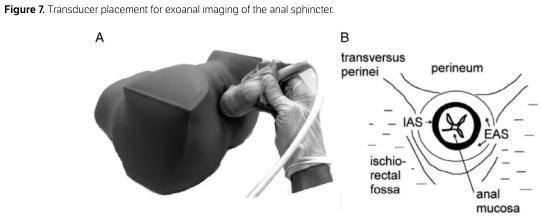
Summary : This figure demonstrates the placement of an ultrasound transducer for exoanal imaging of the anal sphincter, including both a photographic representation and a schematic cross-sectional diagram of the relevant anatomy. photo and anatomical diagram: # Panel A : Transducer Placement Photo • Shows a gloved hand holding an ultrasound transducer. • The transducer is positioned externally against the anal region of a model or patient. • The hand and transducer are oriented perpendicular to the anal opening. • The image is in grayscale, with clear focus on the transducer’s contact with the skin. # Panel B : Cross-sectional Anal Anatomy Diagram • Circular schematic cross-section of the anal region. • Labeled structures: – transversus perinei (top left) – perineum (top right) – ischiorectal fossa (bottom left) – anal mucosa (bottom right) – IAS (internal anal sphincter, inner ring) – EAS (external anal sphincter, outer ring) • The anal canal is depicted centrally, with arrows indicating the orientation of the sphincters. • Surrounding lines represent tissue layers and anatomical boundaries. # Spatial Relationships : • Panel A shows the real-world placement of the transducer, while Panel B provides anatomical context for the imaging plane. • The transducer is placed externally, targeting the sphincter complex as shown in the diagram. # Analysis : • The figure clarifies the correct external placement of the ultrasound transducer for imaging the anal sphincter. • The schematic highlights the anatomical structures visualized in this imaging approach, including both the internal and external anal sphincters and adjacent tissues. • The combination of photo and diagram aids in understanding both technique and anatomical targets for exoanal ultrasound.
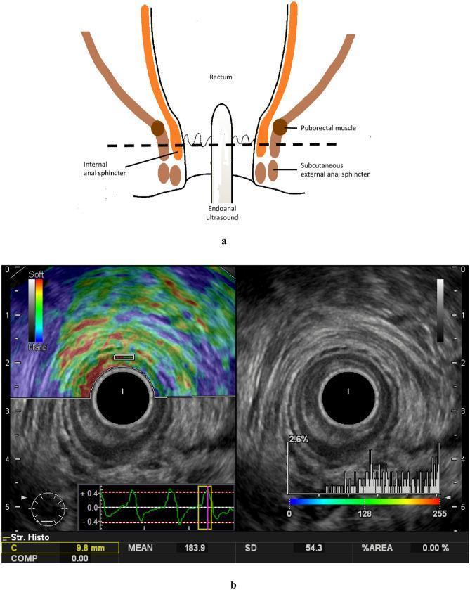
This composite educational image illustrates the application of endoanal ultrasound and real-time tissue elastography (RTE) for assessing anal sphincter anatomy and elasticity. Figure 'a' is a schematic diagram showing an endoanal rectal probe positioned within the anal canal, identifying key landmarks including the rectum, puborectalis muscle, internal anal sphincter (IAS), and subcutaneous external anal sphincter (EAS). Figure 'b' presents dual-panel imaging: the right panel shows a transverse B-mode ultrasound image displaying concentric anatomy of the anal canal. A hypoechoic inner ring (indicated by a white arrow) represents the IAS, surrounded by a more heterogeneous, higher-echogenicity layer (indicated by a yellow arrow) representing the EAS. The left panel shows the corresponding real-time tissue elastography image, where a color-coded strain map is superimposed on the B-mode image. A color scale indicates tissue stiffness (blue/green for softer tissue, red/yellow for harder tissue). Quantifiable data, including a histogram of elasticity values (0–255 range) and mean strain values, are visible at the bottom, demonstrating the methodology for evaluating IAS sclerosis or stiffness changes following clinical interventions like chemoradiotherapy.
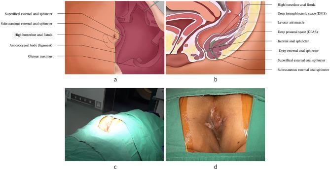
Educational medical composite depicting the anatomy and surgical context of a high horseshoe anal fistula (HHAF). Panel (a) is an anatomical diagram of the perianal region showing external landmarks including the gluteus maximus, anococcygeal body, and the subcutaneous/superficial external anal sphincters, with a dotted line delineating the HHAF scope. Panel (b) presents a sagittal pelvic diagram illustrating the internal anal sphincter, levator ani muscle, and the deep, superficial, and subcutaneous external sphincters. It highlights the deep intersphincteric space (DPIS) and deep postanal space (DPAS) in relation to the fistula tract. Panel (c) is a clinical photograph showing the patient in a prone jackknife position for surgical exposure. Panel (d) provides a close-up clinical photograph of the preoperative surgical field, where a green-shaded overlay identifies the external manifestation of the HHAF. This visual resource is designed for proctological surgical planning and provides clear anatomical orientation of complex perianal fistulas relative to the pelvic floor musculature.
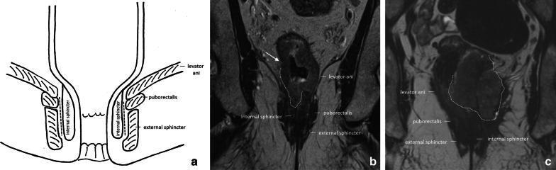
This composite educational image illustrates the pelvic floor and anal canal anatomy and its clinical relevance in rectal cancer staging. Panel (a) is an anatomical diagram depicting the coronal view of the anal canal, highlighting the levator ani, puborectalis muscle, external sphincter, and internal sphincter, with a dashed line marking the intersphincteric plane. Panel (b) is a coronal T2-weighted MRI showing a cT3a rectal tumor in a male patient. An arrow indicates irregular wall thickening with minimal (±1 mm) perirectal extension invading the right internal anal sphincter. Panel (c) is a coronal T2-weighted MRI of a female patient with a cT4b rectal tumor. The tumor (outlined by a dashed line) shows extensive infiltration, invading both internal sphincters and extending into the external sphincter, levator ani, and puborectalis muscles on the left side. Muscles and sphincters appear characteristically hypointense on T2 imaging, while the tumor presents as an intermediate-signal mass disrupting normal anatomical boundaries, essential for determining surgical management such as abdominoperineal resection.
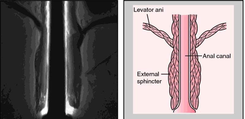
A composite image for medical education comprising a diagnostic radiological image (MRI) on the left and a corresponding anatomical diagram on the right, illustrating the pelvic floor and anal canal. The MRI shows a coronal view of the anorectal region, highlighting the muscular layers of the pelvic outlet. The anatomical diagram provides a schematic representation of the same structures for clarity. It labels the levator ani as a broad, striated muscle group superiorly that forms the pelvic diaphragm and descends to blend with the longitudinal muscle layer of the anal canal. The external sphincter is depicted as a distinct, thicker muscular cuff surrounding the distal portion of the anal canal, located inferior to the levator ani's main attachment points. This visual comparison is designed to teach the spatial relationships between the pelvic floor muscles and the terminal digestive tract, emphasizing the functional anatomy required for fecal continence. The educational focus is on identifying the levator ani and external sphincter during pelvic imaging and understanding their relative morphology.
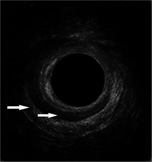
This diagnostic image is an axial cross-section from an endoanal ultrasonography (EAUS) focusing on the mid anal canal. The image demonstrates the characteristic concentric anatomy of the anal sphincter complex with two distinct muscular layers indicated by white arrows. The external anal sphincter (EAS) is visualized as the peripheral, hyperechoic (brighter) ring. A significant structural defect in the EAS is evident, extending from the 9 o'clock to the 3 o'clock position, characterized by a loss of echoic continuity. Internal to this, the internal anal sphincter (IAS) appears as a darker, hypoechoic ring. The IAS also exhibits a pathological defect, visible from the 11 o'clock to the 4 o'clock position. Such findings are clinically significant in the evaluation of obstetric anal sphincter injuries (OASI) and the assessment of fecal or anal incontinence. The image serves as an educational example of how ultrasound can identify localized muscular disruptions and quantify the extent of sphincter damage using clock-face orientation.
external anal hemorrhoids clinical photo thrombosed
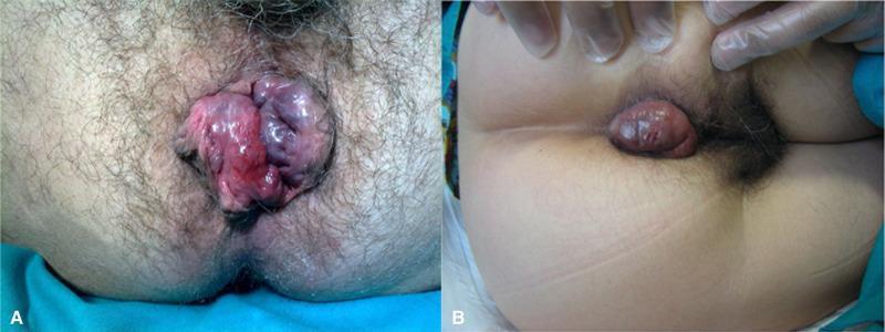
Two clinical photographs (A and B) depicting external anal masses consistent with thrombosed hemorrhoids and perianal lesions. Image A shows a large, multi-lobulated, and irregular mass protruding from the anal verge. The lesion exhibits significant color variation, including areas of deep purple, cyanotic hues, and bright erythema, with a glistening, moist surface texture suggestive of edema or incarceration. The mass largely obscures the anal opening. Image B demonstrates a more localized, ovoid perianal lump. It is characterized by a reddish-brown color that contrasts with the surrounding pale skin. The surface appears tense and smooth but with focal areas of ecchymosis and minor skin abrasions. These images serve as educational examples for differentiating the morphological presentations of acute perianal vascular conditions and external hemorrhoidal disease in a colorectal or primary care clinical context.
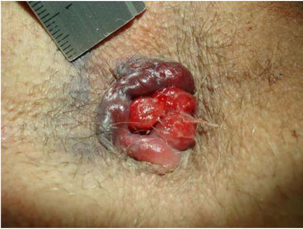
This clinical photograph displays a detailed view of the perianal region demonstrating circular perianal thrombosis and prolapsed hemorrhoids. The central focus shows bright red, lobulated, prolapsing internal hemorrhoids protruding through the anal canal, characterized by a glossy, friable appearance indicative of active or recent bleeding. Surrounding these is a prominent, circular area of dark purple to bluish discoloration consistent with thrombosed external hemorrhoids. Significant edema and swelling are visible in the associated perianal tissue, particularly involving concomitant anal tags on the left-lateral aspect. The surrounding skin is hirsute and shows localized bruising. A medical ruler is partially visible at the top left of the frame for size reference. This image serves as an educational example of advanced hemorrhoidal disease, illustrating the coexistence of grade IV internal prolapse with acute external venous thrombosis and tissue edema.
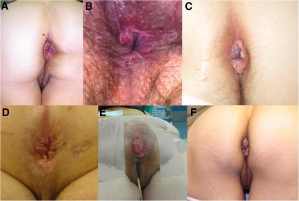
A composite of six clinical photographs (labeled A through F) illustrating various benign anorectal conditions. A: Features prolapsed internal hemorrhoids presenting as reddish-purple, vascular tissue protruding through the anal orifice. B: Demonstrates an anal fissure characterized by erythema, inflammation, and visible mucosal tearing in the perianal region. C: Shows a thrombosed external hemorrhoid, appearing as a smooth, tense, bluish-purple perianal mass. D: Displays an anal fistula with visible scarring, skin induration, and a punctate external opening with purulent drainage. E: A clinical view of a full-thickness rectal prolapse, showing a concentric, erythematous protrusion of the rectal wall with visible circular mucosal folds, captured in a perioperative setting. F: Illustrates anal skin tags (acrochordons), which are hypertrophied, skin-colored, painless connective tissue folds at the anal verge. This collection serves as a visual diagnostic guide for medical education, emphasizing morphology, color, and anatomical location of common proctological pathologies.
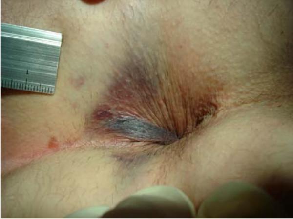
This clinical photograph displays a close-up view of the perianal region, documenting acute tissue trauma. The image shows extensive ecchymosis and subcutaneous hematomas surrounding the anal orifice. The discoloration is multifocal, ranging from deep purple and blue at the anal verge to reddish-purple and brown at the periphery of the affected area. Significant edema is present, resulting in the swelling and distortion of the perianal skin folds. A metal ruler with a metric scale is positioned in the upper left quadrant of the frame for size reference. The visual findings demonstrate an induced subcutaneous hematoma, which serves as a clinical comparison to differentiate mechanical trauma from thrombosed external hemorrhoids (TEH). This material is relevant for proctology and forensic medicine, illustrating the morphology of perianal ecchymosis and the physical manifestations of localized suction or pressure-induced injury to the subcutaneous venous plexus.
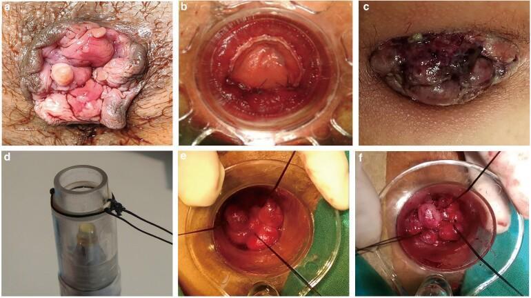
This composite of clinical photographs and procedural frames documents the pathology and surgical management of prolapsed hemorrhoids. Panels (a) and (c) present external views of perianal pathology: (a) shows grade IV prolapsed internal hemorrhoids with characteristic pink, fleshy, nodular protrusions; (c) demonstrates strangulated or thrombosed hemorrhoidal tissue, indicated by a dark purple to black discoloration and significant venous congestion. Panels (b), (e), and (f) depict an intraoperative view using a clear anoscope/proctoscope. These frames illustrate a surgical ligation or mucopexy technique where black suture threads are used to secure and retract the redundant hemorrhoidal mucosa. Panel (d) displays the specialized clear cylindrical surgical instrument used to facilitate the procedure. The series serves as an educational resource for coloproctology, highlighting the clinical presentation of advanced hemorrhoidal disease and the steps of a minimally invasive interventional approach to reduce prolapse and restore anatomical positioning.
External Anal Hemorrhoids - Full Explanation with Images
Anatomy of the Anal Canal
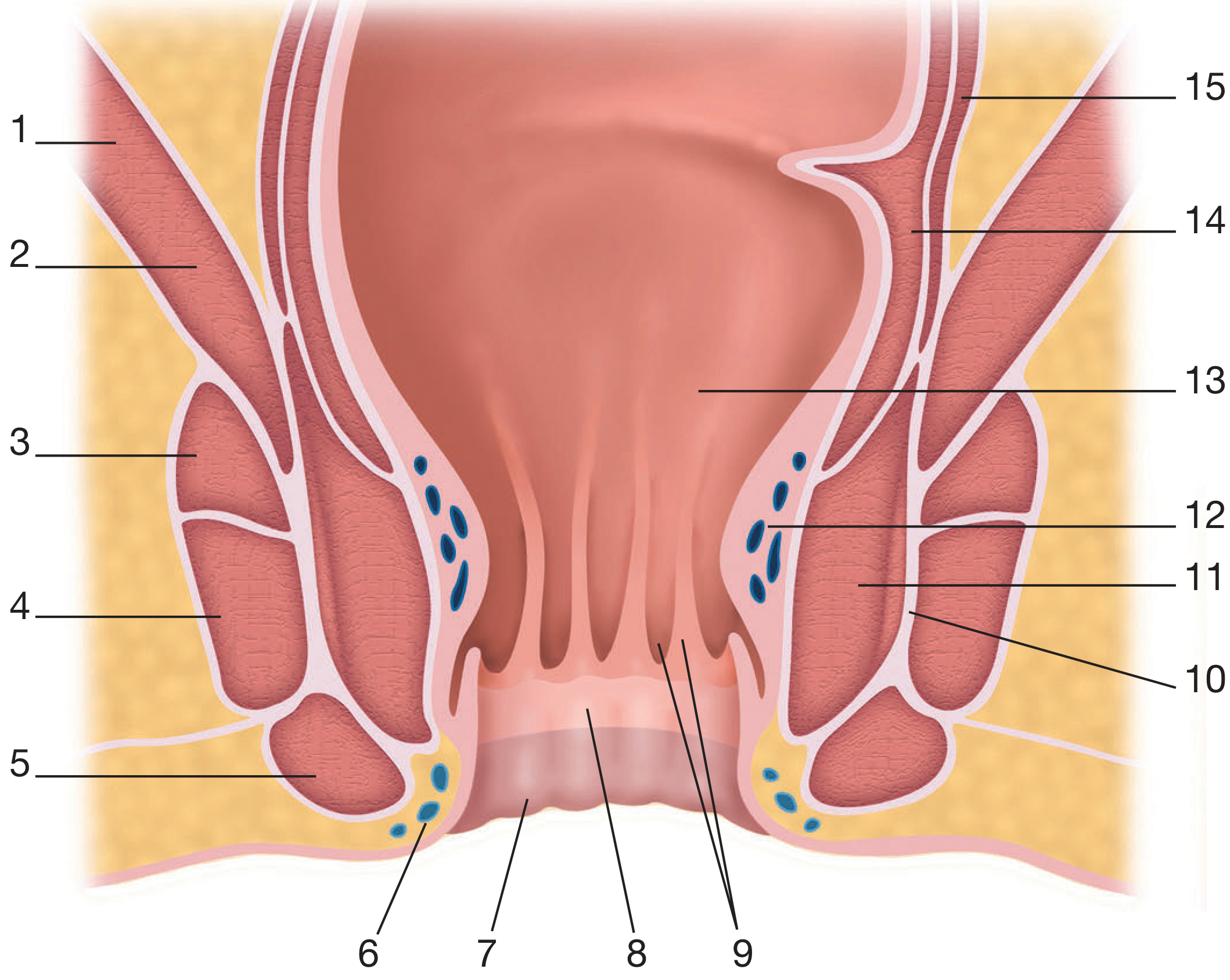
Labeled Anatomy Diagram (Bailey & Love's Surgery):
| Number | Structure |
|---|---|
| 1 | Levator ani (iliococcygeal) |
| 2 | Levator ani (puborectal) |
| 3-5 | External anal sphincter (deep, superficial, subcutaneous parts) |
| 6 | Inferior hemorrhoidal plexus (external hemorrhoids arise here) |
| 7 | Perianal skin |
| 8 | Anoderm |
| 9 | Anal columns and crypts |
| 10 | Conjoined longitudinal muscle |
| 11 | Internal anal sphincter |
| 12 | Superior hemorrhoidal plexus (internal hemorrhoids arise here) |
| 13 | Anorectal junction |
| 14-15 | Circular/longitudinal rectal muscle |
What Are External Hemorrhoids?
- Location: at or below the anal verge (outside the anal canal)
- Covering: perianal skin / anoderm (not mucosa)
- Blood supply: inferior rectal veins
- Innervation: somatic nerves - therefore they are PAINFUL (unlike internal hemorrhoids, which are above the dentate line and covered by insensitive mucosa)
External vs. Internal Hemorrhoids - Key Differences
| Feature | External Hemorrhoids | Internal Hemorrhoids |
|---|---|---|
| Location | Below dentate line | Above dentate line |
| Covering | Squamous skin | Columnar mucosa |
| Sensation | Very painful (somatic nerves) | Usually painless (autonomic) |
| Bleeding | Rare (dark venous) | Common (bright red) |
| Main complication | Thrombosis | Prolapse, bleeding |
| Visible on exam | Yes - perianal lump | Only if prolapsed |
"External hemorrhoids are small venous cushions located at the anal margin. Internal hemorrhoids are covered with mucosa of the anal canal; external hemorrhoids are covered with skin of the anal margin." - Yamada's Textbook of Gastroenterology
Clinical Photos of External Hemorrhoids
1. Thrombosed External Hemorrhoids (Acute)
- Image A (left): Large, multi-lobulated mass at the anal verge. Deep purple-cyanotic color with edema. This is an acute thrombosed external hemorrhoid causing significant pain and swelling
- Image B (right): More localized, ovoid perianal lump - reddish-brown, tense, smooth surface with ecchymosis. Classic single thrombosed external hemorrhoid
2. Mixed and Advanced Hemorrhoidal Disease
3. Diagnostic Guide - Comparison of Anal Conditions
External Anal Sphincter - Anatomy
- Type: Striated (voluntary) skeletal muscle
- Color: Red (high blood supply)
- Nerve supply: Pudendal nerve (S2-S4)
- Parts: Deep, superficial, and subcutaneous (though functionally it acts as a single muscle)
- Attachments: Posteriorly to coccyx; anteriorly fuses with perineal muscles
- Function: Voluntary control of defecation; augments resting anal pressure especially during Valsalva (coughing, sneezing)
Cross-sectional Ultrasound Anatomy:
- IAS (inner ring): Internal anal sphincter - smooth, involuntary, hypoechoic on ultrasound
- EAS (outer ring): External anal sphincter - striated, voluntary, hyperechoic on ultrasound
- Surrounded by ischiorectal fossa, transversus perinei, and perianal skin
Symptoms of External Hemorrhoids
| Symptom | Details |
|---|---|
| Pain | Constant, severe - especially when thrombosed |
| Perianal lump | Visible/palpable at anal verge |
| Swelling / edema | Bluish-purple, tense swelling |
| Itching | Due to skin irritation and moisture |
| Difficulty cleaning | Skin tags after resolved thrombosis |
| Skin tags | Residual fibrotic skin after thrombosis resolves |
"Anal skin tags are discrete folds of skin located at the anal verge. These may be the end result of resolved thrombosed external hemorrhoids." - Maingot's Abdominal Operations
Thrombosed External Hemorrhoid
- A blood clot (thrombus) forms inside the dilated external hemorrhoid vein
- Causes sudden, severe perianal pain
- Appears as a tense, bluish-purple lump at the anal verge
- Peaks in pain at 48-72 hours, then slowly resolves over 1-2 weeks
- Treatment:
- If seen within 72 hours: excision (not just incision) under local anesthetic for fastest relief
- After 72 hours: conservative management (sitz baths, topical anesthetics, stool softeners, analgesia)
Treatment Summary
| Treatment | When Used |
|---|---|
| Sitz baths + fiber + stool softeners | Mild symptoms |
| Topical creams (hydrocortisone, lidocaine) | Itching, mild pain |
| Excision under local anesthetic | Acute thrombosis within 72 hours |
| Hemorrhoidectomy | Large mixed/external hemorrhoids with symptoms |
| Skin tag excision | Residual tags causing hygiene issues |