
Cervical spondolytis
"cervical spondylosis" OR "degenerative cervical myelopathy"
cervical spondylosis MRI myelopathy sagittal T2 cord compression
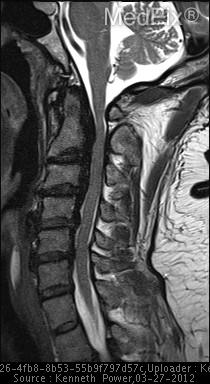
**Imaging Modality:** Sagittal T2-weighted Magnetic Resonance Imaging (MRI) of the cervical spine.
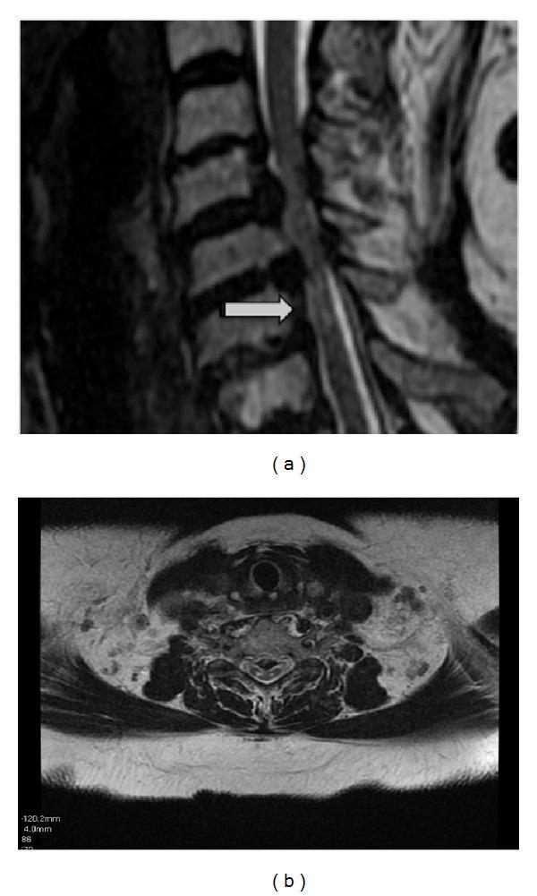
Diagnostic imaging composite of the cervical spine. Image (a) is a sagittal T2-weighted MRI showing degenerative changes including disc space narrowing and spondylosis. A prominent feature is a linear, longitudinal intramedullary high signal intensity lesion within the posterior aspect of the spinal cord (white arrow), extending across multiple vertebral levels. Image (b) is an axial T2-weighted MRI at the level of the cervical spine, demonstrating the cross-sectional anatomy of the spinal cord. In this view, the hyperintensity is localized to the posterior columns. The visual findings are characteristic of chronic spondylotic myelopathy, where repetitive mechanical compression and ischemia lead to signal changes in the cord. The study also depicts narrowing of the spinal canal and degenerative disc disease consistent with advanced cervical spondylosis.
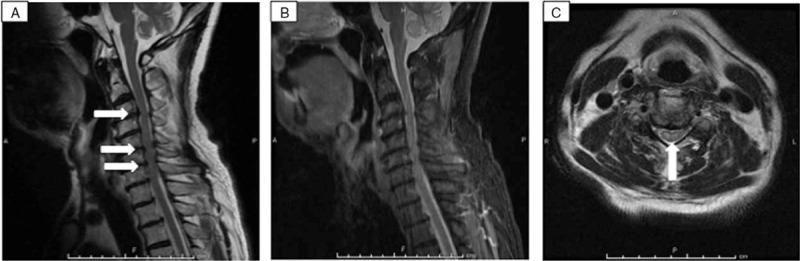
This composite diagnostic image displays MRI sequences of the cervical spine. Image A is a sagittal T2-weighted MRI demonstrating multi-level degenerative changes (spondylosis) at C3/4, C5/6, and C6/7, indicated by white arrows. These changes contribute to narrowing of the spinal canal and ventral cord compression. Image B is a sagittal Short Tau Inversion Recovery (STIR) sequence, which corroborates the findings in Image A by highlighting areas of soft tissue or bone edema and signal abnormalities. Image C is an axial T2-weighted MRI at the C4/5 level, showing a distinct hyperintense (bright) intramedullary signal, highlighted by a white arrow. This signal abnormality involves the central gray matter and extends into the adjacent white matter, a finding consistent with cervical myelopathy or spinal cord ischemia. Collectively, these images illustrate structural cervical spondylosis associated with intrinsic spinal cord signal changes, relevant for clinical diagnosis in patients presenting with acute neurological deficits or quadriplegia.
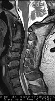
**Imaging Modality:** Sagittal T2-weighted Magnetic Resonance Imaging (MRI) of the cervical spine.
Cervical Spondylosis
Definition
- Discogenic (axial) neck pain
- Radiculopathy (nerve root compromise)
- Myelopathy (cord compression)
- Myeloradiculopathy (both)
Epidemiology and risk factors
- Peak onset is at age 40 to 50, and men are affected more than women.
- C5-6 is the most frequently involved level, followed by C6-7.
- Risk factors are frequent lifting, smoking, and a history of excessive driving.
- People with dystonia and other cervical movement disorders seem predisposed to premature degeneration.
Pathoanatomy
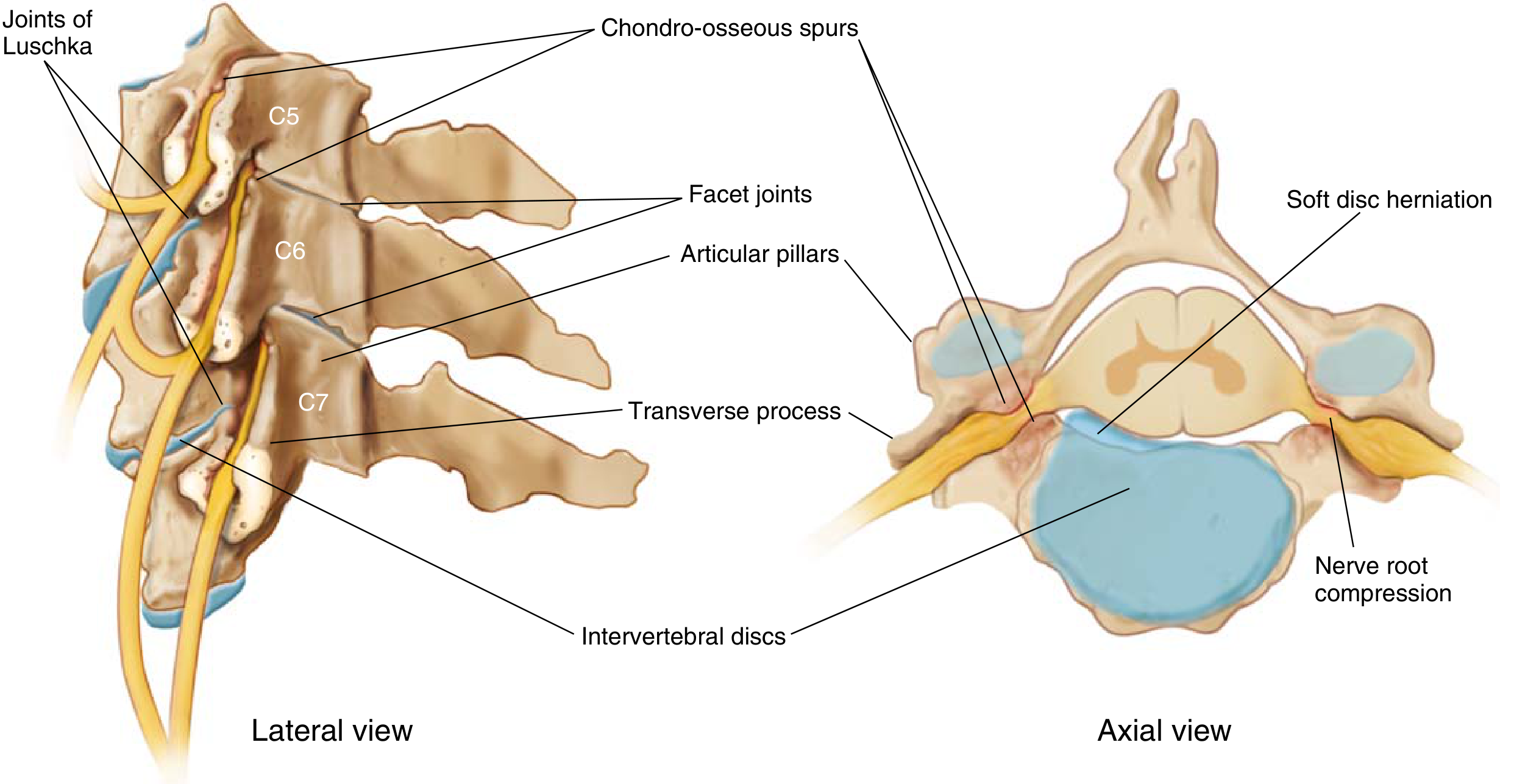
- The intervertebral disc interacts with four other articulations: two uncovertebral joints (of Luschka) and two facet joints.
- Discs progressively collapse and the normal lordosis is lost.
- Disc-osteophyte spurs and a kyphotic alignment cause chronic anterior cord compression.
- Loading of the facet and uncovertebral joints narrows the foramina, which compresses the roots or cord.
- A "soft" disc is a herniation without osteophytes. It is usually posterolateral and causes acute radiculopathy.
- A "hard" disc is a herniation with a disc-osteophyte spur.
- Anterior osteophytes or herniations rarely cause dysphagia.
Clinical features
- Pain and sensory or motor deficit in the affected root distribution.
- C6 and C7 roots are most often involved because of the common levels (C5-6, C6-7).
- Onset is usually insidious. It can progress in a stepwise way or be precipitated by trauma.
- Typical findings are leg spasticity, clumsy or weak hands, and sensory changes in the arms, legs or trunk.
- Spinothalamic or posterior column modalities may be impaired.
- Sphincter dysfunction is usually a late feature.
- Neck pain is often not prominent.
- Lhermitte sign may be present (paresthesia in the legs or trunk on neck flexion).
- (Bradley and Daroff's Neurology in Clinical Practice, p. 2571-2572)
Canal size and dynamic compression
- The cord's anteroposterior diameter is about 10 mm or less.
- CSM rarely develops if the congenital canal diameter exceeds 16 mm.
- On a lateral radiograph, a normal canal is 14 mm or more. Relative stenosis is 10 to 13 mm. Absolute stenosis is less than 10 mm.
- A Pavlov (Torg) ratio below 0.8 is considered abnormal, but its significance is debated.
- In neck extension, the cord is pinched between the anterior spondylotic bar and the posterior hypertrophic facets and infolded ligamentum flavum. Flexion relieves this slightly.
- (Miller's Review of Orthopaedics, 9th ed, p. 759)
Investigations
- Plain films: These show canal diameter, disc space narrowing, osteophytes and alignment.
- MRI: This is the study of choice. It shows cord compression and intramedullary change such as edema or gliosis.
- CT or CT myelography: CT is better for calcified tissue such as osteophytes and OPLL.
- Limits of imaging: Imaging correlates imperfectly with the neurological deficit. Dynamic compression and vascular perfusion contribute.
- Electrophysiology: EMG and nerve conduction studies can help separate radiculopathy from peripheral neuropathy.
Management
- Treatment is conservative: NSAIDs, physiotherapy, activity modification, and a short course of a soft collar.
- Surgery is rarely indicated for neck pain or headache alone without radiculopathy or myelopathy (Bradley and Daroff).
- Epidural steroid injection is sometimes used for radicular pain. This is from my own knowledge, not the retrieved texts.
- The natural history is variable. Some patients stay stable for years and others deteriorate gradually or in steps.
- Collars and bed rest have not been tested in controlled trials.
- Surgical decompression is the usual treatment for progressive or moderate to severe myelopathy.
- Surgery is highly effective at halting progression. Recovery of lost function is less reliable.
- Outcomes are better when symptoms are milder, have lasted less than 12 months, and the patient is under 70.
- Options are anterior cervical discectomy and fusion (ACDF), corpectomy and fusion, laminectomy with or without fusion, and laminoplasty.
- Anterior and posterior approaches both work well. No approach is clearly best, so the choice is individualized.
- (Bradley and Daroff, p. 2572)