

Analyse this , feels itchy
lichen simplex chronicus lichenified plaque wrist dark skin
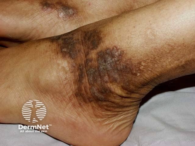
Clinical photograph of the left ankle (dermato) displaying classic features of lichen simplex chronicus (neurodermatitis) in skin of color. Modality: external photography; close-up view of medial/ankle region with oblique lighting reveals thickened, leathery plaques (lichenified skin) with accentuated markings and fissuring. The plaques show variable hyperpigmentation with darker brown to olive tones consistent with pigmented skin; surrounding skin may appear slightly erythematous and dry. Surface scales are fine-to-coarse; there is obvious hyperkeratosis, rugosity, and repeated scratching patterns. The distribution is focal to the ankle region, with potential bilateral symmetry not confirmed in this single image. The morphology includes plaques with well-demarcated borders, rough texture, and accentuated skin lines. Clinically, lichen simplex chronicus arises from chronic scratching or rubbing, leading to thickened epidermis (acanthosis) and hyperkeratosis; pruritus is common and scratching perpetuates the lesion cycle. In skin of colour, post-inflammatory hyperpigmentation can be prominent, and scale may be less conspicuous than in lighter skin. Differential considerations include plaque psoriasis, chronic eczema/atopic dermatitis, contact dermatitis, and less likely fungal infection. This image is valuable for dermatology education, teledermatology, and pattern recognition training, enabling learners to recognize chronic lichenified plaques in darkly pigmented individuals and to correlate clinical features with morphology and distribution.
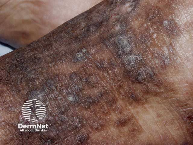
Clinical photography of a cutaneous plaque consistent with lichen simplex chronicus (neurodermatitis) affecting the dorsal hand/forearm. The image demonstrates hyperkeratotic, lichenified plaques with pronounced thickening of the epidermis and accentuated linear skin creases. The plaques are pigmented with variably brown to slate-gray hyperpigmentation and a scaly, rough surface. Dermal inflammation is suggested by peripherally hyperemic borders and faint erythema within the lesion. The distribution appears localized with well-defined margins, and there may be minor fissuring at pressure points. The appearance reflects chronic itch-scratch cycle leading to chronic scratching, which perpetuates the lichenification. The photograph provides a high-resolution, close-up view suitable for dermatology education and differential diagnosis. Notable differentials include atopic dermatitis, discoid (plaque) psoriasis, chronic contact dermatitis, and seborrheic dermatitis; however, the thickened, hyperpigmented plaques with accentuated creases and absence of classic psoriasis scale or orange staining favors lichen simplex chronicus. Clinically, this lesion would correlate with pruritus, focal scratching, and relief after rubbing; management considerations include topical corticosteroids, occlusion, antihistamines, and behavioral modification. This image is relevant for digital dermatology repositories, medical education, and phenotype documentation of neurodermatitis. Documentation should note lesion size, exact location, patient-reported itch intensity, nocturnal scratching, and response to therapy for longitudinal tracking and education.
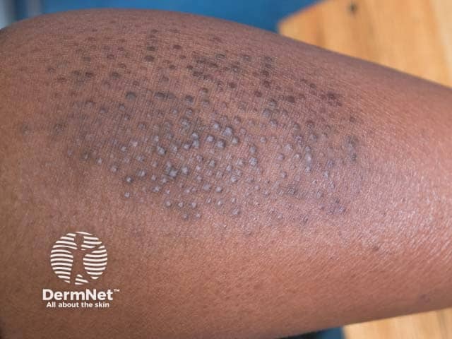
This is a high-resolution clinical photograph of the skin on the shin showing a localized plaque of lichen simplex chronicus with an overlying field of numerous small papules. The image captures a crural region lesion characterized by thickened, lichenified skin with variable hyperpigmentation and surface scale. Interspersed within the plaque are densely packed papules, each approximately 1–3 mm in diameter, with a dome-shaped to flattened contour and a pale or slightly keratotic appearance. The papular component likely reflects an exaggerated pruritus-driven scratching response, producing secondary papulonodular irritation. The overall pattern suggests a chronic itch-scratch cycle rather than an acute inflammatory eruption. From a diagnostic standpoint, the finding corroborates lichen simplex chronicus and supports a differential that includes prurigo nodularis, papular eczema, hypertrophic lichen planus, and eczema with papules. Clinically, this image is valuable for education and comparison in dermatology, dermoscopy training (if performed), and case-based review for chronic dermatitis. Potential clinical uses include documenting lesion evolution, guiding biopsy or topical therapy, and informing patient counseling about itch suppression and skin barrier restoration. This image is most relevant to dermatology, dermatopathology correlation, and educational resources about chronic pruritic dermatoses. This image fosters practical understanding of chronic dermatitis. Useful for clinical teaching and exam case discussions.
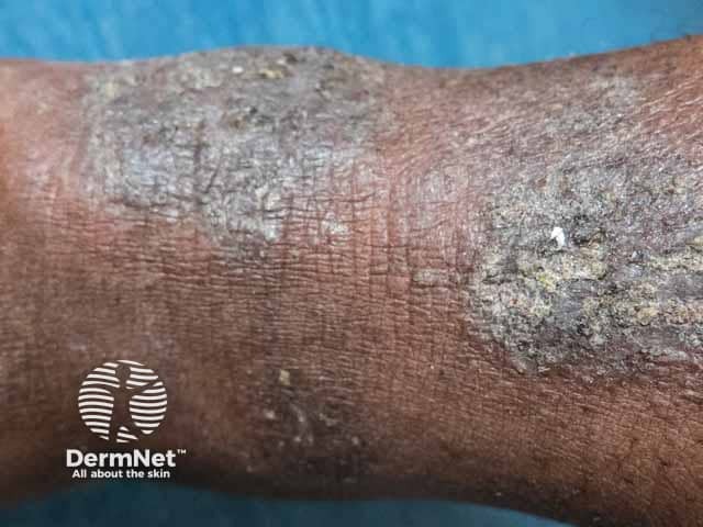
This is a high‑resolution clinical dermatology photograph of the forearm illustrating features of lichen simplex chronicus (neurodermatitis). The image shows thickened, hyperpigmented plaques with a coarse, leathery, lichenified surface and accentuated skin markings. Hyperkeratosis is evident as scale at the plaque margins, and shallow fissures may be present in the most involved areas. Erythema is mild to absent, consistent with a chronic scratching process rather than an acute dermatitis. The distribution is localized to the forearm with well‑defined, oval to elongated plaques that merge into a single broader involvement in places. The color spectrum ranges from brown to slate gray in the hyperpigmented zones, with surrounding normal or subtly inflamed adjacent skin. The morphological pattern—lesions with uniform thickness, crisp borders, and rough surface—aligns with lichenification caused by repetitive scratching and chronic itch. Clinically, this finding supports a diagnosis of lichen simplex chronicus and indicates pruritus with behavioral components. Management considerations include potent topical corticosteroids or calcineurin inhibitors, occlusion therapy, moisturizers, and itch control. Differential diagnoses include eczema/dermatitis, psoriasis with postinflammatory changes, lichen planus, and chronic contact dermatitis. This image supports education, diagnosis, and teledermatology discussions, and may accompany clinical history to guide treatment decisions and follow‑up.
Skin Analysis - Itchy Wrist Lesion
What the image shows
Most Likely Diagnosis: Lichen Simplex Chronicus (LSC)
- Something triggers an itch (stress, dry skin, contact irritant)
- Repeated scratching causes the skin to thicken
- Thickened skin produces more itch signals
- The scratching habit persists even after the original trigger is gone
Other Possibilities to Consider
| Condition | Why it fits | Why it might not |
|---|---|---|
| Contact dermatitis | Wrist location - could be reaction to bracelet metal (nickel), soap, or perfume | Usually more acutely inflamed/weepy unless chronic |
| Atopic eczema (localized) | Dry, itchy, lichenified patch is very common in eczema | Would expect other sites involved or personal/family history |
| Tinea (ringworm) | Can appear as a scaly patch | Usually has a clearer ring-shaped border, not as lichenified |
What to Do
- Stop scratching - this is the single most important step. Cover with a soft cotton sleeve or bandage to prevent habitual scratching, especially at night.
- Moisturize - apply a thick, fragrance-free moisturizer (petroleum jelly / Vaseline works well) several times a day to the dry patch.
- Remove potential irritants - try going without the bracelet for 2 weeks and see if it improves; this will help rule out contact dermatitis.
- A mild-to-moderate potency topical corticosteroid (e.g., hydrocortisone 1% or betamethasone) applied once or twice daily for 1-2 weeks is the standard first-line treatment.
- For stubborn patches, topical calcineurin inhibitors (tacrolimus ointment or pimecrolimus cream) are a good steroid-sparing option.
- If very thick, intralesional triamcinolone (steroid injection) may be needed.
- Oral antihistamines at night can help break the itch-scratch cycle by reducing nighttime scratching.
Important disclaimer: This is an educational analysis only and is not a medical diagnosis. The lesion looks benign and consistent with a chronic itch-scratch condition, but you should see a dermatologist for a proper in-person examination, especially if the patch grows, becomes painful, bleeds, or does not improve with moisturizing. If you also have patches elsewhere on your body, that would support a diagnosis of atopic eczema and warrants evaluation.