
Pathogenesis of tuberculosis refer just robins
Reading File
Now let me search for tuberculosis pathogenesis in Robbins.
Finding Sources
Finding Sources
Finding Sources
I have two Robbins books. Let me read the main tuberculosis section from Robbins & Kumar Basic Pathology (block5) which has the most content, and also from Robbins Cotran & Kumar (the larger pathology book).
Reading File
Reading File
Excellent content. Let me now read more from both sources to get the complete pathogenesis, primary vs secondary TB, and morphology sections.
Reading File
Reading File
Let me fetch the key pathogenesis diagram and continue reading the remaining content about Primary and Secondary TB.
Reading File
Reading File
Loading Image
I now have comprehensive content from both Robbins books. Let me also fetch the Ghon complex image and get secondary TB content.
Reading File
Loading Image
I now have all the information needed. Let me compile a comprehensive answer from Robbins.
Pathogenesis of Tuberculosis
Source: Robbins & Kumar Basic Pathology and Robbins, Cotran & Kumar Pathologic Basis of Disease
Causative Organism and Transmission
Tuberculosis (TB) is a communicable chronic granulomatous disease caused by Mycobacterium tuberculosis. It most often affects the lungs but can involve any organ. Transmission is primarily by airborne droplets from an individual with active disease. The source of M. bovis infection (historically via contaminated milk) is now rare in countries with pasteurized milk.
Key Concept: Infection vs. Disease
Infection implies seeding of a tissue focus, which may or may not progress to disease. Most newly infected, immunocompetent individuals develop an asymptomatic, self-limited focus that heals as a tiny fibro-calcific nodule. Viable organisms remain dormant for decades. If immune defenses are lowered, reactivation can produce communicable, life-threatening disease.
Pathogenesis - Step by Step
The course of TB is dictated by the development of cell-mediated (Th1) immunity, which both confers resistance and causes tissue hypersensitivity and damage. The effector cells for protective immunity and damaging hypersensitivity are the same - so the appearance of tissue hypersensitivity also signals acquisition of immunity.
Phase A: Before Activation of Cell-Mediated Immunity (First ~3 Weeks)
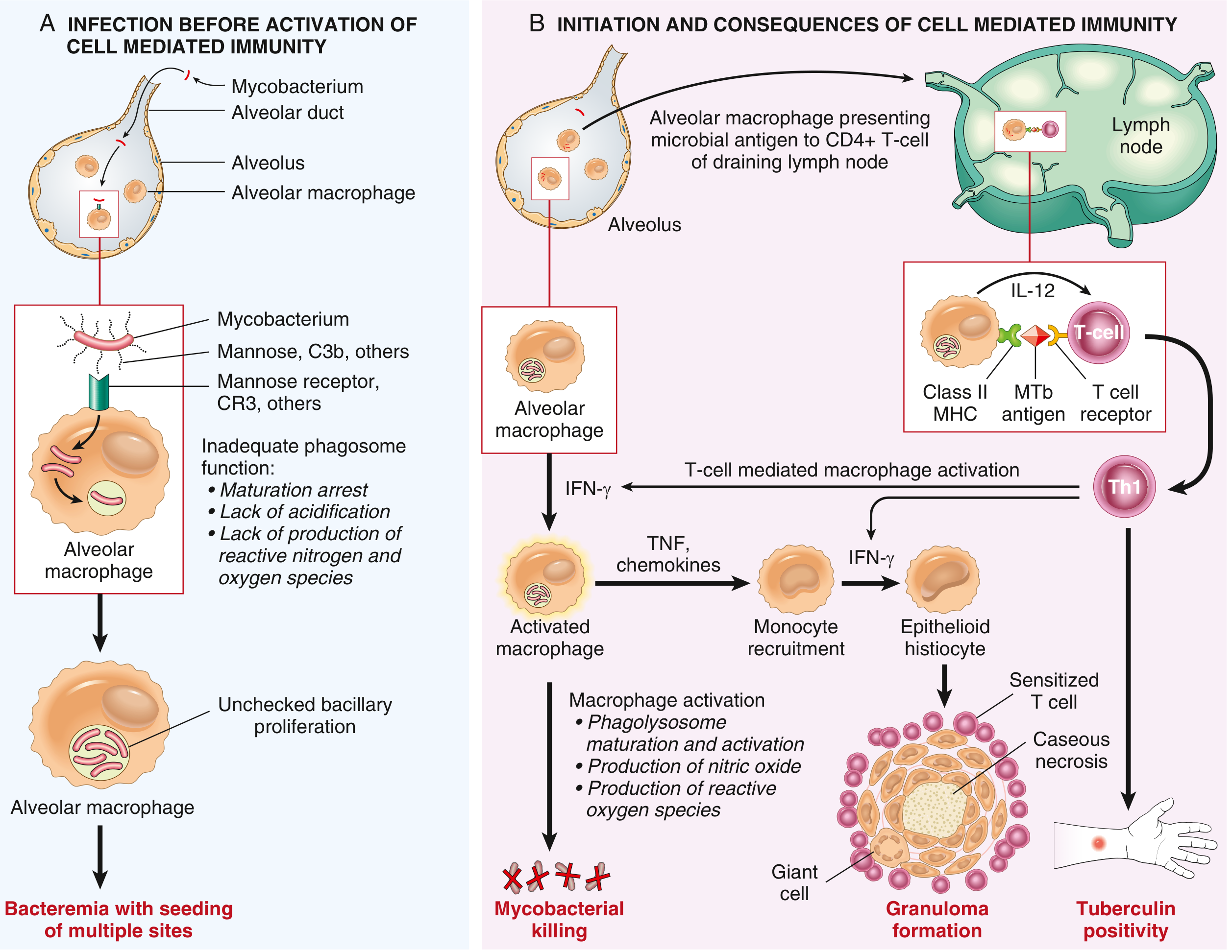
1. Entry into Macrophages
- M. tuberculosis is inhaled and reaches alveoli
- It enters alveolar macrophages by phagocytosis mediated by several receptors:
- Mannose receptor (recognizes mannose-capped lipoarabinomannan on the mycobacterial wall)
- Complement receptor CR3 (Mac-1/CD11b/CD18) - binds C3b-opsonized bacteria
- Other pattern-recognition receptors (TLRs)
2. Replication Within Macrophages (Phagosome Escape)
- Once phagocytosed, M. tuberculosis inhibits phagosome maturation and blocks phagolysosome fusion
- Mechanism: The bacterium recruits a host protein called coronin to the phagosome membrane, which activates the phosphatase calcineurin, leading to inhibition of phagosome-lysosome fusion
- Result: No acidification, no reactive nitrogen/oxygen species - bacteria replicate unchecked within the phagosome
- Bacteria also infect neutrophils, resist killing, induce death of infected cells
3. Unchecked Bacillary Proliferation and Bacteremia
- During the first 3 weeks, bacilli proliferate freely in pulmonary alveolar macrophages and air spaces
- This results in bacteremia and seeding of multiple sites (lymph nodes, liver, spleen, bone marrow, kidneys, meninges, etc.)
- Most individuals are asymptomatic or have a mild flu-like illness at this stage
Phase B: Initiation and Consequences of T Cell-Mediated Immunity (~3 Weeks Onward)
4. Development of Cell-Mediated Immunity
- Dendritic cells migrate from lungs to draining lymph nodes, displaying mycobacterial antigens via Class II MHC to CD4+ T cells
- IL-12 (and IL-18) secreted by antigen-presenting cells drive differentiation of CD4+ Th1 cells
- TLR2 stimulation by mycobacterial ligands promotes IL-12 production
5. Th1-Mediated Macrophage Activation
- Th1 cells produce IFN-γ, the critical mediator that activates macrophages
- IFN-γ activates macrophages via three mechanisms:
- iNOS upregulation → produces nitric oxide (NO) and reactive nitrogen intermediates → kills mycobacteria in the acidic phagolysosome
- Antimicrobial peptides (defensins/cathelicidins) are mobilized against bacteria
- Autophagy is stimulated → sequesters and destroys intracellular bacteria
- TNF is also released → responsible for recruitment of monocytes → these develop into activated macrophages that form granulomas
6. Granulomatous Inflammation and Tissue Damage
- Macrophages activated by IFN-γ differentiate into "epithelioid histiocytes"
- These aggregate to form granulomas; some fuse to form Langhans giant cells
- TNF and chemokines recruit more monocytes
- In many individuals, this response halts infection before significant tissue destruction
- In others (due to age, immunosuppression), ongoing immune response results in caseous necrosis - sometimes without granuloma formation
7. Caseous Necrosis
- The central area of granulomas undergoes caseous (cheese-like) necrosis due to:
- Delayed-type hypersensitivity reaction
- Cytotoxic T cell (CD8+) killing of infected macrophages
- Important: The necrotic material is relatively inhospitable to bacterial growth, but viable bacilli can persist
8. Tuberculin Positivity (Delayed Hypersensitivity)
- The development of Th1 immunity is detected by the tuberculin (PPD/Mantoux) skin test or IFN-γ release assays (IGRAs)
- A positive test = T cell-mediated immunity to mycobacterial antigens
- Loss of tuberculin positivity (anergy) in a known TB-infected patient is an ominous sign of fading resistance and heralds severe disease
Summary Diagram
| Stage | Timing | Key Events |
|---|---|---|
| Entry | Day 0 | Inhaled bacilli phagocytosed by alveolar macrophages via mannose receptor & CR3 |
| Phagosome escape | Days 1-3 | Coronin-calcineurin pathway blocks phagolysosome fusion; unchecked replication |
| Bacteremia | Week 1-3 | Bacilli spread to hilar nodes and hematogenous seeding of multiple organs |
| CMI onset | ~Week 3 | Dendritic cells activate CD4+ T cells in lymph nodes; IL-12 drives Th1 differentiation |
| Macrophage activation | Week 3+ | IFN-γ → iNOS → NO; defensins; autophagy → mycobacterial killing |
| Granuloma formation | Week 3+ | Epithelioid histiocytes + Langhans giants + caseous necrosis → wall off infection |
| Containment (95%) | Weeks-months | Ghon focus calcifies; latent infection established |
| Failure (5%) | Variable | Progressive primary or later reactivation disease |
Critical Mediators
| Mediator | Source | Role |
|---|---|---|
| IL-12 | Macrophages, DCs | Drives Th1 differentiation |
| IFN-γ | Th1 cells | Activates macrophages; most critical mediator |
| TNF | Activated macrophages | Monocyte recruitment; granuloma maintenance |
| NO (via iNOS) | Activated macrophages | Key bactericidal molecule |
| Defensins/cathelicidin | Macrophages | Direct antimycobacterial toxicity |
Defects in any component of this Th1 pathway (IL-12, IFN-γ, TNF, or NO production) result in poorly formed granulomas, absent resistance, and progressive disease.
Primary Tuberculosis
- Develops in a previously unexposed, unsensitized individual
- Only ~5% of newly infected people develop significant disease
- Ghon focus: 1-1.5 cm gray-white area of caseous consolidation, typically in the lower upper lobe or upper lower lobe, close to the pleura
- Ghon complex: Ghon focus + ipsilateral hilar/mediastinal lymph node caseation (connected by lymphangitis)
Gross pathology - Ghon Complex (Robbins, Cotran & Kumar):
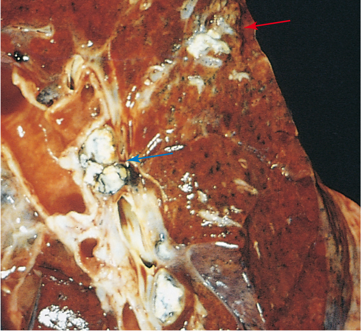
- In ~95% of cases, cell-mediated immunity controls infection → Ghon complex undergoes progressive fibrosis and calcification (Ranke complex)
- Hematogenous seeding occurs in all primary infections, but is controlled by immunity
Progressive primary TB occurs in:
- Overtly immunocompromised patients
- Severe malnutrition
- HIV-positive with CD4+ T cells <200 cells/μL (granulomatous reaction may be absent)
Secondary (Reactivation) Tuberculosis
- Arises in a previously sensitized host - from reactivation of dormant primary lesions (decades later) or reinfection
- Only <5% of patients with primary TB develop secondary TB
Key features distinguishing from primary TB:
| Feature | Primary TB | Secondary TB |
|---|---|---|
| Host | Unsensitized | Previously sensitized |
| Location | Lower upper lobe / upper lower lobe (subpleural) | Apex of upper lobes (both lungs) |
| Lymph node involvement | Prominent (hilar caseation) | Less prominent |
| Cavitation | Rare | Common - important source of infectivity |
| Bacteremia/seeding | Common | Less common (walled off rapidly) |
- Apical location in secondary TB is thought to relate to high oxygen tension at the apices (favorable for M. tuberculosis)
- Because of pre-existing hypersensitivity, bacilli elicit a prompt tissue response that walls off the focus
- Cavitation occurs when caseous material liquefies and drains into bronchi → patients produce infectious sputum
In HIV patients:
- Mildly immunocompromised (CD4+ >300): "usual" secondary TB with apical cavitation
- Severely immunocompromised (CD4+ <200): resembles progressive primary TB (lower/middle lobe consolidation, hilar adenopathy, no cavitation); extrapulmonary involvement >50%
Why TNF Matters Clinically
Patients with rheumatoid arthritis or IBD treated with TNF antagonists are at markedly increased risk for TB reactivation, because TNF is essential for maintaining granuloma integrity and monocyte recruitment.
Robbins & Kumar Basic Pathology (9780323790185), p. 475-477; Robbins, Cotran & Kumar Pathologic Basis of Disease (9780443264528), p. 351-354
This is a shared conversation. Sign in to Orris to start your own chat.