
Cillopathies of kidney
kidney ciliopathies cilia
primary cilium structure ciliopathy polycystic kidney disease nephronophthisis
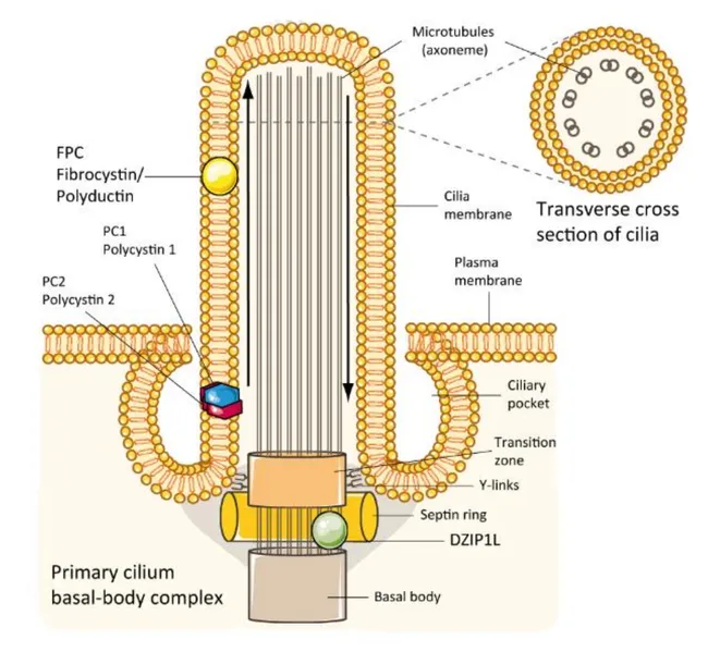
This digital schematic illustration depicts the molecular pathophysiology of autosomal recessive polycystic kidney disease (ARPKD). The image emphasizes a renal tubular epithelial cell with its primary cilium emanating from the apical membrane of a collecting duct/loop of Henle segment. The left side labels the PKHD1-encoded fibrocystin (fibrocystin/polyductin) protein localized to the ciliary membrane and apical plasma membrane, illustrating its role in the ciliopathy pathway alongside polycystin-1 (PC1) and polycystin-2 (PC2). The diagram highlights the basal body–transition zone complex, with DZIP1L positioned at centrioles and the distal basal body, underscoring its necessity for trafficking gene products into the ciliary axoneme. A transverse section inset shows microtubule axonemes and the ciliary pocket, connecting to the primary cilium’s sensory function. Arrows denote intracellular trafficking and-lumen orientation. Dysfunctions in fibrocystin and DZIP1L disrupt ciliary signaling, promoting cystogenesis in ARPKD and reflecting a ciliopathy mechanism shared with other cystic diseases. The illustration also notes that fibrocystin is variably expressed in pancreatic and bile duct epithelium, consistent with multisystem involvement. This visualization serves educational purposes for genetics, nephrology, and cell biology, and supports research into targeted therapies addressing ciliary assembly and trafficking defects. Clinicians and students can correlate genotype–phenotype concepts with this concise framework for learning.
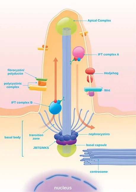
Educational diagram of the renal nephron focusing on the cilia-centrosome complex in tubular epithelium and its role in Autosomal Dominant Polycystic Kidney Disease (ADPKD). The image depicts a non-motile primary cilium projecting into the tubular lumen, housing the polycystin-1 and polycystin-2 heterodimer, and associated IFT (intraflagellar transport) components at the basal body and centriole. In healthy kidneys, the PC1/PC2 complex mediates mechanosensory calcium influx in response to tubular flow, regulating downstream signaling pathways. In PKD, PKD gene mutations abolish or alter this complex, disrupting intracellular Ca2+ homeostasis and shifting balance toward cAMP-driven proliferation and enhanced secretory activity. The diagram notes downstream pathways such as ERK and mTOR, which contribute to cyst initiation and progressive enlargement of cysts from renal tubular epithelium. Additional features illustrate basal body, transition zone, and basal capsule architecture, along with Wnt/Hedgehog signaling crosstalk and ciliary transport (IFT A/B). The educational utility spans medical education, nephrology, genetics, and cell biology; potential applications include teaching ciliary biology, pathophysiology of PKD, and therapeutic targets aimed at mitigating cyst growth. This representation serves as a conceptual reference rather than a diagnostic radiograph, and supports discussions of genotype-phenotype correlations, experimental models, and drug development strategies in ciliopathies.
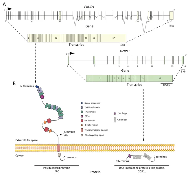
Genetic/genomic illustration detailing autosomal recessive polycystic kidney disease (ARPKD) genetics, with emphasis on PKHD1 and DZIP1L gene architecture. The image portrays the PKHD1 locus on chromosome 6p12, including gene structure, exons, and transcript organization, alongside a schematic depiction of the fibrocystin/polyductin (FPC) protein and its membrane-spanning and cytoplasmic domains. It also shows the DAZ interacting protein 1-like (DZIP1L) gene and transcript, encoding the DZIP1L protein localized to the primary cilium. The diagram underscores inheritance: ARPKD follows autosomal recessive transmission, with unaffected parents and a 25% risk to siblings; the majority of cases arise from PKHD1 mutations, with over 300 reported variants. Truncating PKHD1 mutations predict severe, perinatal or neonatal disease, while missense variants are associated with milder phenotypes and extended survival. A subset of patients carries DZIP1L mutations, often resulting in a milder clinical course and reduced perinatal mortality. The illustration integrates exonic mapping, transcript lengths, and domain architecture to connect genotype to protein function and cystogenesis mechanisms. Clinically, this image supports genetic counseling, diagnostic test design, and interpretation of sequence variants; it informs differential diagnosis among polycystic kidney diseases and renal ciliopathies; it is a valuable educational resource for medical genetics, nephrology, pediatrics, and basic science research into kidney development and tubulogenesis.
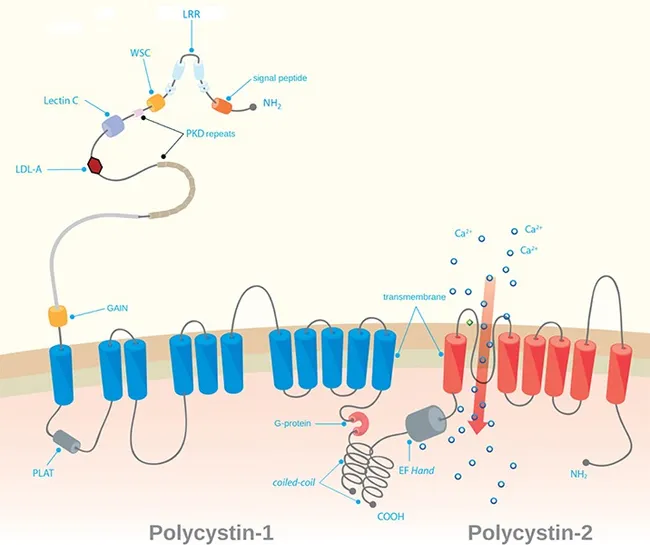
Comprehensive educational illustration showing the molecular architecture of autosomal dominant polycystic kidney disease (ADPKD). This schematic depicts two key PKD gene products, polycystin-1 (polycystin-1) and polycystin-2 (polycystin-2), embedded in the surface membranes of renal tubular epithelial cells and within the primary cilium. The image emphasizes that PKD1 mutations (chromosome 16p13.3) and PKD2 mutations (chromosome 4q21) disrupt a calcium-regulated signaling complex, leading to dysregulated tubular cell proliferation and cystogenesis. The left side highlights polycystin-1 features, including extracellular domains and transmembrane segments, while the right side shows polycystin-2 as a calcium-permeable ion channel. The transmembrane linkage and cytosolic signaling components suggest how altered calcium homeostasis influences downstream pathways governing cell adhesion, polarity, and fluid secretion. The illustration conveys the concept of haploinsufficiency with somatic second-hit events required for cyst formation, consistent with autosomal dominant inheritance with variable expressivity and high penetrance by age decades. Though clinically heterogeneous, patients commonly present with bilateral renal cysts, progressive loss of function, hypertension, and possible extrarenal manifestations. Used for genetics education, biomedical research, and training in molecular renal pathophysiology. The image also supports conceptual learning of gene dosage effects, allelic heterogeneity, and genotype-phenotype correlations essential for genetic counseling, risk assessment, and targeted molecular therapies in nephrology practice.
Ciliopathies of the Kidney
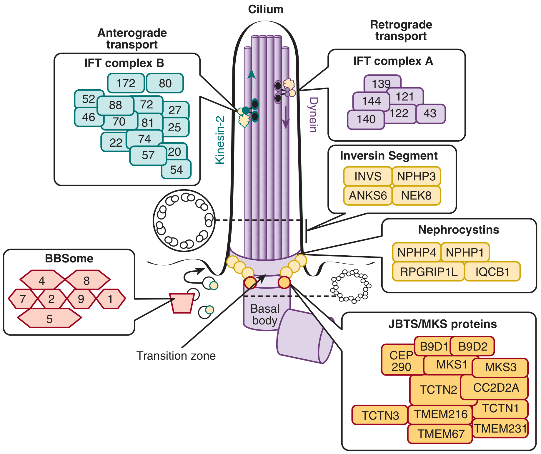
1. Normal Ciliary Machinery
| Component | Location | Role |
|---|---|---|
| Basal body | Base of cilium (mother centriole) | Cilium assembly, anchoring |
| Transition zone | Proximal cilium | Gating of proteins in/out of cilium |
| Inversin compartment | Proximal axoneme | Unknown; INVS, NPHP3, NEK8, ANKS6 |
| IFT complex B (+ Kinesin-2) | Anterograde | Cargo transport toward ciliary tip |
| IFT complex A (+ Dynein-2) | Retrograde | Cargo transport toward base |
| BBSome | Ciliary membrane | Trafficking of membrane-associated proteins |
| Nephrocystins | Transition zone | Epithelial morphogenesis, cell-cell contacts |
- Brenner and Rector's The Kidney, p. 2010-2011
2. Classification of Renal Ciliopathies
A. Cystic Ciliopathies (Polycystic Kidney Diseases)
Autosomal Dominant Polycystic Kidney Disease (ADPKD)
- Genes: PKD1 (85-90%) on chromosome 16p; PKD2 (10-15%) on chromosome 4q
- Proteins: Polycystin-1 (PC1) and Polycystin-2 (PC2), localized to the primary cilia of tubular cells
- Mechanism: PC1 is a mechanosensor; PC1/PC2 heterodimers sense fluid flow and regulate calcium influx. Loss of polycystin function (two-hit model) reduces mechanosensing below a critical threshold, disrupting downstream signaling leading to cell polarity loss, increased proliferation, and fluid secretion into forming cysts. mTOR and ERK pathways are upregulated.
- Epidemiology: 1 in 500-1,000 individuals; accounts for ~10% of chronic kidney disease
- Morphology: Bilateral massively enlarged kidneys (up to 4 kg each); innumerable cysts 1-4 cm from all nephron segments; compressed normal parenchyma between cysts
- Clinical Features:
- Symptoms onset typically in the 4th decade
- Flank pain, dragging sensation, intermittent gross hematuria
- Hypertension in ~75%
- Liver cysts in 1/3; saccular (berry) aneurysms of Circle of Willis in 10-30%; mitral valve prolapse in 20-25%
- Progressive to ESKD; PKD1 mutations progress faster than PKD2
- Robbins & Kumar Basic Pathology, p. 525-526
Autosomal Recessive Polycystic Kidney Disease (ARPKD)
- Gene: PKHD1 (chromosome 6p12) encoding fibrocystin/polyductin; a minor subset caused by DZIP1L mutations
- Mechanism: Fibrocystin localizes to the ciliary membrane and apical plasma membrane; its deficiency disrupts ciliary signaling. DZIP1L localizes to the distal basal body and is required to traffic gene products into the cilium.
- Clinical features:
- Presents in infancy/childhood; severe disease causes death in neonatal period
- Kidneys enlarged with cortical and medullary cysts oriented radially (perpendicular to cortical surface)
- Obligatory hepatic involvement: congenital hepatic fibrosis leading to portal hypertension
- Renal failure in surviving children/adolescents
B. Autosomal Recessive Ciliopathies with Interstitial Nephritis and/or Renal Cystic Disease
- Brenner and Rector's The Kidney, p. 2011
Nephronophthisis (NPHP)
- Genetics: Autosomal recessive; ~20 genes; homozygous NPHP1 deletion in ~21% of cases
- Epidemiology: 1 in 50,000 live births; most common genetic cause of ESKD in children (~5% of pediatric ESKD in North America)
- Types by age of renal failure:
- Infantile NPHP: INVS (NPHP2) mutations - renal failure birth to age 3 years; often + situs inversus, cardiac VSD, hepatic fibrosis
- Juvenile NPHP: Most other genes (NPHP1, NPHP3, NPHP4...) - renal failure in 1st-3rd decade
- Morphology:
- Juvenile form: Small kidneys with granular surface; thin cortex and medulla; indistinct corticomedullary junction; small, thin-walled cysts at the corticomedullary junction from distal tubules/collecting ducts; thickened tubular basement membranes; interstitial fibrosis with scant chronic inflammation
- Infantile form: Enlarged, cystic kidneys; less prominent TBM thickening
- Imaging: Cysts often too small for ultrasound; contrast CT/MRI more sensitive; medullary streaking on excretory urography
- Clinical onset: Insidious; polyuria and polydipsia (tubular dysfunction), growth retardation, normochromic normocytic anemia, progressive CKD
- Extrarenal manifestations (gene-specific):
| Feature | Associated Gene |
|---|---|
| Retinitis pigmentosa (Senior-Loken syndrome) | IQCB1 (NPHP5), CEP290 (NPHP6), RPGRIP1L (NPHP8) |
| Oculomotor apraxia (Cogan syndrome) | NPHP1, NPHP4 |
| Cerebellar aplasia/hypoplasia (JBTS overlap) | CEP290, RPGRIP1L |
| Situs inversus, cardiac VSD, hepatic fibrosis | INVS (NPHP2) |
| Hepatic fibrosis (COACH syndrome) | TMEM67 (NPHP11) |
- Brenner and Rector's The Kidney, p. 2013
Joubert Syndrome (JBTS)
- Genetics: Autosomal recessive; 34 genes identified
- Cardinal feature: "Molar tooth" sign on MRI - midbrain hypoplasia with abnormally deep interpeduncular fossa, elongated and thickened superior cerebellar peduncles, vermis hypoplasia
- Renal involvement: Nephronophthisis-like interstitial nephritis; renal cysts
- Other features: Neonatal hyperpnea/apnea, oculomotor apraxia, ataxia, retinal dystrophy, hepatic fibrosis, polydactyly
- Grainger & Allison's Diagnostic Radiology
Meckel Syndrome (MKS)
- Genetics: Autosomal recessive; 13 genes; lethal in utero/neonatally
- Features: Occipital encephalocele, polydactyly, renal cystic dysplasia (large multicystic kidneys), hepatic bile duct proliferation
- Note: Represents the most severe end of the NPHP-JBTS-MKS spectrum
Pathogenetic Overlap in NPHP-JBTS-MKS
-
Module 1 (NPHP1, NPHP4, RPGRIP1L): Transition zone + cell-cell contacts; needed for apical surface organization
-
Module 2 (IQCB1/NPHP5, CEP290/NPHP6): Centrosomes + transition fibers; indispensable for ciliogenesis
-
Module 3 (MKS proteins, TCTN1/2/3, TMEM216, TMEM67, CC2D2A): Transition zone; connected to Hedgehog signal transduction
-
Inversin module (INVS, NPHP3, NEK8): Inversin compartment; function uncertain
-
Brenner and Rector's The Kidney, p. 2012
C. Bardet-Biedl Syndrome (BBS)
- Genetics: Autosomal recessive; mutations in 21+ genes encoding proteins of the BBSome complex at the basal body; an "archetypal ciliopathy"
- Renal features: Multiple calyceal clubbing, calyceal diverticula, parenchymal dysplasia, fetal lobulation, renal cysts, horseshoe kidneys, vesicoureteral reflux; renal failure in early adult life is common
- Classic pentad:
- Retinitis pigmentosa
- Polydactyly (postaxial)
- Obesity
- Hypogonadism
- Learning difficulties
- Renal anomalies in ~80% of patients
- Comprehensive Clinical Nephrology, 7th Edition, p. 740
D. Skeletal Ciliopathies with Renal Involvement (SRTD Spectrum)
- Features: Constricted thorax, short ribs, short tubular bones, "trident" acetabular roof, polydactyly
- Renal involvement: NPHP-like interstitial nephritis
- Mechanism: Defects in IFT-A subunits/DYNC2H1 (retrograde), IFT80/IFT172 (IFT-B), NEK1 (ciliogenesis kinase), EVC/EVC2 (Hedgehog signaling at basal body)
- Includes: Ellis-van Creveld syndrome, Cranioectodermal dysplasia (Sensenbrenner syndrome), Mainzer-Saldino syndrome, RHYS syndrome (retinitis pigmentosa + hypopituitarism + NPHP + skeletal dysplasia)
- Brenner and Rector's The Kidney, p. 2016
E. Oro-Facial-Digital Syndrome Type 1 (OFD1)
- Genetics: X-linked dominant; OFD1 gene on Xp22; lethal in males in utero
- Renal features: Renal cystic disease in 60% of female patients after age 18; kidneys may be indistinguishable from ADPKD on imaging; liver and pancreatic cysts also occur
- Extrarenal: Oral (cleft tongue/palate, hyperplastic frenula), facial (broad nasal root), digital (brachydactyly, polydactyly, syndactyly), CNS anomalies (agenesis of corpus callosum, Dandy-Walker)
- Protein: OFD1 protein is a core component of the human centrosome throughout the cell cycle (contains N-terminal LisH motif important in microtubule dynamics)
- Brenner and Rector's The Kidney
F. Alström Syndrome (ALMS)
- Gene: ALMS1 (1 gene); autosomal recessive
- Renal involvement: Progressive nephropathy
- Features: Retinal dystrophy, sensorineural hearing loss, obesity, type 2 diabetes, cardiomyopathy; phenotypically overlaps with BBS
3. Molecular Pathogenesis - Summary
Primary cilium dysfunction
        ↓
Impaired mechanosensing (flow sensing by PC1/PC2)
Impaired signal gating (transition zone defects)
Defective Wnt & Hedgehog signaling
Disrupted IFT → abnormal ciliary composition
        ↓
↓ Intracellular Ca²⁺ influx
↑ cAMP signaling
↑ mTOR / ERK activation
↑ Cell proliferation + ↑ fluid secretion
↓ Cell polarity (loss of oriented cell division)
        ↓
Cyst initiation → progressive cyst enlargement
Interstitial fibrosis and tubular atrophy (NPHP-type)
Renal dysplasia (BBS, MKS)
- Robbins & Kumar Basic Pathology; Brenner and Rector's The Kidney
4. Comparison Table of Major Renal Ciliopathies
| Disease | Inheritance | Key Gene(s) | Ciliary Location | Renal Phenotype | Key Extrarenal Features |
|---|---|---|---|---|---|
| ADPKD | AD | PKD1, PKD2 | Ciliary membrane | Large bilateral cysts; ESKD 4th-6th decade | Berry aneurysm, liver cysts, mitral valve prolapse |
| ARPKD | AR | PKHD1, DZIP1L | Ciliary membrane | Radially oriented cysts; enlarged kidneys | Congenital hepatic fibrosis, portal hypertension |
| NPHP (juvenile) | AR | NPHP1-20 | Transition zone | Small kidneys; corticomedullary cysts; TBM thickening | Retinitis pigmentosa, oculomotor apraxia |
| Senior-Loken | AR | IQCB1, CEP290 | Centrosome/trans. zone | NPHP | Severe early retinitis pigmentosa |
| Joubert (JBTS) | AR | 34 genes | Transition zone | NPHP-like | Molar tooth sign, ataxia, neonatal apnea |
| Meckel (MKS) | AR | 13 genes | Transition zone | Large multicystic dysplastic kidneys | Occipital encephalocele, polydactyly; lethal |
| Bardet-Biedl | AR | 21 BBS genes | BBSome/basal body | Calyceal clubbing, cysts, dysplasia | Retinitis pigmentosa, obesity, polydactyly |
| Skeletal SRTD | AR | WDR19, IFT80... | IFT machinery | NPHP-like | Short ribs, short limbs, polydactyly |
| OFD1 | X-linked dominant | OFD1 | Centrosome | PKD-like cysts (60%) | Oral/facial/digital anomalies |
| Alström | AR | ALMS1 | Centrosome | Progressive nephropathy | Blindness, deafness, T2DM, cardiomyopathy |
5. Clinical Approach
- Genetic testing (NGS panel) is the definitive diagnostic tool; classic markers like homozygous NPHP1 deletion can be detected by MLPA
- Ultrasound may miss small NPHP cysts; CT/MRI more sensitive for corticomedullary cysts
- MRI brain shows molar tooth sign in JBTS
- Kidney biopsy shows interstitial fibrosis, tubular atrophy, TBM thickening (NPHP-type); bilateral large cysts (PKD-type)
- Treatment is largely supportive; tolvaptan (V2 receptor antagonist, slows cyst growth) is approved for ADPKD; mTOR inhibitors under study
- Genetic counseling is essential given the high genetic heterogeneity and allelic overlap in these disorders
- Brenner and Rector's The Kidney, 2-Volume Set (Chapters 45-46) - primary reference
- Robbins & Kumar Basic Pathology, pp. 524-527
- Comprehensive Clinical Nephrology, 7th Edition
- National Kidney Foundation Primer on Kidney Diseases, 8e
- Grainger & Allison's Diagnostic Radiology