
Teratoma testis handwritten
- Handwritten-style short notes
- Handwritten-style detailed notes
- A handwritten-style image/poster
- Exam answer format
Create handwritten-style exam notes on testicular teratoma
testicular teratoma
testicular teratoma pathology management clinical overview
https://www.ncbi.nlm.nih.gov/books/NBK567728
A single clean handwritten medical exam-notes page titled “TESTICULAR TERATOMA”. Use realistic legible blue-ink handwriting on faint ruled white notebook paper. Include compact headings and short bullets: Definition; Types (prepubertal benign vs postpubertal malignant potential); Clinical features; Tumour markers; Gross and microscopy; Spread and diagnosis; Management; Key exam points. Add a small simple hand-drawn cross-section of the testis in the margin. The spelling must be exact and all text highly readable.
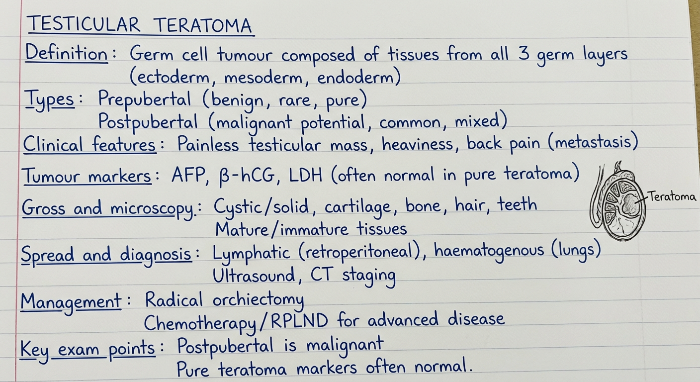
Testicular teratoma - exam notes
Definition
Types and significance
| Feature | Prepubertal type | Postpubertal type |
|---|---|---|
| Typical setting | Infants and children | Adolescents and adults |
| Relationship to GCNIS | Not associated | Usually GCNIS-derived |
| i(12p) abnormality | Absent | Usually present |
| Biological behaviour | Usually benign | Malignant potential, even if histologically mature |
| Occurrence | Often pure | Often part of a mixed NSGCT |
Robbins & Kumar Basic Pathology, p. 901.
Pathology
- Well-circumscribed, often heterogeneous mass
- Solid and cystic areas
- May contain cartilage, bone, teeth, hair, skin-like tissue, or mucinous material
- Hemorrhage/necrosis may occur in mixed malignant tumours
- Tissues from two or three germ layers, arranged haphazardly:
- Ectoderm: skin, neural tissue, squamous epithelium
- Mesoderm: cartilage, bone, muscle, fat
- Endoderm: respiratory or gastrointestinal epithelium, glands
- May be mature or contain immature, commonly neuroectodermal, tissue.
- Rarely develops somatic-type malignancy, for example sarcoma, adenocarcinoma, or squamous carcinoma.
Robbins & Kumar Basic Pathology, p. 901.
Clinical features
- Painless unilateral testicular swelling or mass
- Heaviness or dull ache in the scrotum
- Occasionally acute pain due to hemorrhage/infarction
- Retroperitoneal nodal disease or distant metastases may cause back pain, cough, or constitutional symptoms.
Tumour markers
- Pure teratoma: usually does not elevate AFP or beta-hCG.
- Raised AFP suggests an associated yolk-sac tumour component.
- Raised beta-hCG may indicate associated choriocarcinoma or syncytiotrophoblastic cells.
- Obtain AFP, beta-hCG, and LDH before orchiectomy and use serial values for staging/follow-up.
Diagnosis and staging
- Scrotal ultrasonography: intratesticular, often heterogeneous cystic-solid lesion.
- Serum AFP, beta-hCG, LDH.
- Radical inguinal orchiectomy for diagnosis and primary treatment.
- Do not perform trans-scrotal biopsy or scrotal orchiectomy.
- CT chest/abdomen/pelvis for staging.
- Histology determines pure versus mixed GCT and prepubertal versus postpubertal type.
Treatment
- Testis-sparing surgery may be considered for a benign-appearing lesion with normal markers and adequate residual normal testis.
- Orchiectomy is used if malignancy cannot be excluded.
- Radical inguinal orchiectomy first.
- Stage I disease: risk-adapted surveillance or retroperitoneal lymph-node dissection (RPLND) in selected cases.
- Metastatic NSGCT: cisplatin-based chemotherapy when indicated, followed by resection of residual masses where appropriate.
- Teratoma is relatively chemotherapy-resistant, so residual teratoma requires complete surgical excision when feasible.
- Radiotherapy has no routine role.
Complication: growing teratoma syndrome
- Enlarging metastatic masses during/after chemotherapy
- Tumour markers normalize or fall
- Resection specimen shows mature teratoma
- Treatment: complete surgical excision, not additional chemotherapy.
One-line viva answer
Testicular teratoma is a nonseminomatous germ-cell tumour containing differentiated tissues from multiple germ layers; it is usually benign in the prepubertal type but has malignant potential in postpubertal patients regardless of whether the tissue appears mature.
Seminoma handwritten
testicular seminoma
testicular seminoma diagnosis treatment overview NCI
A single clean handwritten medical exam-notes page titled “SEMINOMA (Testis)”. Use realistic highly legible blue-ink handwriting on faint ruled white notebook paper. Include compact correct exam bullets: Definition: malignant germ-cell tumour, most common testicular GCT, peak 30–40 years. Risk factors: cryptorchidism, gonadal dysgenesis, family history. Clinical: painless testicular enlargement. Gross: homogeneous pale grey-white fleshy mass, no hemorrhage or necrosis. Microscopy: sheets/lobules of uniform large cells with clear glycogen-rich cytoplasm, central nuclei and prominent nucleoli; fibrous septa with lymphocytes; may have granulomas. Markers: AFP normal; beta-hCG mildly elevated in some cases due to syncytiotrophoblasts; LDH. Spread: lymphatic to para-aortic nodes. Diagnosis: ultrasound, markers, radical inguinal orchiectomy, CT staging. Treatment: stage I surveillance preferred; radiotherapy or carboplatin selected cases; advanced disease cisplatin chemotherapy. Prognosis excellent, highly radiosensitive and chemosensitive. Add a small neat hand-drawn testis and a small microscopic cell sketch in margins. All text must be readable and spelling exact.
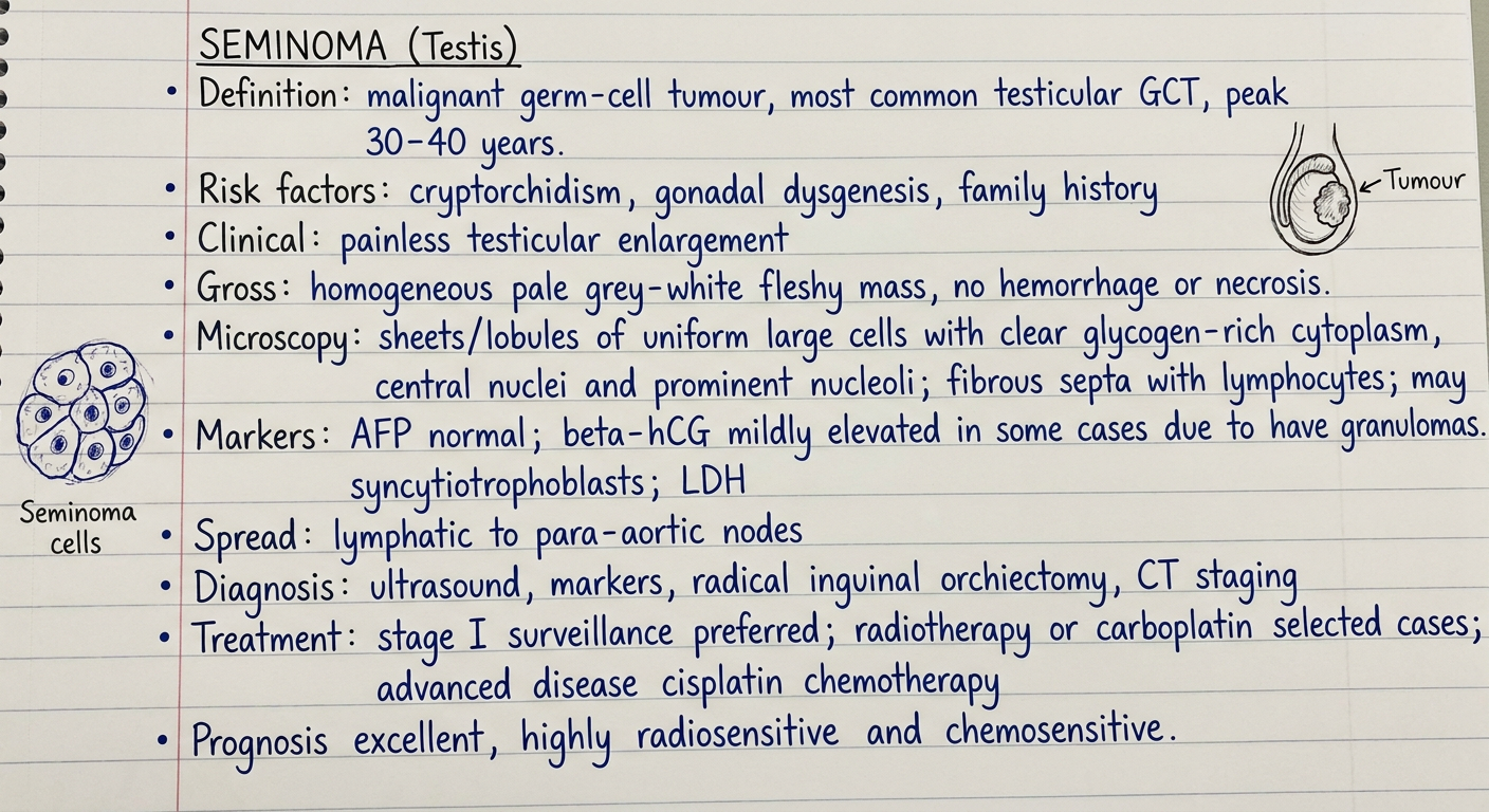
Seminoma - handwritten-style exam notes
Definition
- Seminoma is the most common pure testicular germ-cell tumour.
- It is a malignant tumour arising from germ-cell neoplasia in situ (GCNIS).
- Typically affects men aged 30-40 years.
Risk factors
- Cryptorchidism
- Gonadal dysgenesis
- Family history of testicular germ-cell tumour
- Prior germ-cell tumour in the opposite testis
Clinical features
- Painless enlargement or a firm testicular mass
- Feeling of testicular heaviness or dull ache
- Occasionally back pain from retroperitoneal lymph-node metastasis
- Gynecomastia is uncommon and may occur if beta-hCG is elevated.
Gross morphology
- Well-defined, homogeneous, pale gray-white, fleshy mass
- Usually no hemorrhage or necrosis
- Large lesions may rarely show necrosis
Microscopy
- Sheets or lobules of large uniform polygonal cells
- Clear, glycogen-rich cytoplasm with distinct cell borders
- Central round nucleus and prominent nucleolus
- Fibrous septa containing dense lymphocytic infiltrate
- Granulomatous reaction may occur
- Occasional syncytiotrophoblasts can be present.
Tumour markers
- AFP: always normal in pure seminoma
- Raised AFP indicates an associated nonseminomatous component until proved otherwise.
- beta-hCG: mildly elevated in about 10%-15% due to syncytiotrophoblasts.
- LDH: may reflect tumour burden.
Spread
- Spreads mainly through lymphatics.
- First involves para-aortic/retroperitoneal lymph nodes.
- Hematogenous spread occurs later, commonly to lungs, liver, bone, or brain.
Diagnosis
- Scrotal ultrasound
- Serum AFP, beta-hCG, and LDH
- Radical inguinal orchiectomy for diagnosis and primary treatment
- CT of chest, abdomen, and pelvis for staging
- Avoid trans-scrotal biopsy.
Treatment
- Stage I: radical inguinal orchiectomy followed mainly by active surveillance.
- Selected stage I cases: adjuvant carboplatin or para-aortic radiotherapy.
- Stage IIA/IIB: radiotherapy or cisplatin-based chemotherapy, depending on nodal burden and clinical factors.
- Advanced or bulky disease: cisplatin-based combination chemotherapy.
Prognosis
- Highly radiosensitive and chemosensitive.
- Excellent cure rate, including in advanced disease.
- Stage I disease has a cure rate approaching 100% with appropriate management, as summarized by the NCI treatment guidance.
A homogeneous gray-white testicular tumour with clear cells, fibrous septa, and lymphocytes is seminoma. AFP must remain normal in pure seminoma.