

IMG-20260706-WA0002.jpg
একবার একটু কি আমি একটা নতুন suggestion পাঠিয়েছি। সেটা হচ্ছে ওখানে একটা neuro টানো। একটা neuro বাদ দিয়ে যে abdomen আছে, abdomen এতে অনেকগুলো topic
আপনার suggestion বুঝতে পেরেছি। ছবিতে তিনটি section আছে:
- Abdomen - অনেক বেশি topic (২০+ topics)
- Head & Neck - মাঝারি topics
- Neuroanatomy - কম topics
আপনি বলছেন যে Neuroanatomy থেকে একটি topic বাদ দিয়ে, সেই জায়গায় Abdomen-এর একটি topic সরিয়ে দিতে চান - কারণ Abdomen-এ অনেক বেশি topic আছে, load কমাতে।
একটু clarify করুন:
Asking for Preferences
আপনি কি করতে চাইছেন?
আমি বলছি, এখান থেকে শুধু abdomen-এ যেই যেই question গুলো আছে, সেই সেই question গুলোর ছবি দিয়ে ভালো করে আমাকে হচ্ছে করিয়ে দে। কালকে পরীক্ষা তো তার আ গে।
বুঝতে পেরেছি! কালকে পরীক্ষা - Abdomen section-এর সব topics ভালো করে পড়িয়ে দেব। ছবি সহ।
Abdomen-এর topics হলো:
- Anterior abdominal wall
- Rectus sheath
- Inguinal canal
- Spermatic cord
- Testis
- Peritoneum
- Greater/Lesser sac
- Greater omentum
- Epiploic foramen
- Portal vein
- Stomach
- Duodenum
- Caecum
- Appendix
- Liver
- Biliary apparatus
- Pancreas
- Kidney
- Ureter
- Ischiorectal fossa
- Urinary bladder
- Male urethra
- Uterus
- Supports of uterus
- Anal canal
এগুলো একসাথে cover করতে গেলে অনেক বড় হবে। কোনটা থেকে শুরু করব?
Asking for Preferences
কোথা থেকে শুরু করায় ভালো?
Hi, সব প্রশ্নের answer গুলো একে একে দিই। সূচনা ইংরেজি দিদি, ডায়াগ্রাম দিবি, যেটা পরীক্ষায় আঁকা যায়। খুব একটা বড় দিবি না। আর হচ্ছে WBHS হচ্ছে যেটা হচ্ছে লিখিতে নাম্বার দেবে, সেই টপিক।
Reading File
Finding Sources
Finding Sources
Finding Sources
Reading File
Reading File
ভালো! Library থেকে ভালো content এবং diagrams পেয়েছি। এখন একে একে শুরু করি। প্রথমে Topic 1 দিয়ে শুরু করি।
Loading Image
Loading Image
চলো শুরু করি! একে একে সব topic cover করব। কালকের পরীক্ষার জন্য সবচেয়ে দরকারি points দেব।
📚 WBHS Anatomy - Abdomen Topics
✅ Topic 1: Anterior Abdominal Wall
Layers (superficial to deep) - "Some Incredible Fat Finds Extra Tissue Plainly":
| Layer | Details |
|---|---|
| Skin | |
| Superficial fascia | Camper's (fatty) + Scarpa's (membranous) |
| External oblique | Fibers run downward & medially |
| Internal oblique | Fibers run upward & medially |
| Transversus abdominis | Horizontal fibers |
| Transversalis fascia | |
| Extraperitoneal fat | |
| Parietal peritoneum |
Key points:
- Rectus abdominis = vertical muscle, enclosed in rectus sheath
- Linea alba = fusion of all 3 aponeuroses in midline
- Linea semilunaris = lateral border of rectus
- Arcuate line = midway between umbilicus & pubic symphysis
✅ Topic 2: Rectus Sheath
Formation:
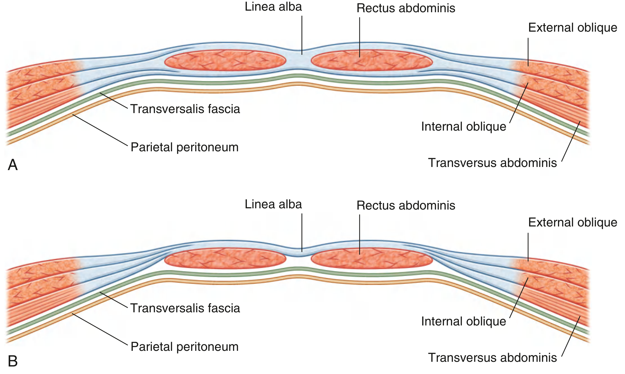
Above arcuate line (upper 3/4):
- Anterior wall = External oblique aponeurosis + Anterior half of Internal oblique aponeurosis
- Posterior wall = Posterior half of Internal oblique + Transversus abdominis aponeurosis
Below arcuate line (lower 1/4):
- Anterior wall = ALL THREE aponeuroses (Ext. oblique + Int. oblique + Transversus)
- Posterior wall = ABSENT - only Transversalis fascia
Exam tip: "Below arcuate line - posterior wall absent, rectus touches transversalis fascia directly."
Contents of rectus sheath:
- Rectus abdominis muscle
- Pyramidalis muscle
- Superior & inferior epigastric vessels
- Lower 5 intercostal nerves (T7-T11) + subcostal nerve (T12)
✅ Topic 3: Inguinal Canal
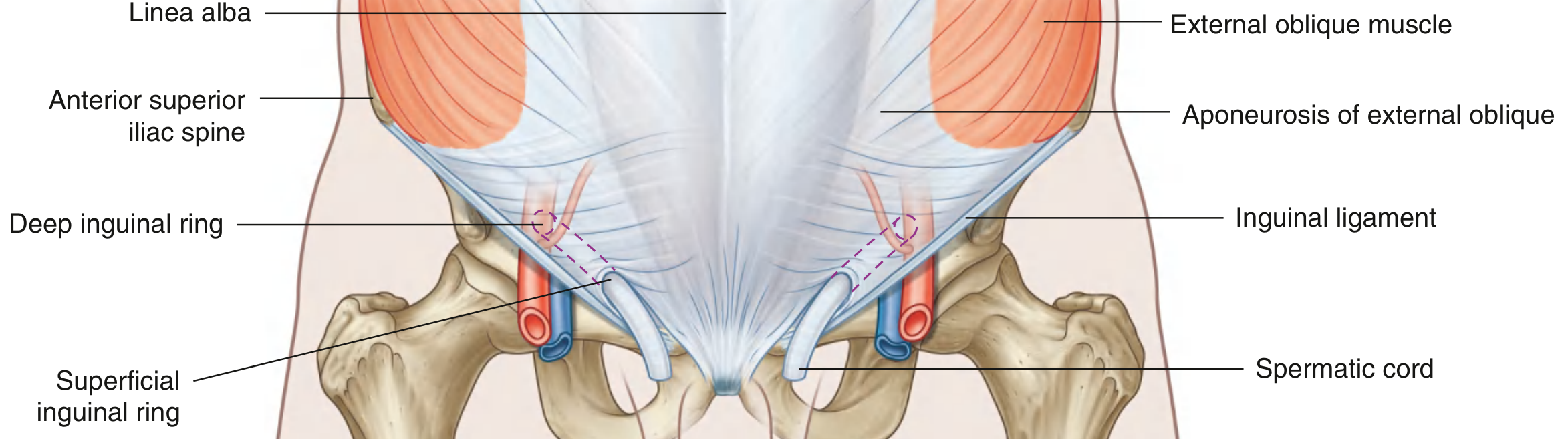
Definition: Oblique passage, ~4 cm long, above & parallel to medial half of inguinal ligament.
Boundaries - Draw this in exam:
Deep ring ----4 cm----> Superficial ring
(ASIS midpoint) (above pubic tubercle)
↓ lateral medial ↓
| Wall | Structures |
|---|---|
| Anterior | External oblique aponeurosis (whole length) + Internal oblique (lateral 1/3) |
| Posterior | Transversalis fascia (whole length) + Conjoint tendon (medial 1/3) |
| Roof | Arching fibers of Internal oblique + Transversus abdominis |
| Floor | Inguinal ligament + Lacunar ligament (medially) |
Contents:
- Male: Spermatic cord + Ilioinguinal nerve + Genital branch of genitofemoral nerve
- Female: Round ligament of uterus + Ilioinguinal nerve + Genital branch of genitofemoral nerve
Deep ring: Lateral to inferior epigastric vessels
Superficial ring: Triangular gap in external oblique aponeurosis, above pubic tubercle
Exam trick - Hernia:
- Indirect hernia = enters through deep ring (lateral to inferior epigastric) - congenital
- Direct hernia = pushes through posterior wall (medial to inferior epigastric) - acquired, older men
এটুকু পড়ে নাও। এরপর বলো "Next" - আমি পরের topics দেব:
Topic 4: Spermatic Cord → Topic 5: Testis → Topic 6: Peritoneum...
তোমার পরীক্ষার জন্য সবগুলো একসাথে না দিয়ে batch করে দিচ্ছি যাতে ভালো করে মাথায় ঢোকে। 💪
ঠাই, কিন্তু এটা এত কম লিখলে তো হবে না। কারণ মানুষ যে এত কম লিখলে নাম্বার দেবে পরীক্ষায়? তোমাকে বললাম।
ঠিক আছে! WBHS লিখিত পরীক্ষায় ভালো নম্বর পেতে হলে detailed answer দরকার। একটা একটা topic বিস্তারিত করে দিই।
প্রথমে বলো - কোন topic থেকে শুরু করব? নাকি একটা একটা করে সব দিই?
Asking for Preferences
কোন topic দিয়ে শুরু করব? · কতটা detail চাও?
ঠিক আছে! Anterior Abdominal Wall থেকে শুরু করি, 300-400 words করে। Library থেকে content টেনে দিচ্ছি।
Finding Sources
Finding Sources
Finding Sources
Finding Sources
Reading File
Loading Image
Reading File
চমৎকার! এখন সব তথ্য পেয়েছি। বিস্তারিত উত্তর লিখছি।
📚 WBHS Anatomy — Topic 1
ANTERIOR ABDOMINAL WALL
Boundaries:
- Superiorly: Xiphoid process and costal margins (7th-10th costal cartilages)
- Posteriorly: Vertebral column (lumbar vertebrae)
- Inferiorly: Upper parts of pelvic bones (iliac crests, inguinal ligaments, pubic symphysis)
Layers of Anterior Abdominal Wall (Superficial to Deep)
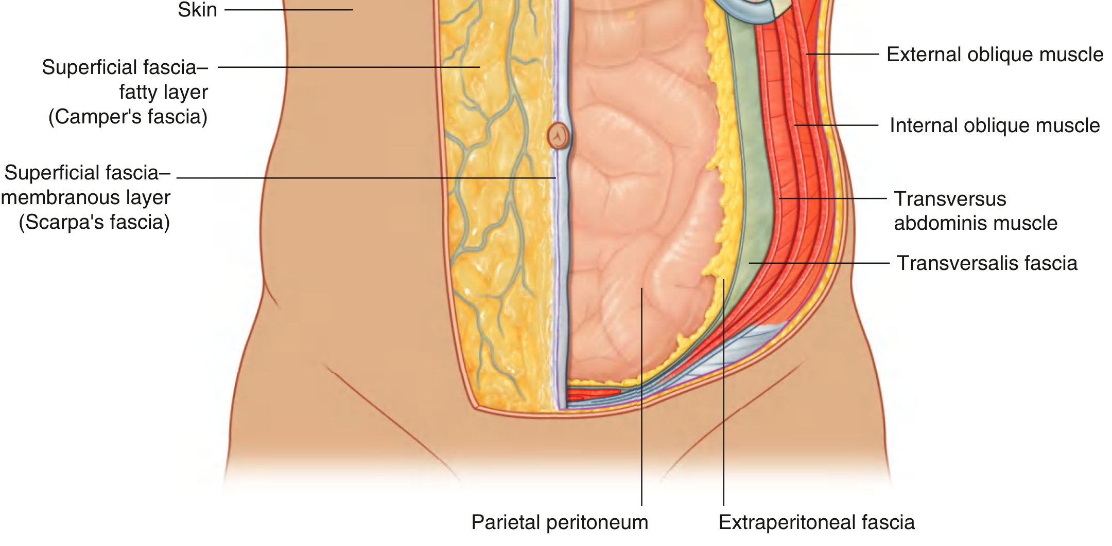
(1) Skin
Thin and mobile, attached loosely to the underlying layers except at the umbilicus where it is firmly attached.
(2) Superficial Fascia (Subcutaneous tissue)
Has TWO layers below the umbilicus:
- Superficial layer = Camper's fascia - fatty layer, contains fat, continuous with superficial fascia of thigh. In males continues over penis and into scrotum forming dartos fascia. In females forms a component of labia majora.
- Deep layer = Scarpa's fascia - membranous layer, contains little or no fat. Inferiorly fuses with fascia lata of thigh (just below inguinal ligament). In midline firmly attached to linea alba and symphysis pubis. Continues into perineum as Colles' fascia.
(3) Muscles (Anterolateral group - 5 muscles)
There are 3 flat muscles and 2 vertical muscles:
Flat Muscles:
| Muscle | Origin | Insertion | Fiber Direction |
|---|---|---|---|
| External oblique | Lower 8 ribs | Linea alba, pubic tubercle, ASIS | Inferomedially ("hands in pocket") |
| Internal oblique | Inguinal lig., iliac crest, thoracolumbar fascia | Lower 3 ribs, linea alba, pubic crest | Superomedially (at right angles to external oblique) |
| Transversus abdominis | Inguinal lig., iliac crest, lower 6 costal cartilages | Linea alba, pubic crest | Horizontal (transverse) |
Vertical Muscles:
- Rectus abdominis - from pubic symphysis/crest to xiphoid process and 5th-7th costal cartilages. Has 3-4 tendinous intersections. Enclosed in rectus sheath.
- Pyramidalis - small triangular muscle, may be absent. Pubic crest to linea alba. Enclosed in rectus sheath anterior to rectus.
Functions of anterolateral muscles:
- Maintain abdominal viscera within cavity and protect them
- Help maintain erect posture against gravity
- Contraction assists expiration (quiet and forced) by pushing viscera upward
- Increase intra-abdominal pressure during: coughing, vomiting, defecation, micturition, childbirth
(4) Transversalis fascia
Deep fascia lining the inner surface of transversus abdominis. Forms the internal spermatic fascia at the deep inguinal ring.
(5) Extraperitoneal fascia
Connective tissue layer deep to transversalis fascia. Contains variable amounts of fat. Separates transversalis fascia from peritoneum. Viscera located here are called retroperitoneal (e.g., kidneys, ureters, aorta).
(6) Parietal peritoneum
Innermost layer. Lines the abdominal cavity. Sensitive to pain, temperature, touch.
Important Structures:
- Linea alba - fibrous band in midline formed by fusion of all 3 aponeuroses. From xiphoid to pubic symphysis.
- Linea semilunaris - curved line at lateral border of rectus abdominis
- Arcuate line (Douglas) - curved line midway between umbilicus and pubic symphysis, marks lower border of posterior rectus sheath
- Inguinal ligament - lower free border of external oblique aponeurosis, from ASIS to pubic tubercle
Blood Supply:
- Superior epigastric artery (from internal thoracic artery) - enters rectus sheath superiorly
- Inferior epigastric artery (from external iliac artery) - enters rectus sheath inferiorly
- They anastomose within the sheath
- Also: lower intercostal arteries, deep circumflex iliac artery
Nerve Supply:
- T7-T11 (lower intercostal nerves) - segmental supply
- T12 (subcostal nerve)
- L1 via iliohypogastric and ilioinguinal nerves
Applied Anatomy:
- McBurney's incision - muscle-splitting incision through anterior abdominal wall for appendicectomy
- Urine extravasation after urethral rupture tracks in Scarpa's fascia plane (does NOT go to thigh due to fusion with fascia lata; goes to scrotum and perineum)
- Haematoma of rectus sheath - inferior epigastric artery injury; presents as painful abdominal mass
Next topic - Topic 2: Rectus Sheath দেব? নাকি অন্য topic চাও? বলো "Next" দিলেই দিচ্ছি। 💪
This is a shared conversation. Sign in to Orris to start your own chat.