
BERA ENT in detaill
Reading File
BERA (Brainstem Evoked Response Audiometry) is a key electrophysiological test in ENT used to assess the auditory pathway from cochlea to brainstem. Let me search the medical library for detailed coverage.
Searching sources...
Finding Sources
Finding Sources
Finding Sources
Finding Sources
Good. I have three strong sources. Let me read them in parallel now.
Reading File
Reading File
Reading File
Excellent content. Let me now read more from Cummings (the most detailed ABR section) and from Scott-Brown's on clinical applications.
Reading File
Reading File
Reading File
Reading File
I now have extensive, high-quality information. Let me also fetch the ABR waveform image and check clinical applications/neonatal screening from KJ Lee, then compile the full answer.
Finding Sources
Loading Image
I have more than enough detailed content from three authoritative ENT textbooks. Here is the comprehensive answer:
BERA (Brainstem Evoked Response Audiometry) - Detailed Notes for ENT
BERA is synonymous with ABR (Auditory Brainstem Response). It is an electrophysiological test that records electrical activity of the auditory pathway from the cochlea to the brainstem in response to auditory stimuli.
1. Definition
BERA records surface-averaged electrical potentials generated by sequential activation of the auditory nerve and brainstem nuclei following acoustic stimulation. Because the brainstem is relatively impervious to sedatives, anaesthetics, and CNS depressants, it is an objective, reliable test in uncooperative or sleeping patients.
- Cummings Otolaryngology, p. 2577
2. Historical Background
- First described by Jewett and Williston who identified a complex occurring under 10 ms post-onset of an auditory stimulus, comprising five distinct potentials (waves I-V).
- Scott-Brown's Otorhinolaryngology, p. 694
3. Basis / Principle
An auditory click stimulus causes synchronized activation of thousands of auditory nerve fibres. The summed electrical activity is recorded from scalp electrodes, averaged across 1000-3000 sweeps, and produces a characteristic waveform with five positive peaks (waves I-V) within 10 ms.
4. Wave Generators ("EECOL" Mnemonic)
| Wave | Generator | Approximate Normal Absolute Latency |
|---|---|---|
| I | Distal (peripheral) end of 8th nerve (cochlear nerve) | ~1.5 ms |
| II | Proximal end of 8th nerve (near porus acusticus) | ~2.5 ms |
| III | Cochlear nucleus (ventral acoustic stria) | ~3.5 ms |
| IV | Superior olivary complex + lateral lemniscus | ~4.5 ms |
| V | Lateral lemniscus (+ possibly inferior colliculus) | ~5.5 ms |
Mnemonic: EECOL - Eighth nerve (distal), Eighth nerve (proximal), Cochlear nucleus, Olivary complex, Lateral lemniscus
- KJ Lee's Essential Otolaryngology, p. 1211
- Cummings Otolaryngology, p. 2578
- Scott-Brown's Otorhinolaryngology, p. 694
Note: Wave V is the most robust and reliable wave. Waves IV and V often fuse into a complex (IV/V complex).
5. Technique / Recording Parameters
Electrodes
- Non-inverting (active): High forehead below the hairline or vertex (Cz)
- Inverting (reference): Medial surface of the ipsilateral earlobe (Ai) - emphasises Wave I
- Contralateral inverting: Medial surface of the contralateral earlobe - emphasises separation of waves IV and V
- Ground: Centre of the forehead
This gives a 2-channel montage.
Stimulus
- Click stimulus: 100 microsecond rectangular pulse - most common for retrocochlear testing
- Tone burst/pip: Frequency-specific (used for threshold estimation, e.g. 500 Hz, 1000 Hz, 2000 Hz, 4000 Hz)
- Stimulus rate: 20-30 clicks/second (non-integer rate such as 17.7/sec to avoid periodic noise)
- Fast-rate ABR uses 77.7 clicks/second
- Polarity: Constant or alternating
Signal Averaging
- 1000-3000 sweeps are averaged to extract the evoked potential from background noise
- Each condition is replicated (acquired twice or three times) to confirm reliability
Filtering
- High-pass filter: 100 Hz
- Low-pass filter: 1000-3000 Hz
- For 500 Hz tone burst: lower high-pass to ~30 Hz
- Notch filter (60 Hz) only used sparingly when electrical artefact cannot be eliminated
Recording Window
-
10-15 ms post-stimulus onset - to capture all five waves
-
Scott-Brown's Otorhinolaryngology, p. 694-695
-
Cummings Otolaryngology, p. 2577-2578
6. Normal Interpeak Latency Values
| Interval | Normal Limit |
|---|---|
| Wave I absolute latency | ~1.5 ms |
| Wave V absolute latency | < 6.2 ms |
| I-III interwave interval | < 2.5 ms (KJ Lee: 2.3 ms) |
| III-V interwave interval | < 2.4 ms (KJ Lee: 2.1 ms) |
| I-V interwave interval | < 4.4 ms |
| Inter-ear latency difference (ILD) of wave V | < 0.5 ms |
| Fast-rate latency shift | < 1.0 ms |
- KJ Lee's Essential Otolaryngology, p. 1211
- Scott-Brown's Otorhinolaryngology, Table 52.1, p. 695
7. Clinical Indications
A. Neonatal / Paediatric Hearing Screening
- Brainstem function is identifiable on ABR at approximately 28 weeks gestational age - waves I, III and V appear.
- ABR "maturity" (adult-like latencies) is not reached until approximately 18 months after birth - neonatal latencies are prolonged compared to adults.
- BERA is the gold standard test for Universal Newborn Hearing Screening (UNHS) when OAE refer.
- It is the first-choice test for infants in the at-risk registry: birth asphyxia, ototoxic drugs, NICU, family history, congenital infections, craniofacial anomalies, hyperbilirubinaemia.
- KJ Lee's Essential Otolaryngology, p. 1210-1211
B. Threshold Estimation
- Used in children, neonates, and malingerers who cannot or will not cooperate with behavioural audiometry.
- Tone-burst BERA gives frequency-specific thresholds at 500 Hz, 1000 Hz, 2000 Hz, 4000 Hz.
- ABR threshold correlates well with behavioural auditory threshold.
C. Retrocochlear Lesion Detection - Acoustic Neuroma (Vestibular Schwannoma)
- The primary neurodiagnostic indication.
- An acoustic neuroma delays conduction time along the cochlear nerve, prolonging absolute and interwave latencies.
- Diagnostic sensitivity of ABR for acoustic neuroma:
- I-III interval: ~90% sensitivity, 100% specificity
- I-V interval: ~75% sensitivity
- III-V interval: ~45% sensitivity
- Inter-ear latency difference (ILD): >90% sensitivity and specificity
- Scott-Brown's Otorhinolaryngology, p. 695
D. Neurological / Brainstem Disease
- Multiple sclerosis (demyelination prolongs interwave latencies)
- Brainstem tumours, haemorrhage, infarction
- Coma and brain death evaluation
E. Intraoperative Monitoring
- Used during posterior fossa surgery (e.g. acoustic neuroma resection, microvascular decompression) to monitor auditory nerve integrity in real time.
- Decline or loss of wave V amplitude or marked latency prolongation warns the surgeon of potential cochlear nerve injury.
- Cummings Otolaryngology, p. 2578-2579
8. Interpretation - Patterns of Abnormality
Retrocochlear Lesion (e.g. Acoustic Neuroma)
Suspect if ANY of the following:
- Interpeak latency I-V > 4.4 ms
- Interaural wave V latency difference > 0.2 ms (KJ Lee) or > 0.5 ms (Scott-Brown normative table)
- III-V interval > 2.4 ms
- Absent wave I with present wave V (suggests peripheral cochlear nerve involvement)
- Absent or poorly formed ABR waveform
- Fast-rate latency shift > 1.0 ms
Cochlear (Sensory) Hearing Loss
- All absolute latencies are prolonged equally (latency-intensity shift)
- Interwave intervals remain normal
- ABR threshold elevated corresponding to degree of hearing loss
Auditory Neuropathy / Dyssynchrony (AN/AD)
- Normal OAEs (outer hair cells intact)
- Absent or severely abnormal ABR (lack of neural synchrony)
- This dissociation is the hallmark finding.
Conductive Hearing Loss
- All wave latencies shifted to the right (prolonged) by the amount of conductive loss
- Interwave intervals remain normal
- ABR improves with bone conduction stimulation
Normal ABR
- All five waves (I-V) present with normal absolute and interwave latencies
- Symmetrical bilaterally
9. BERA vs. OAE - Comparison
| Feature | BERA/ABR | OAE |
|---|---|---|
| Tests | Auditory nerve + brainstem | Outer hair cells (cochlea) |
| Result | Objective threshold + neural path | Cochlear function only |
| Sedation needed | Often (especially neonates) | No |
| Frequency specific | Yes (tone burst) | Yes |
| Screening cost | Higher | Lower (preferred 1st screen) |
| Auditory neuropathy | Abnormal | Normal |
10. Advantages of BERA
- Objective - does not require patient cooperation or response
- Unaffected by sedation/anaesthesia - brainstem is impervious to CNS depressants
- Frequency-specific thresholds possible with tone bursts
- Neurodiagnostic - detects retrocochlear and brainstem pathology
- Intraoperative monitoring capability
- Useful from neonatal period onwards
11. Limitations
- Time-consuming compared to OAE
- Requires quiet, often sedated patient in infants
- A good response requires synchronous neural firing - absent in auditory neuropathy, but this is also diagnostically useful
- Does not test cortical/central auditory processing (needs AMLR or P300 for that)
- Cannot assess hearing above the brainstem level
12. Aural Atresia and BERA
BERA is part of the audiological workup in congenital aural atresia at birth (along with OAE) with formal audiometry at 18 months to plan management.
- KJ Lee's Essential Otolaryngology
Summary Table
| Feature | Details |
|---|---|
| Also called | ABR, BAER, BAEP |
| Stimulus | Click (100 µs) or tone burst |
| Number of sweeps | 1000-3000 |
| Recording window | 10-15 ms |
| Waves | I-V (5 peaks) |
| Most reliable wave | Wave V |
| Normal I-V interval | < 4.4 ms |
| Normal wave V latency | < 6.2 ms |
| Key diagnostic use | Acoustic neuroma, neonatal screening |
| Intraoperative use | Yes |
| Affected by anaesthesia | No |
Sources:
- Cummings Otolaryngology Head and Neck Surgery, pp. 2577-2579
- Scott-Brown's Otorhinolaryngology Head & Neck Surgery Vol 2, pp. 694-695
- KJ Lee's Essential Otolaryngology, p. 1211
Retinoblastoma
Reading File
Retinoblastoma is the most common intraocular malignancy of childhood. Let me search the medical library for detailed coverage.
Searching sources...
Finding Sources
Finding Sources
Excellent. Kanski's Clinical Ophthalmology and Robbins Pathology are the best sources. Let me read all key sections in parallel.
Reading File
Reading File
Reading File
Excellent data from Kanski's and Thompson genetics. Let me read more of Kanski's for clinical features, staging, and treatment, and also fetch the key images.
Reading File
Reading File
Now let me read more from Kanski's for clinical features, staging (International Classification), and treatment details, and fetch the key clinical images.
Reading File
Loading Image
I now have comprehensive data from Kanski's Ophthalmology, Robbins Pathology, and Thompson Genetics. Let me read the remaining treatment section and fetch one more clinical image.
Reading File
Loading Image
I now have comprehensive content. Let me fetch the Flexner-Wintersteiner rosette histology image and the leukocoria image from Kanski's, then compile the full answer.
Reading File
Loading Image
I now have all the information needed for a comprehensive answer. Here it is:
Retinoblastoma - Detailed Notes
1. Overview
Retinoblastoma is the most common primary intraocular malignancy of childhood, occurring in up to 1:18,000 live births. It accounts for approximately 3% of all childhood cancers. After uveal melanoma, it is the second most common malignant intra-ocular tumour overall. Survival rates exceed 95% in specialised centres, but are much lower in the developing world.
- Kanski's Clinical Ophthalmology, p. 891
2. Genetics - The "Two-Hit Hypothesis" (Knudson)
Retinoblastoma is the prototype of a tumour suppressor gene (TSG) disease. The responsible gene is RB1 on chromosome 13q14.
Two-Hit Model
- First hit: mutation in one RB1 allele
- Second hit: somatic mutation in the second allele → loss of both copies → malignant transformation
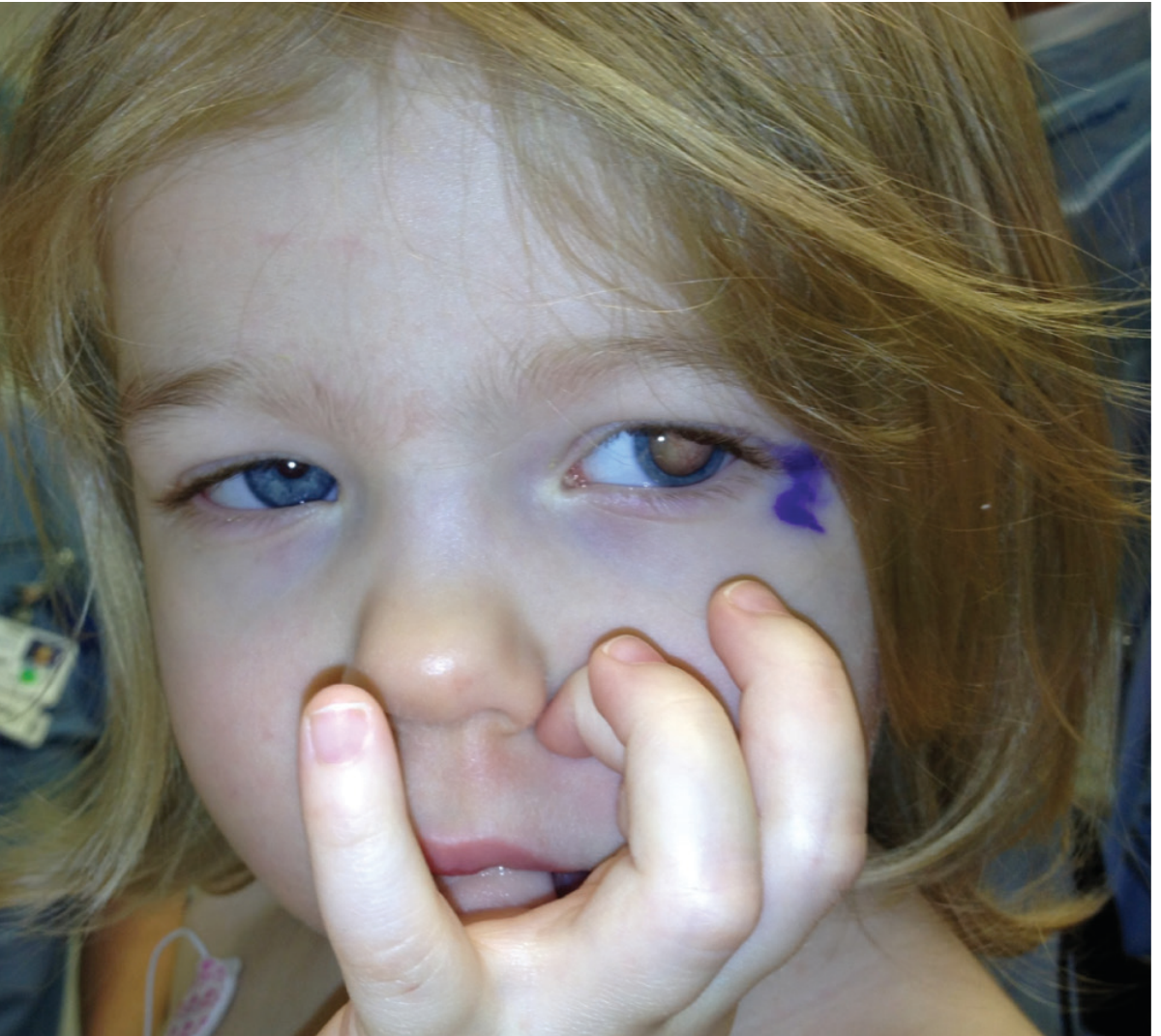
Retinoblastoma presenting as leukocoria (white reflex) in the left eye of a young child (Thompson & Thompson Genetics)
Hereditary vs. Sporadic
| Feature | Hereditary (Germline) | Sporadic (Somatic) |
|---|---|---|
| Proportion | 40% | 60% |
| First hit | Germline (all cells) | Somatic (single retinal cell) |
| Laterality | Usually bilateral, multifocal | Unilateral, unifocal |
| Age of onset | Earlier (< 1 year) | Later (~2 years) |
| Inheritance | Autosomal dominant (apparent) | Not inherited |
| Second malignancies | Yes (osteosarcoma, etc.) | No |
| Penetrance | >90% but not complete | - |
- Over 900 different RB1 mutations have been reported.
- In a small proportion of sporadic cases, NMYC proto-oncogene amplification is observed instead of RB1 mutation.
- Thompson & Thompson Genetics, p. 370; Kanski's, p. 891; Robbins Pathology, p. 1924
Second Malignancies in Heritable Retinoblastoma
- Pinealoblastoma ("trilateral retinoblastoma") - up to 10%, usually before age 5
- Osteosarcoma
- Soft tissue sarcoma
- Melanoma
- Overall risk ~6%, but increases 5-fold if external beam radiotherapy was used
- Kanski's Clinical Ophthalmology, p. 891
3. Pathology
Gross Appearance
Growth patterns:
- Endophytic - grows into the vitreous; seeding of tumour cells throughout the eye
- Exophytic - grows into the subretinal space; causes retinal detachment
- Mixed
- Diffuse infiltrating - rare pattern; flat infiltration of retina, often older children, mimics uveitis
Optic nerve invasion may occur with spread along the subarachnoid space to the brain. Metastases go to regional lymph nodes, lung, brain, and bone.
Histology
- Small, round, basophilic cells (retinoblasts) with large hyperchromatic nuclei and scanty cytoplasm
- Many tumours are undifferentiated
- Differentiation is shown by rosette formation:
| Rosette Type | Significance |
|---|---|
| Flexner-Wintersteiner rosettes | Most characteristic; show photoreceptor differentiation; columnar cells around a central lumen |
| Homer-Wright rosettes | Less specific; pseudorosettes; no central lumen |
| Fleurettes | Most differentiated; represent photoreceptor-like elements |
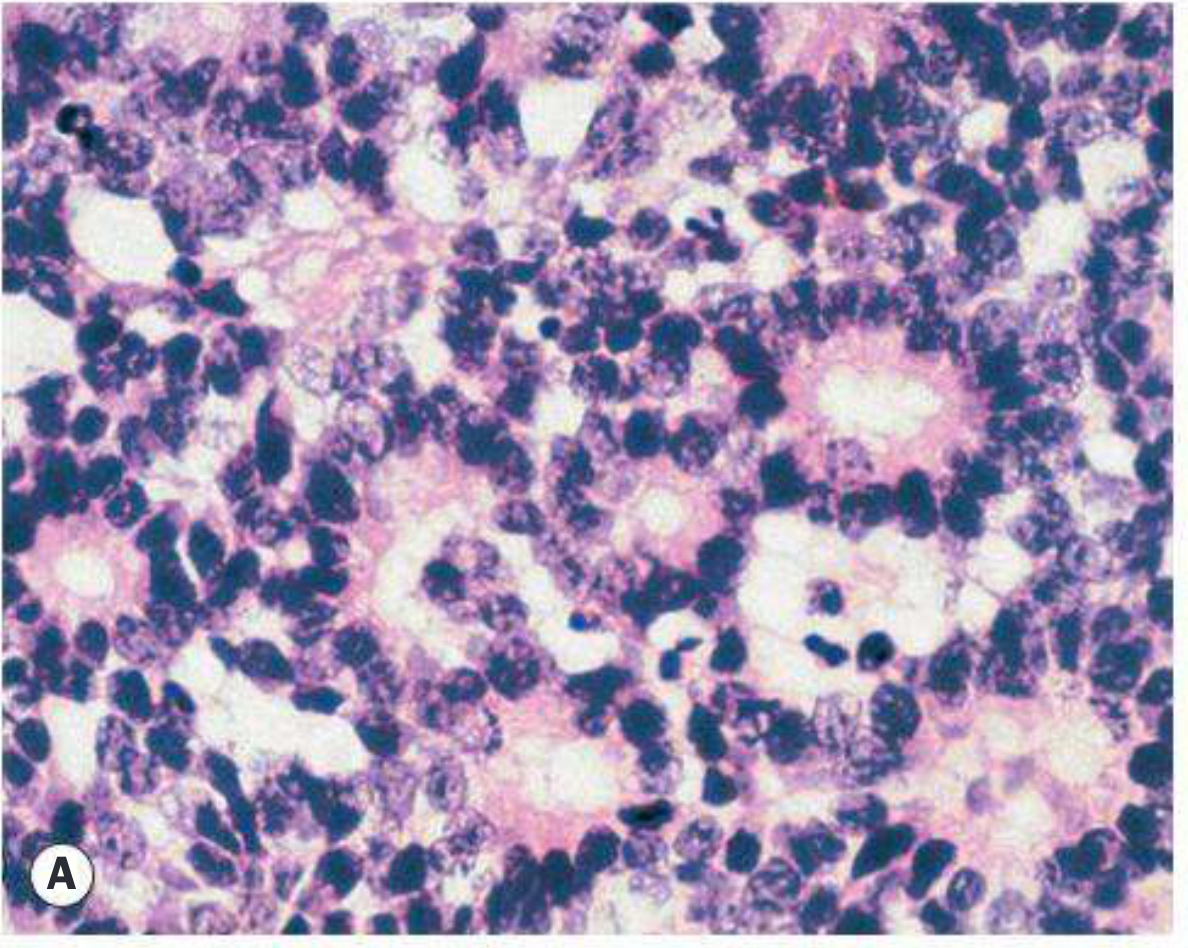
Histology: Dense clusters of small basophilic cells with large hyperchromatic nuclei - undifferentiated retinoblastoma (Robbins Pathology)
- Focal zones of dystrophic calcification are characteristic
- Viable tumour cells encircle blood vessels; zones of necrosis in relatively avascular areas
- Degree of differentiation does not correlate with prognosis
- Robbins & Cotran Pathologic Basis of Disease, p. 1924
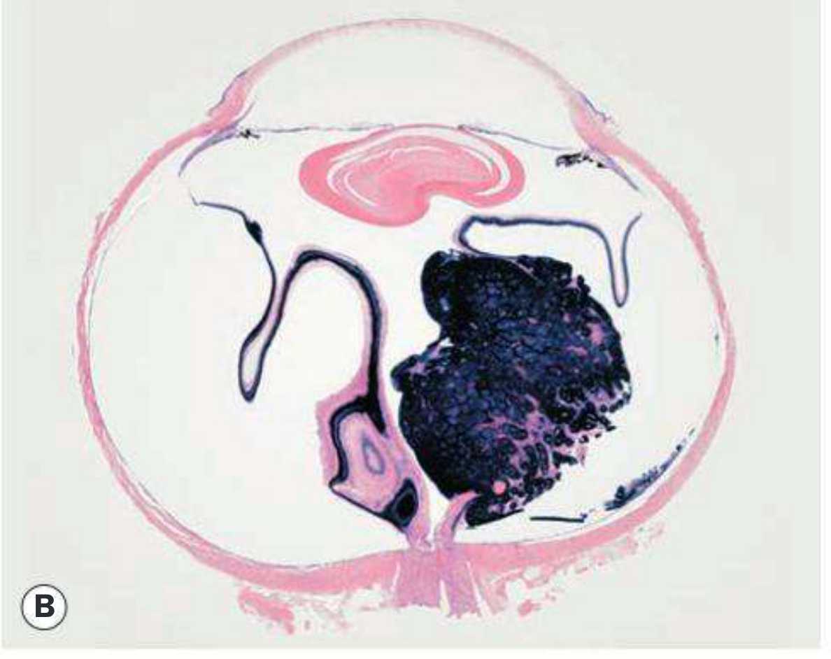
Gross section of enucleated globe showing the dark tumour mass filling most of the vitreous cavity (Kanski's Ophthalmology)
4. Clinical Features
Age of Presentation
- Bilateral cases: within the first year of life
- Unilateral cases: around 2 years of age
Presenting Signs (in order of frequency)
| Feature | Frequency |
|---|---|
| Leukocoria (white pupillary reflex) | ~60% - most common |
| Strabismus | ~20% - second most common |
| Painful red eye (neovascular glaucoma) | Less common |
| Heterochromia iridis | Rare |
| Orbital cellulitis appearance | Rare (masquerade) |
| Proptosis | Advanced disease |
| Reduced vision | May be noted by parents |
TIP: Retinoblastoma must be excluded in any young child with leukocoria or a squint. Fundus examination is mandatory in ALL cases of childhood squint. - Kanski's, p. 892
Leukocoria may first be noticed in family photographs when flash photography causes a white rather than red reflex in the affected eye.
5. International Classification of Retinoblastoma (IIRC)
| Group | Description |
|---|---|
| A | Small intraretinal tumours (<3 mm) away from foveola and disc |
| B | Tumours >3 mm, or macular/juxtapapillary location, or with subretinal fluid |
| C | Tumour with focal subretinal or vitreous seeding within 3 mm of tumour |
| D | Tumour with diffuse subretinal or vitreous seeding >3 mm from tumour |
| E | Extensive tumour occupying >50% of globe ± neovascular glaucoma, haemorrhage, optic nerve/anterior chamber extension |
- Kanski's Clinical Ophthalmology, p. 892
6. Investigations
Examination Under Anaesthesia (EUA)
- General examination for congenital abnormalities
- Tonometry
- Corneal diameter measurement
- Anterior chamber exam (hand-held slit lamp)
- Wide-field ophthalmoscopy with documentation (colour drawings or photography)
- Cycloplegic refraction
Imaging
- Ultrasound (B-scan): First-line; assesses tumour size, detects calcification (characteristic), excludes differential diagnoses (e.g. Coats disease)
- CT scan: Also detects calcification but delivers radiation; avoided by many practitioners especially in heritable cases due to second malignancy risk
- MRI: Does not detect calcification; useful for optic nerve evaluation, extraocular extension, pinealoblastoma detection, and differentiating from simulating conditions
- Plain X-ray: Detects calcification in resource-poor settings
Systemic Assessment
- Physical examination
- MRI of orbit and skull (in high-risk cases)
- If metastases suspected: bone scan, bone marrow aspiration, lumbar puncture
Genetic Studies
- Tumour tissue + blood samples from patient and relatives
- RB1 mutation detection approaches 95% sensitivity
7. Differential Diagnosis of Leukocoria
| Condition | Key Differentiating Feature |
|---|---|
| Coats disease | Telangiectatic retinal vessels; no calcification on US |
| Persistent foetal vasculature (PHPV) | Microphthalmia; no calcification |
| Toxocara | History of exposure to dogs; unilateral |
| Cataract | Lens opacity on slit-lamp |
| ROP | Premature infant, bilateral |
| Norrie disease | X-linked, bilateral |
| Retinal astrocytic hamartoma | Associated with tuberous sclerosis |
8. Screening
- Germline mutations are transmitted with autosomal dominant inheritance (50% transmission; ~40% offspring affected due to incomplete penetrance)
- Siblings at risk should be screened by:
- Prenatal ultrasonography
- Ophthalmoscopy soon after birth
- Regular examinations until age 5-7 years
- With effective screening and early treatment: final visual acuity of 6/6 to 6/12 achievable in up to 90%
- Risk to siblings:
- 2% if both parents are healthy
- 40% if one parent is affected
- Kanski's Clinical Ophthalmology, p. 892
9. Treatment
Treatment is highly individualised and requires a collaborative team (ophthalmologist, paediatric oncologist, ocular pathologist, geneticist, allied health).
Chemotherapy (Mainstay)
1. Intravenous (systemic) chemotherapy
- CEV regimen: Carboplatin + Etoposide + Vincristine
- 3-6 cycles depending on group
- Used for chemo-reduction to shrink tumour before focal consolidation
2. Selective Ophthalmic Artery Infusion (Intra-arterial chemotherapy, IAC)
- Catheter via femoral artery → ophthalmic artery
- Drugs: Melphalan or topotecan
- Significantly better globe salvage vs. IV chemotherapy, especially for Group D eyes
- Risks: chorioretinal ischaemia, visual loss
- Kanski's, p. 894
3. Intravitreal chemotherapy
- Intravitreal melphalan for vitreous seeding
- Small risk of extraocular dissemination
Focal/Local Consolidation
| Modality | Indication |
|---|---|
| Transpupillary thermotherapy (TTT) | Focal consolidation post-chemo; direct treatment of small tumours |
| Cryotherapy | Pre-equatorial tumours; triple freeze-thaw technique; no deep invasion or vitreous seeding |
| Brachytherapy (radioactive plaque) | Anterior tumour without vitreous seeding; resistance to chemotherapy |
External Beam Radiotherapy (EBRT)
- Avoided especially in heritable retinoblastoma due to 5-fold increased risk of second malignancy within the irradiated field
- Reserved for residual/relapsed disease after other treatments
- Complications: cataract, radiation neuropathy, radiation retinopathy, orbital hypoplasia
Enucleation (Surgical Removal of the Eye)
Indicated when:
- Neovascular glaucoma
- Anterior chamber infiltration
- Optic nerve invasion
- Tumour occupies >50% of vitreous volume (Group E)
- Failure of chemo-reduction
- Diffuse retinoblastoma
- Technique: minimal manipulation; optic nerve section must be ≥10 mm from the globe
- Kanski's Clinical Ophthalmology, p. 894
10. Prognosis and Spread
Favourable Factors
- Small tumour (Groups A-B)
- No optic nerve involvement
- No choroidal invasion
- Early diagnosis and treatment
Adverse Prognostic Factors
- Extraocular extension
- Optic nerve invasion (especially beyond the lamina cribrosa)
- Massive choroidal invasion
- Anterior chamber involvement
Pattern of Spread
- Direct: along optic nerve → subarachnoid space → brain
- Haematogenous: to bone, bone marrow
- Lymphatic: to regional lymph nodes
- Metastases tend to go to brain and bone marrow; seldom to the lungs
- Robbins & Cotran Pathologic Basis of Disease, p. 1924
Survival
-
95% in specialised centres (developed world)
- Much lower in developing world due to late presentation
11. Key Points Summary
| Feature | Detail |
|---|---|
| Most common intraocular tumour | In children |
| Gene | RB1 on chromosome 13q14 |
| Inheritance | Autosomal dominant (40% hereditary) |
| Bilaterality | Hereditary = bilateral; Sporadic = unilateral |
| Most common presentation | Leukocoria (60%) |
| Second most common sign | Strabismus (20%) |
| Characteristic histology | Flexner-Wintersteiner rosettes |
| Characteristic imaging finding | Calcification on B-scan/CT |
| Mainstay of treatment | Chemotherapy (CEV; intra-arterial) |
| Avoid | External beam radiotherapy (hereditary cases) |
| Enucleation indication | Group E, failed chemo, optic nerve invasion |
| Trilateral retinoblastoma | + Pinealoblastoma (hereditary) |
| Prototype of | Tumour suppressor gene / two-hit hypothesis |
Sources:
- Kanski's Clinical Ophthalmology: A Systematic Approach, 10th ed., pp. 891-895
- Robbins & Cotran Pathologic Basis of Disease, pp. 1924-1925
- Thompson & Thompson Genetics and Genomics in Medicine, 9th ed., pp. 369-371
- Robbins & Kumar Basic Pathology, p. 146
This is a shared conversation. Sign in to Orris to start your own chat.