
White matter of cerebrum and corpus callosum anatomy detailed explanation in simple way
Reading File
Now let me search the medical library for white matter and corpus callosum anatomy.
Finding Sources
Finding Sources
Finding Sources
Finding Sources
Good finds. Let me now read the most relevant anatomy sections - Gray's Anatomy for white matter and corpus callosum, plus Neuroanatomy through Clinical Cases.
Finding Sources
Reading File
Let me now read the key sections - White Matter Tracts from Kaplan & Sadock and Gray's Anatomy white matter sections, along with the Neuroanatomy clinical cases book.
Reading File
Reading File
Reading File
Excellent content. Let me now read the corpus callosum section from Kaplan & Sadock and fetch relevant anatomy images.
Reading File
Reading File
Now let me fetch the key anatomy images and read the corpus callosum projections section from Kaplan:
Loading Image
Loading Image
Reading File
Now I have enough detailed content to write a comprehensive, well-illustrated explanation. Let me also fetch one more relevant image showing the white matter tracts/association fibers.
Loading Image
I now have rich content from multiple authoritative textbooks and excellent diagrams. Here is the full, detailed explanation:
White Matter of the Cerebrum & Corpus Callosum - A Simple Explanation
What Is White Matter?
Think of the brain as a city. The gray matter (the cortex) is where all the buildings are - offices, homes, factories. The white matter is the road and highway network connecting them all. Without those roads, none of the buildings can communicate.
White matter gets its name from the white, fatty myelin sheath that wraps around the axons (nerve fibers) running through it. Myelin makes signal transmission fast - like upgrading from dirt roads to motorways.
In the cerebrum, white matter occupies about 42% of the total volume of the hemispheres. The vast majority of these fibers serve cortex-to-cortex connectivity, not just connections to subcortical sites.
- Kaplan & Sadock's Comprehensive Textbook of Psychiatry, p. 1437
The Three Types of White Matter Fibers
All white matter tracts in the cerebrum fall into one of three categories:
1. Projection Fibers - The "Highways In and Out of the City"
These fibers connect the cortex to subcortical structures - the thalamus, brainstem, and spinal cord.
- Corticofugal fibers go from the cortex outward (e.g., corticospinal tract going down to the spinal cord to move muscles)
- Corticopetal fibers go into the cortex from below (e.g., thalamocortical fibers bringing sensory information up)
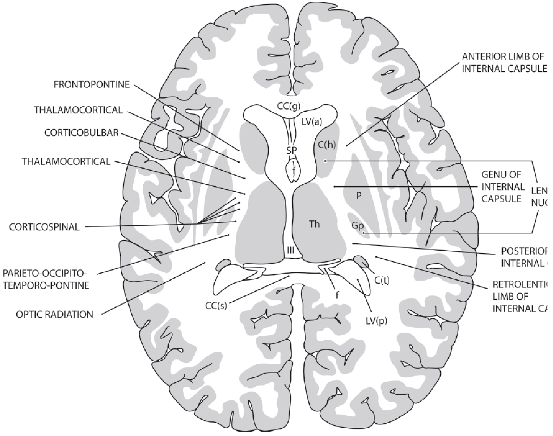
All projection fibers converge into a tight bundle called the internal capsule, which then fans out upward as the corona radiata - like a bouquet of flowers spreading from its stem.
Internal Capsule - 5 Key Parts:
| Part | Location | What runs through it |
|---|---|---|
| Anterior limb | Between caudate & lenticular nucleus | Frontopontine fibers, frontal-thalamic connections |
| Genu | Bend between anterior & posterior limbs | Corticobulbar fibers (to cranial nerve nuclei) |
| Posterior limb | Between lenticular nucleus & thalamus | Corticospinal fibers (to spinal cord) |
| Retrolenticular limb | Behind the lenticular nucleus | Optic radiation (visual pathway) |
| Sublenticular limb | Below the lenticular nucleus | Auditory radiation |
2. Commissural Fibers - The "Bridges Between the Two Cities"
These fibers cross the midline and connect the left hemisphere to the right hemisphere. The biggest and most important of these is the corpus callosum.
3. Association Fibers - The "Local Roads Within the City"
These fibers connect cortical areas within the same hemisphere. They come in two sizes:
Short association fibers (U-fibers):
- Also called arcuate fibers
- Connect adjacent gyri, looping in a "U" shape just under the cortex
- These are notably spared in diseases like Binswanger disease and CADASIL
Long association tracts (the 6 major ones):
| Tract | Connection | Clinical Note |
|---|---|---|
| Superior longitudinal fasciculus | Frontal - parietal - occipital cortices (lateral hemisphere) | Language, spatial awareness |
| Arcuate fasciculus | Frontal - temporal lobes | Damage causes conduction aphasia |
| Uncinate fasciculus | Orbital frontal - anterior temporal | Memory, behavior |
| Inferior occipitofrontal fasciculus | Occipital - frontal (running ventrally) | Visual-frontal integration |
| Inferior longitudinal fasciculus | Temporal - occipital lobes | Visual recognition |
| Cingulum | Under the cingulate gyrus; frontal - parahippocampal | Limbic connections, memory |
- Kaplan & Sadock's Comprehensive Textbook of Psychiatry, p. 202
The Corpus Callosum - The Brain's Main Bridge
What is it?
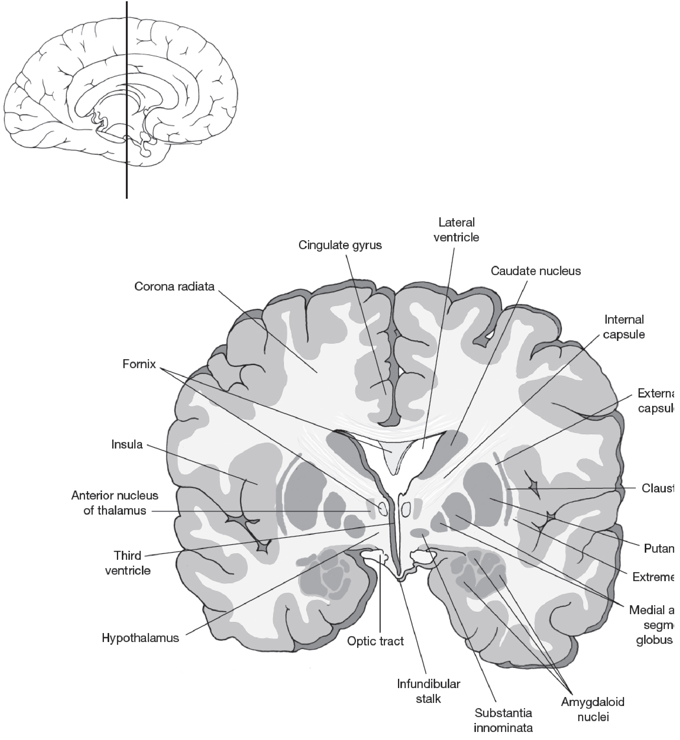
The corpus callosum is the largest white matter structure in the entire brain, containing approximately 300 million axons. It is a thick, C-shaped band lying in the midline, forming the roof of the lateral ventricles. It also supports the septum pellucidum hanging from its underside.
Think of it as the internet cable connecting two computers (the two hemispheres) so they share information seamlessly.
Structure - 4 Main Parts (from front to back)
ROSTRUM → GENU → BODY (TRUNK) → SPLENIUM
  ↑          ↑         ↑              ↑
tip at    curved    long flat       rounded
very front  front    middle part    back end
| Part | Location | What it connects |
|---|---|---|
| Rostrum | Thin, most anterior tip | Basal frontal lobes |
| Genu | The "knee" - curved front part | Prefrontal cortices (bilateral) |
| Body (trunk) | Middle section | Motor, premotor, and parietal cortices |
| Splenium | Thickened posterior end | Occipital and posterior parietal cortices; also carries optic radiations |
What does it do?
Most corpus callosum fibers connect homotopic cortical regions - meaning the same area on the left is connected to the same area on the right (e.g., left motor cortex to right motor cortex). Some fibers are heterotopic, connecting different areas across hemispheres.
Important exceptions: The hand area of the primary motor cortex and the entire primary visual cortex do NOT receive major callosal connections - these areas work more independently.
Development
The corpus callosum grows in a rostrocaudal wave (front to back) between ages 3 and 15 years. White matter volume increases roughly linearly throughout childhood and adolescence as myelination progresses. The most dramatic growth occurs in the dorsolateral prefrontal cortex between ages 5-17.
- Kaplan & Sadock's Comprehensive Textbook of Psychiatry, pp. 10861-10862
The Anterior Commissure - The Corpus Callosum's Smaller Sibling
The other main commissure is the anterior commissure - a much smaller bundle that connects structures in the two temporal lobes and the olfactory system. It is located at the inferior boundary of the interventricular foramina (Foramina of Monro).
- Neuroanatomy through Clinical Cases, 3rd Edition, p. 57
Summary: White Matter Fiber Types at a Glance
WHITE MATTER FIBERS
│
├── PROJECTION FIBERS (cortex ↔ subcortical)
│   ├── Internal capsule (compact bundle)
│   └── Corona radiata (fan above internal capsule)
│
├── COMMISSURAL FIBERS (left ↔ right hemisphere)
│   ├── Corpus callosum (LARGEST - 300 million axons)
│   │   ├── Rostrum
│   │   ├── Genu → prefrontal
│   │   ├── Body → motor/parietal
│   │   └── Splenium → occipital
│   └── Anterior commissure (temporal lobes)
│
└── ASSOCIATION FIBERS (within same hemisphere)
    ├── Short: U-fibers (adjacent gyri)
    └── Long:
        ├── Superior longitudinal fasciculus
        ├── Arcuate fasciculus
        ├── Uncinate fasciculus
        ├── Inferior occipitofrontal fasciculus
        ├── Inferior longitudinal fasciculus
        └── Cingulum
Clinical Relevance (Why This Matters)
- Stroke in the internal capsule (especially posterior limb) - causes contralateral hemiplegia because all corticospinal fibers for the opposite side of the body run through a small area
- Corpus callosum damage (trauma, tumors, multiple sclerosis) - causes "split-brain" syndrome: the left hand literally does not know what the right hand is doing; alien hand syndrome
- Agenesis of the corpus callosum - a congenital absence; patients may have seizures, intellectual disability, or occasionally be asymptomatic
- Arcuate fasciculus damage - causes conduction aphasia: patient understands speech and can speak fluently, but cannot repeat words
- Binswanger disease / small vessel disease - damages deep white matter and long association tracts, causing subcortical dementia with slowed processing and poor executive function (U-fibers are typically spared)
- Multiple sclerosis - demyelination affects white matter tracts throughout the brain, causing a wide range of motor and sensory deficits depending on lesion location
Sources:
- Kaplan & Sadock's Comprehensive Textbook of Psychiatry (White Matter Tracts, pp. 197-204; White Matter and Cerebral Connectivity, p. 1437)
- Neuroanatomy through Clinical Cases, 3rd Edition (Anatomical Study Guide, pp. 46-57)
- Junqueira's Basic Histology, 17e (Central Nervous System)
- Gray's Anatomy for Students (Corpus Striatum & White Matter)
This is a shared conversation. Sign in to Orris to start your own chat.