
Mandibular nerve branches
Reading File
The mandibular nerve (V3) is the largest division of the trigeminal nerve and the only one carrying both sensory and motor fibers. It exits through the foramen ovale.
Searching sources...
Finding Sources
Finding Sources
Good results from multiple textbooks. Let me read the most comprehensive sources - Gray's Anatomy and Scott-Brown's.
Reading File
Reading File
Excellent content. Let me also fetch the nerve diagram from Scott-Brown's and check the Gray's Anatomy section on facial branches of V3.
Loading Image
Reading File
I now have comprehensive information from multiple textbooks. Here is the full answer:
Mandibular Nerve (V3) - Branches
The mandibular nerve is the largest division of the trigeminal nerve and the only division carrying both sensory and motor fibers. The motor root of the trigeminal nerve passes through the foramen ovale and unites with the sensory component outside the skull, forming V3. It then enters the infratemporal fossa, where it divides into an anterior and posterior division, after giving off collateral branches from the main trunk.
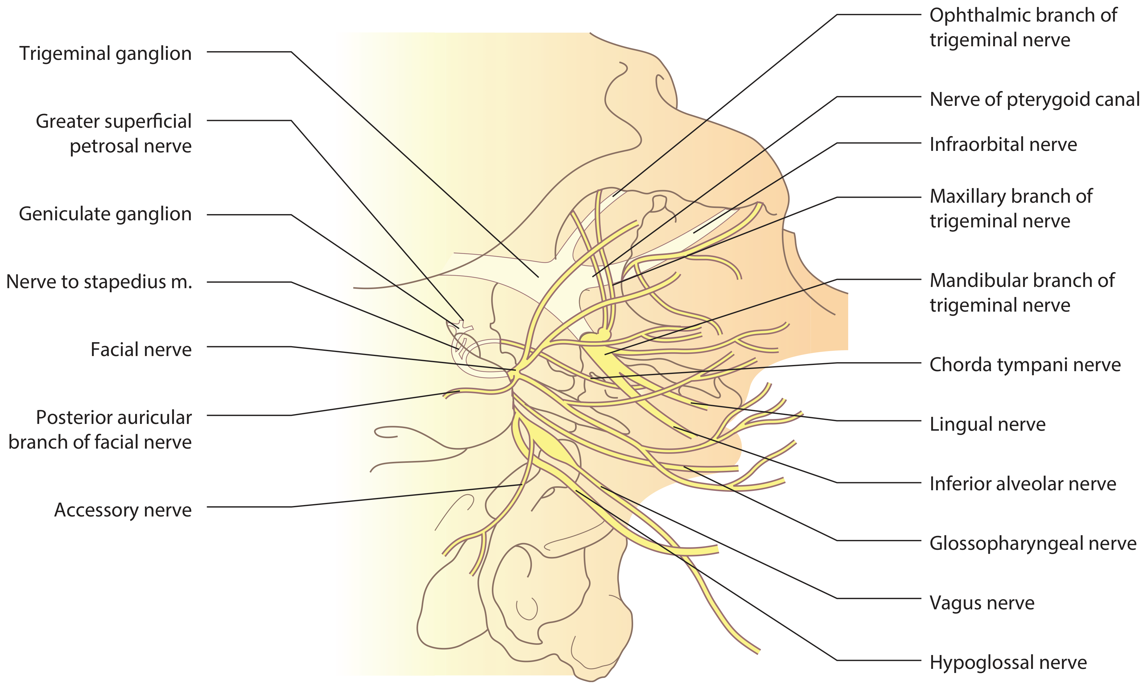
Branches from the Main Trunk (before division)
| Branch | Type | Distribution |
|---|---|---|
| Nervus spinosus (meningeal branch) | Sensory | Re-enters skull through foramen spinosum; supplies dura mater of middle cranial fossa |
| Nerve to medial pterygoid | Motor | Medial pterygoid; also gives twigs to tensor tympani and tensor veli palatini |
Anterior Division (mostly motor, one sensory branch)
| Branch | Type | Distribution |
|---|---|---|
| Deep temporal nerves (anterior & posterior) | Motor | Temporalis muscle |
| Nerve to masseter (masseteric nerve) | Motor | Masseter; passes through mandibular notch |
| Nerve to lateral pterygoid | Motor | Lateral pterygoid muscle |
| Long buccal nerve (buccal nerve) | Sensory | Passes between heads of lateral pterygoid, pierces buccinator; supplies mucous membrane of cheek and buccal gingiva |
Posterior Division (mostly sensory, one motor branch)
| Branch | Type | Distribution |
|---|---|---|
| Auriculotemporal nerve | Sensory | Arises by two roots encircling the middle meningeal artery; passes posterior to the TMJ through the parotid gland; supplies the external acoustic meatus, tympanic membrane, TMJ, and the skin of the temple and scalp. Also carries postganglionic parasympathetic fibers from the otic ganglion to the parotid gland |
| Lingual nerve | Sensory | Descends on medial pterygoid; joined by the chorda tympani (branch of CN VII) about 2 cm below the skull base; supplies anterior 2/3 of tongue (general sensation), floor of mouth, and lingual gingiva. Carries taste fibers (via chorda tympani) and preganglionic parasympathetics to submandibular ganglion |
| Inferior alveolar nerve | Sensory + motor | Descends on medial pterygoid, enters mandibular foramen; gives off mylohyoid nerve (motor to mylohyoid and anterior belly of digastric) before entering the canal; supplies lower teeth and gingiva; terminal branch exits as mental nerve through mental foramen to supply skin and mucosa of lower lip and chin |
Summary: Muscles innervated by V3 (Motor)
All four muscles of mastication plus four others:
- Temporalis
- Masseter
- Medial pterygoid
- Lateral pterygoid
- Mylohyoid
- Anterior belly of digastric
- Tensor tympani
- Tensor veli palatini
Sensory territories
- Skin of the lower face, cheek, lower lip, chin
- Anterior part of the external ear and temporal region
- External acoustic meatus and outer surface of tympanic membrane
- Anterior 2/3 of tongue (general sensation)
- Lower teeth, lower jaw, and gingiva
- Mucous membrane of the cheek
- Mastoid air cells
- Dura of the middle cranial fossa
Associated ganglion - Otic Ganglion
The otic ganglion lies just below the foramen ovale, medial to V3. It receives preganglionic parasympathetic fibers via the lesser superficial petrosal nerve (from CN IX via the tympanic plexus) and relays secretomotor fibers to the parotid gland through the auriculotemporal nerve.
Sources: Gray's Anatomy for Students, Scott-Brown's Otorhinolaryngology Head & Neck Surgery Vol 2, Localization in Clinical Neurology 8e
This is a shared conversation. Sign in to Orris to start your own chat.