
This is my forensic pathogenesis plz help me to learn it
python3 -c " import pdfplumber pdf_path = '/tmp/workspace/attachments/7baef118-ebeb-463e-9aa5-9455fc0001cc/23.04.24----Chapter-2 Death-Sowdi Sir.pdf.pdf' with pdfplumber.open(pdf_path) as pdf: print(f'Total pages: {len(pdf.pages)}') for i, page in enumerate(pdf.pages[:5]): print(f'\n--- PAGE {i+1} ---') text = page.extract_text() if text: print(text[:2000]) else: print('[No text extracted]') "
python3 -c " import pdfplumber pdf_path = '/tmp/workspace/attachments/7baef118-ebeb-463e-9aa5-9455fc0001cc/23.04.24----Chapter-2 Death-Sowdi Sir.pdf.pdf' with pdfplumber.open(pdf_path) as pdf: all_text = [] for i, page in enumerate(pdf.pages): text = page.extract_text() if text and text.strip(): all_text.append(f'=== PAGE {i+1} ===') all_text.append(text.strip()) full = '\n'.join(all_text) print(full) "
📚 Forensic Pathology - Chapter 2: THANATOLOGY (Death)
🔷 1. DEFINITION OF DEATH
| Type | Definition |
|---|---|
| Somatic / Clinical Death | Permanent and irreversible damage to Brain, Heart, and Lungs |
| Molecular Death | Death of individual tissues and cells; completes 2-3 hours after somatic death |
Key Tip: Molecular death involves changes in the eye, skin, and muscles. This is why organs must be harvested quickly after somatic death.
🔷 2. DIFFICULTIES IN DIAGNOSING SOMATIC DEATH
- Warmness of body - body may still be warm
- Suspended Animation - vital functions are at minimum compatible with life
- Coma due to sedatives/hypnotics (e.g., Barbiturates)
- Hypothermia
🔷 3. IMPORTANCE OF DETERMINING DEATH
- Disposal of the dead body
- Issuing the Death Certificate
- Conducting a Post-mortem examination
- Organ Transplantation (time-critical!):
| Organ | Maximum Time for Transplant |
|---|---|
| Liver | Within 15 minutes |
| Kidney | 30-40 minutes |
| Heart | 1 hour |
🔷 4. BRAIN STEM DEATH
What is Brain Stem Death?
- Mid brain
- Pons
- Medulla
Tests / Criteria for Confirmation:
- Coma for more than 6 hours (or 24 hours if cardiac arrest is the cause)
- No abnormal decorticate or decerebrate postures
- No spontaneous respiration
- No epileptic movements
- All brain stem reflexes absent (see below)
Brain Stem Reflexes (must all be ABSENT):
| Reflex | Afferent (Input) | Efferent (Output) |
|---|---|---|
| Pupillary reflex | Optic nerve | Oculomotor nerve |
| Vestibulo-ocular reflex | Auditory nerve | Abducent nerve |
| Corneal reflex | Trigeminal nerve | Facial nerve |
| Gag reflex | Glossopharyngeal nerve | Vagus nerve |
| Doll's eye reflex | Auditory nerve | Abducent nerve |
Diagnosis Steps:
- Establish positive diagnosis of coma and its cause
- Confirm irremediable structural brain damage
- Attempt and fail to remedy the cause:
- Correct low blood pressure
- Correct hypoxia
- Remove blood clot
Brain Stem Death CANNOT be declared in:
- Absence of coma
- Children below 5 years of age
- Coma due to drugs, hypothermia, metabolic disorders, or shock
🔷 5. SUDDEN DEATH
Causes:
A. UNNATURAL Causes:
- Violence
- Poisoning
- Combination of both
B. NATURAL Causes:
- Coronary disease
- Congenital heart diseases
- Valvular heart diseases
- Hypertensive heart diseases
- Infection
- Cardiac tamponade
- Aortic aneurysm
- Pulmonary embolism
- Haemoptysis
- Infections
- Chronic asthma
- Anaphylaxis
- Obstruction to air passage
- Intracerebral haemorrhage
- Subarachnoid haemorrhage
- Cerebral thrombosis
- Embolism
- Infections
- Brain tumour
- Haemorrhage in GI tract
- Rupture of abdominal aneurysm
- Liver diseases
- Acute pancreatic bleeding
- Adrenal haemorrhage
- Diabetic coma
- Myxoedemic and parathyroid crisis
- Drug abuse
- Sudden withdrawal of steroids
- Anaesthesia complications
- Mismatched blood transfusion
- Bacteraemic shock
- Shock due to fear/emotion
- Malaria
- In children: SIDS (Cot death), congenital abnormalities, concealed puncture wounds
🔷 6. SUSPENDED ANIMATION
- Apparent death
- Voluntary act (Death Trance)
- Involuntary - due to Hypothermia, Drowning, in newborns, Electric shock
🔷 7. MODES, MANNER, MECHANISM, AND CAUSE OF DEATH
A. Modes of Death (Bichat's Classification)
| Mode | System Failing | Examples |
|---|---|---|
| Coma | Brain failure | Brain diseases, injuries, opium/alcohol poisoning, uraemia |
| Syncope | Heart failure | Heart disease, exhausting diseases, digitalis/tobacco/aconite poisoning |
| Asphyxia | Lung failure | Pneumonia, opium, CO/CO₂, mechanical interference |
B. Gordon's Classification (Anoxia)
| Type of Anoxia | Mechanism | Examples |
|---|---|---|
| Anoxic Anoxia | Mechanical interference to air passage | Smothering, Hanging, Strangulation, Drowning, Stampede |
| Anaemic Anoxia | Reduced O₂ carrying capacity of blood | Acute haemorrhage, CO poisoning, nitrite poisoning |
| Histotoxic Anoxia | Tissues cannot utilize O₂ | Cyanide poisoning |
| Stagnant Anoxia | Inefficient circulation | Shock, CCF (congestive cardiac failure) |
Memory Trick for Anoxia types: "AHHS" - Anoxic, Histotoxic, Haemic (anaemic), Stagnant
C. Manner of Death
| Manner | Description |
|---|---|
| Natural | Death due to disease |
| Unnatural/Violent | Death due to violence - can be Accidental, Suicidal, or Homicidal |
D. Mechanism of Death
- Metabolic acidosis/alkalosis
- Sepsis
- Toxaemia
- Paralysis
E. Cause of Death
🔷 QUICK REVISION TABLE
| Term | One-line Definition |
|---|---|
| Somatic Death | Irreversible failure of Brain + Heart + Lungs |
| Molecular Death | Cell death, completes 2-3 hrs after somatic death |
| Brain Stem Death | Irreversible loss of midbrain + pons + medulla functions |
| Suspended Animation | Appears dead but alive at minimal vital function |
| Sudden Death | Unexpected death raising suspicion of foul play |
| Mode of Death | Physiological state at time of death (Coma/Syncope/Asphyxia) |
| Manner of Death | Natural vs. Unnatural (Accidental/Suicidal/Homicidal) |
| Mechanism of Death | Biochemical disturbance (acidosis, sepsis, etc.) |
| Cause of Death | Disease/injury triggering the fatal chain of events |
🔷 COMMON EXAM QUESTIONS FROM THIS CHAPTER
- What are the types of death? Define each.
- What are the criteria for brain stem death?
- Why can't brain stem death be declared in children under 5?
- What are the organ transplant time limits?
- Classify sudden death with examples.
- What is Suspended Animation? Give causes.
- Classify anoxia (Gordon) with examples.
- Differentiate: Mode / Manner / Mechanism / Cause of death.
- What is Histotoxic anoxia? Give an example.
- What poisons cause death by syncope? (Digitalis, Tobacco, Aconite)
Answer the 3 question
Question 3: Why Can't Brain Stem Death Be Declared in Children Under 5?
1. Immature and Still-Developing Brain
- The brain of a child under 5 is still in active neurological development
- The brain stem has greater plasticity (ability to recover) at this age
- What appears to be permanent, irreversible damage may actually be reversible in a developing brain
2. Unreliable Clinical Tests
- The standard brain stem reflex tests were designed and validated for adults
- Reflexes in young children are physiologically different - some reflexes normally absent in adults may be present, and vice versa
- This makes interpretation of test results unreliable and inaccurate in children under 5
3. Higher Tolerance to Hypoxia
- Young children, especially infants, have greater tolerance to hypoxic injury compared to adults
- Their brain cells can survive longer periods of oxygen deprivation
- A child who appears brain dead may still have recoverable neurological function
4. Difficulties in Establishing the Cause of Coma
- In young children it is often harder to confirm the exact cause of coma
- Metabolic, genetic, or congenital causes may mimic irreversible brain damage
- Misdiagnosis risk is significantly higher
5. Ethical and Legal Safeguards
- The declaration of brain stem death has direct implications for organ harvesting
- Extra caution is mandated to protect vulnerable pediatric patients
- The medical and legal system requires a higher threshold of certainty before declaring death in a child
Summary Table
| Reason | Why It Matters |
|---|---|
| Immature brain | May recover even from severe damage |
| Unreliable reflex tests | Tests not validated for this age group |
| Higher hypoxia tolerance | Brain cells can survive longer |
| Difficult cause confirmation | Reversible causes may be missed |
| Ethical/legal protection | Organ harvesting safeguard |
Key Exam Point: Brain stem death is also excluded when coma is due to drugs, hypothermia, metabolic disorders, or shock - not just age. These must all be ruled out before declaration.
Answer 6 question
Question 6: What is Suspended Animation? Give Causes.
Definition
- Also called "Apparent Death"
- The person is NOT dead - they only appear so
- This is why it is a difficulty in diagnosing somatic death
Key Characteristics
| Feature | Description |
|---|---|
| Appearance | Person looks dead |
| Vital functions | Present but at extremely low level |
| Reversibility | Fully reversible - person can recover |
| Medico-legal risk | Premature certification of death |
Types of Suspended Animation
1. Apparent Death (Involuntary)
- Occurs due to external or pathological causes
- The person has no control over the condition
2. Voluntary Act / Death Trance
- Practiced by certain yogis and ascetics
- Person voluntarily slows down all body functions to an undetectable level
- Historically documented in Indian subcontinent
Causes of Suspended Animation
A. Environmental Causes
| Cause | Mechanism |
|---|---|
| Hypothermia | Extreme cold slows all metabolic and physiological processes drastically |
| Drowning | Cold water immersion + oxygen deprivation drops all vital signs to minimum |
B. Physiological / Age-related
| Cause | Mechanism |
|---|---|
| Newborns | Neonates, especially premature ones, have minimal detectable vital signs at birth |
| Electric shock | Temporary stunning of the nervous system and heart activity |
C. Voluntary
| Cause | Mechanism |
|---|---|
| Death Trance | Self-induced through deep meditation / yogic practices |
Medico-Legal Importance
- Premature burial risk - A person in suspended animation may be wrongly certified dead and buried alive
- Wrong death certificate - Issuing a death certificate to a living person is a serious medico-legal offense
- Organ harvesting danger - Organs must NOT be harvested from a person in suspended animation
- Related to Brain Stem Death - Suspended animation must be ruled out before declaring brain stem death
- Conditions like hypothermia and drug coma (barbiturates) must be excluded before death is confirmed
How to Differentiate from True Death
| Sign | Suspended Animation | True Death |
|---|---|---|
| Body warmth | May be present | Lost gradually |
| Pupil reaction | May be sluggish but present | Absent |
| Heartbeat | Very faint, detectable by ECG | Absent |
| Respiration | Extremely slow/shallow | Absent |
| Response to stimuli | May respond faintly | No response |
| Reversibility | Yes | No |
One-line exam answer: Suspended animation is a state of apparent death where vital functions are at minimum compatible with life, caused by hypothermia, drowning, electric shock, neonatal state, or voluntary death trance - and is medico-legally important to distinguish from true death to prevent premature burial or wrongful organ harvesting.
Describe the features of asphyxia
🫁 Features of Asphyxia
Definition
Important: Asphyxia indicates a mode of dying, NOT a cause of death.
Pathophysiology - The Vicious Cycle
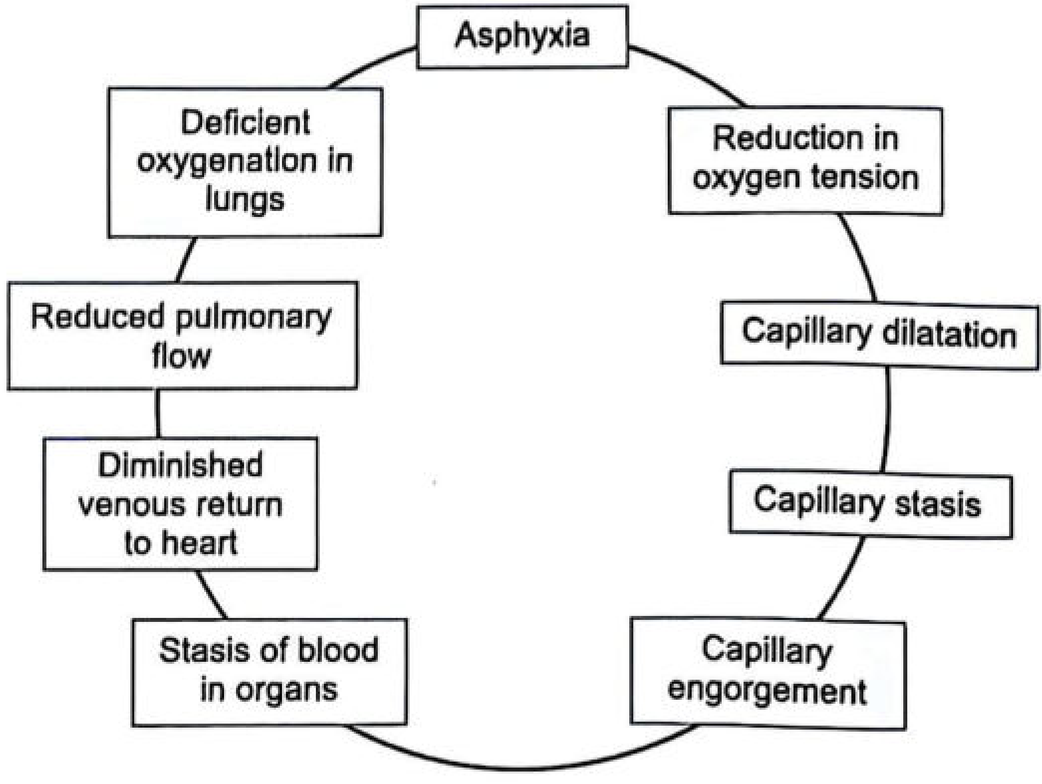
- Asphyxia → Reduction in O₂ tension
- → Capillary dilatation
- → Capillary stasis → Capillary engorgement
- → Stasis of blood in organs
- → Diminished venous return to heart
- → Reduced pulmonary flow → Deficient oxygenation in lungs
- → Back to worsening asphyxia (cycle repeats)
Key Facts (Numbers to Remember)
| Fact | Value |
|---|---|
| Normal arterial O₂ (young adults) | 90-100 mm Hg |
| Normal arterial O₂ (above 60 yrs) | 60-85 mm Hg |
| O₂ level in fatal asphyxia | 20-40 mm Hg |
| Brain uses of total body O₂ | 20% (only 1.4% of body weight) |
| Cerebral cortex - nerve function stops | After 8-15 seconds of ischemia |
| Brainstem ganglia - function stops | After 25-30 seconds |
| Cortex - irreparable damage | After ~3 minutes |
| Basal ganglia - irreparable damage | After 6-7 minutes |
| Vagal centre - irreparable damage | After 9-10 minutes |
| Breathing stops after cardiac arrest | Within 20 seconds |
| Heart stops after breathing stops | Within 20 minutes |
Cardinal (Classic) Signs of Asphyxia
1. 🔴 Petechial Hemorrhages (Most Important Sign)
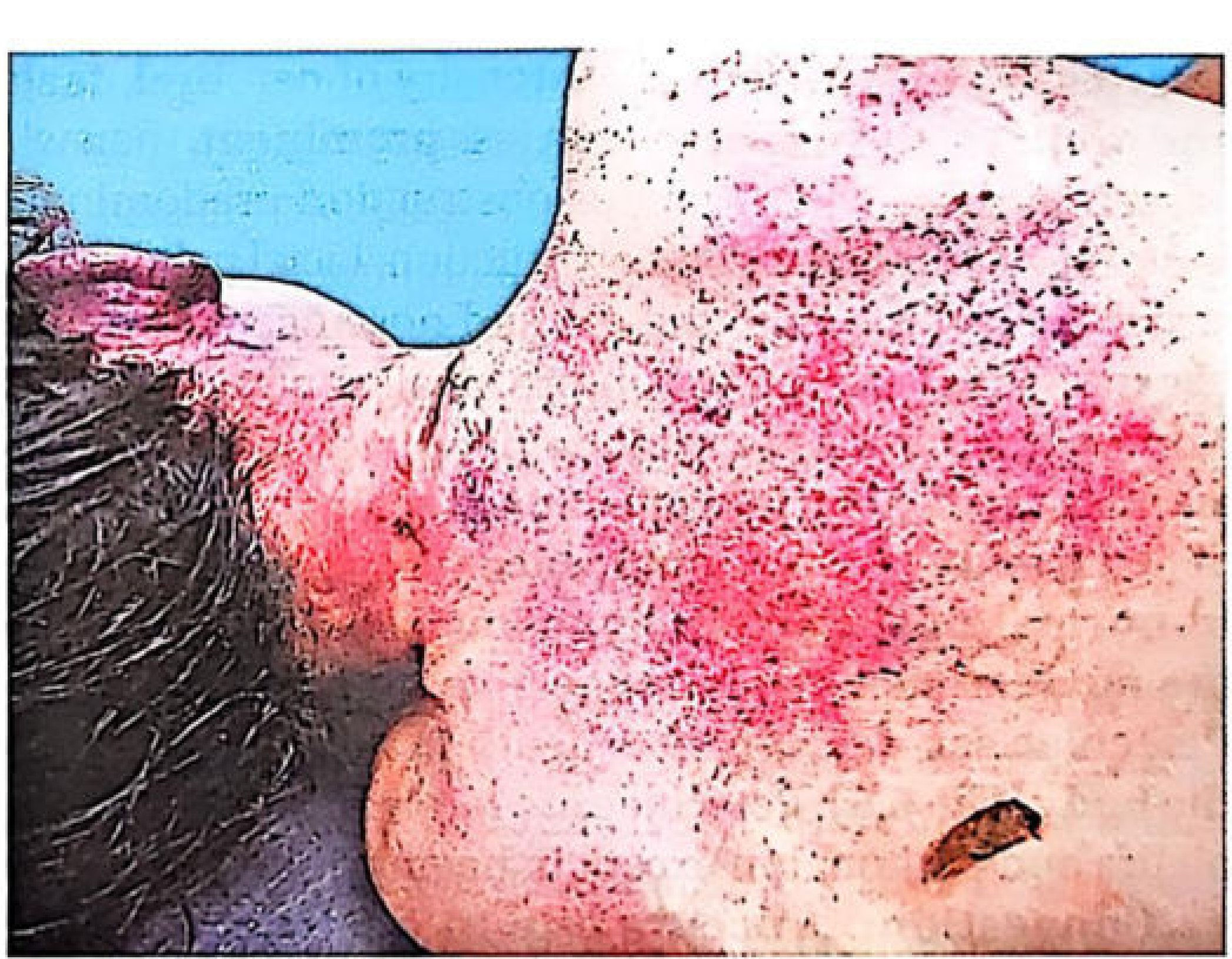
- Cause: Raised venous pressure → overdistension and rupture of venules (NOT due to hypoxia of vessel walls)
- Minimum time to produce: 15-30 seconds
- Size: 0.1 to 2 mm (if larger = ecchymoses)
- Location:
- Skin of face, neck
- Sclerae and conjunctivae (highly characteristic)
- Outer and inner surfaces of eyelids
- Mucosal surfaces of mouth
- Viscera - brain, liver, lungs
- Serous membranes (visceral pleura - called Tardieu spots)
2. 🔵 Cyanosis
- Cause: Reduced O₂ tension → rise in reduced haemoglobin (>5 g/100 ml)
- Blood turns purplish-blue when oxygen is given up
- Locations: Lips, ears, tip of nose, fingernails, cheeks, skin (hypostatic stains)
- Internally: Lungs, liver, spleen, kidneys, meninges
- Also caused by methaemoglobin and sulphaemoglobin
- Note: After 24 hours, oxygen dissociates from blood - cyanosis appears in any dead body, so it must be assessed early
3. 🫀 Congestion of Organs (Visceral Congestion)
- All organs appear engorged with dark blood
- Due to stasis of blood in dilated capillaries and venules
- Particularly marked in brain, lungs, liver, spleen, kidneys
4. 🩸 Fluidity of Blood
- In asphyxia, blood flow slows → tendency to clot
- As a defence mechanism, fibrinolysin is released from endothelium
- This keeps blood in a fluid state even after death
- Blood remains fluid for a minimum of 4-6 hours (until post-mortem clotting)
- Blood is already dark blue in colour
Autopsy (Post-Mortem) Findings
External Findings:
| Feature | Description |
|---|---|
| Face | Congested, swollen, cyanosed |
| Eyes | Petechiae in sclerae/conjunctivae, congested |
| Lips | Cyanosed, bluish-purple |
| Skin | Petechiae, cyanosis, livid hypostasis |
| Froth | At mouth and nostrils (especially in drowning) |
Internal Findings:
Lungs:
- Heavier than normal (450-500 g or more) - indicates respiration stopped while heart continued beating
- Normal right lung weight (~300 g) - compatible with sudden cardiac arrest
- Congested, edematous
- Tardieu spots - petechiae on visceral pleura (subpleural petechiae)
Histological Signs:
- Partial disruption of alveolar septa with hemorrhage
- Intra-alveolar hemorrhage
- Pulmonary edema
Heart:
- Right side dilatation - NOTE: This is now considered obsolete as a specific sign of asphyxia (seen in any congestive death)
- Blood is dark and fluid
Brain:
- Congested
- Petechial hemorrhages in brain substance
- Cerebral edema
Stages of Asphyxia (Phases)
| Stage | What Happens |
|---|---|
| Stage 1 - Dyspnoea | Increased respiratory efforts, rising CO₂, HR increases, BP rises |
| Stage 2 - Convulsions | Loss of consciousness, convulsions due to cerebral hypoxia |
| Stage 3 - Apnoea | Respiratory centre fails, breathing stops |
| Stage 4 - Terminal | Heart continues briefly, then final cardiac arrest |
Tardieu Spots (Special Feature)
- Subpleural petechial hemorrhages on visceral pleura of lungs
- Named after French physician Auguste Ambroise Tardieu
- Considered classic but not exclusive to asphyxia
- Also seen on pericardium and thymus
- Caused by raised intrathoracic pressure rupturing small venules
Summary of All Features (Quick Revision)
| Feature | Key Point |
|---|---|
| Petechiae | Sclerae, conjunctivae, skin - due to raised venous pressure |
| Cyanosis | Bluish discolouration - >5g/dL reduced Hb |
| Visceral congestion | All organs engorged and dark |
| Fluid dark blood | Fibrinolysin keeps blood liquid for 4-6 hrs |
| Heavy lungs | >450-500g = asphyxial death |
| Tardieu spots | Subpleural petechiae on visceral pleura |
| Cerebral edema | Brain hypoxia and congestion |
| Froth at mouth | Especially in drowning/strangulation |
Top Exam Point: The 4 cardinal signs of asphyxia are: (1) Petechial hemorrhages, (2) Cyanosis, (3) Visceral congestion, (4) Fluidity of blood. Tardieu spots are a special variant of petechiae on serous membranes and are highly characteristic.
Obscure atopsy
🔬 Obscure Autopsy
Definition
"These are a source of confusion to any pathologist." - Essentials of Forensic Medicine & Toxicology
Key Points About Obscure Autopsy
- Usually seen in a younger, healthy person who dies suddenly and unexpectedly
- Non-medical persons (including police) believe autopsy can always determine cause of death - this is NOT always true
- The doctor must admit inability to give a positive opinion when findings are absent
- Mild natural disease must NOT be implicated unless other possibilities are carefully eliminated
Causes of Obscure Autopsy
1. 🫀 Natural Diseases
| Sub-type | Examples |
|---|---|
| With obscure / microscopic findings | Myocarditis, early infarction |
| Precipitated by emotion / work stress | Emotional excitement → cardiac arrest |
| Functional failure | Epilepsy, paroxysmal atrial fibrillation |
2. ⚗️ Biochemical Disturbances
- Uraemia, Diabetes, Potassium deficiency (hyperkalemia)
- Respiratory pigment disorders - Anaemic anoxia, Porphyria
- Hypoglycaemia - very common obscure death
3. 🧪 Endocrine Dysfunction
- Adrenal insufficiency
- Thyrotoxicosis or Myxoedema
4. 🤕 Concealed Trauma
a) Cerebral Concussion
- Blunt force injury to head → transient unconsciousness
- Autopsy may show NO external or internal marks
- Petechiae may be seen in some cases
- Even microscopic examination shows NO axonal injury up to 12 hours
b) Neck Injury (Self-Reducing)
- Cervical spinal fracture-dislocation (in diving, falls, head impacts)
- Dislocation is often self-reducing - no external injury visible
- Death may be instantaneous - cord contusion/laceration with no obvious signs
- Complete dissection of spine is essential; spinal cord cut longitudinally may show internal bruising
c) Blunt Injury to the Heart
- Contusion of chest (steering-wheel impact, blast, heavy punching)
- Heart may stop without much visible evidence of trauma
- Coronary spasm or functional cardiac inhibition → sudden death
d) Reflex Vagal Inhibition ⭐ (Most Important)
Case Example: A soldier was dancing with his girlfriend. He playfully "tweaked" (pinched) her neck. She dropped dead on the spot. No injuries or signs of asphyxia were found. Death was due to vagal inhibition.
- Pressure on baroreceptors in carotid sinuses/carotid body
- → Reflex stimulation of vagus nerve
- → Sudden cardiac arrest
- Can be triggered by:
| Trigger Site | Example |
|---|---|
| Neck | Blow to larynx, pinching, cold water immersion |
| Chest | Punch, blast |
| Abdomen | Blow to abdomen |
| Genital organs | Blow to genitals |
| Ear | Manipulation of external auditory meatus |
| Drowning | Cold water + sudden immersion |
5. ☠️ Poisoning (Without Macroscopic Change)
- Delayed suboxic or narcotic poisoning
- Anaesthetic overdosage or maladministration
- Neurotoxic or cytotoxic poisons
- Plant poisoning
- Snake bite
6. 🔀 Miscellaneous
- Allergy / Drug idiosyncrasy
- Anaphylaxis (see below)
Anaphylactic Deaths (Special Obscure Cause)
- Acute immunologic reaction → cutaneous, GI, respiratory, cardiovascular collapse
- Most deaths occur within 1-2 hours
- Mechanism: Bronchospasm + vasodilation + laryngeal oedema
- Mediators: histamine, leukotrienes, bradykinin, kallikrein
- Drugs: Penicillin, Aspirin, horse-serum products, vaccines, iodine agents
- Insect bites
- Foods: Fish, shellfish, eggs, nuts
- Hormones: Insulin, ACTH
What to Do in an Obscure Autopsy?
| System | What to Look For |
|---|---|
| Airways | Foreign body in respiratory tract |
| Cardiovascular | Cardiac hypertrophy, coronary artery narrowing, thrombosis, atherosclerosis, coronary ostia patency |
| Brain | Sickle cell thrombi, fat embolism |
| Blood | Pancytopenia, leukaemia |
| Other | Tongue bite (epilepsy), needle marks (drug abuse), small electrical burns, myocarditis, electrolyte imbalance |
Laboratory Investigations Required:
- Bacteriological
- Virological
- Histological (microscopy - often reveals what naked eye misses)
- Biochemical (electrolytes, glucose, tryptase)
- Toxicological
Clinical Investigation:
- Interview witnesses who observed the deceased before death
- Review available history carefully
How to Certify Cause of Death in Obscure Cases?
| Situation | Certification |
|---|---|
| Senile atrophy, no other finding, natural death likely | "Myocardial degeneration due to senility" |
| Unequivocally natural death, but no specific cause | "Undetermined natural circumstances" |
| Equivocal circumstances | "Cause and manner of death: Undetermined" |
Key Point: Even when the cause of death cannot be established, a negative autopsy is still valuable because it excludes injury or poisoning that may have been wrongly alleged.
Natural Causes Commonly Producing Obscure Autopsy
| Condition | Why Obscure |
|---|---|
| Hypoglycaemia | No structural lesion - biochemical only |
| Hyperkalemia | Cardiac arrhythmia with no structural change |
| Cardiac arrhythmias | No anatomical finding |
| Conduction system disorders | Requires special staining to detect |
| Epilepsy | May die in fit - tongue bite only clue |
| Vagal inhibition | Absolutely NO autopsy findings |
| Snake bite | Small inconspicuous fang marks |
| Acute myocarditis | May need Aschoff bodies on histology |
| Early myocardial infarction | Changes appear only after 12-24 hours |
| Anaphylaxis | Only laryngeal oedema and lung overexpansion |
Summary Table
| Feature | Detail |
|---|---|
| Definition | Autopsy with no definite, minimal, or obscure cause of death |
| Common victim profile | Young, healthy person, sudden unexpected death |
| Most common obscure causes | Vagal inhibition, epilepsy, hypoglycaemia, cardiac arrhythmia |
| Autopsy finding in vagal inhibition | None |
| Key investigation | Histology + toxicology + biochemistry |
| Certification when truly unknown | "Undetermined" |
| Medico-legal importance | Negative autopsy still excludes foul play |
Top Exam Answer: An obscure autopsy is one where no definite cause of death is found. The most classic example is vagal inhibition where autopsy shows absolutely nothing - death is diagnosed only by exclusion. The doctor must not yield to police pressure to give a positive opinion when findings are absent.
Tardieus spots
🔴 Tardieu's Spots
Definition & History
Named after Dr. Auguste Ambrose Tardieu - a French Police Surgeon who first described them in 1866.
Mechanism of Formation
| Mechanism | How It Works |
|---|---|
| 1. Raised venous pressure | Obstruction to venous drainage → increased intracapillary pressure → overdistension → rupture of venules |
| 2. Increased capillary permeability | Anoxia → pores between endothelial cells enlarge → blood transudes into tissues → capillary rupture |
Minimum time required to form = 15-30 seconds of mechanical obstruction
Gross Appearance
| Feature | Description |
|---|---|
| Shape | Round, well-defined |
| Colour | Dark red / dark purple |
| Size | Pinhead sized (0.1 - 2 mm) |
| Variation | May be scanty (seen only under low magnification) OR numerous (like a measles rash) |
| If > 2 mm | Called ecchymoses, not petechiae |
Locations (Where They Are Found)
External (on body surface):
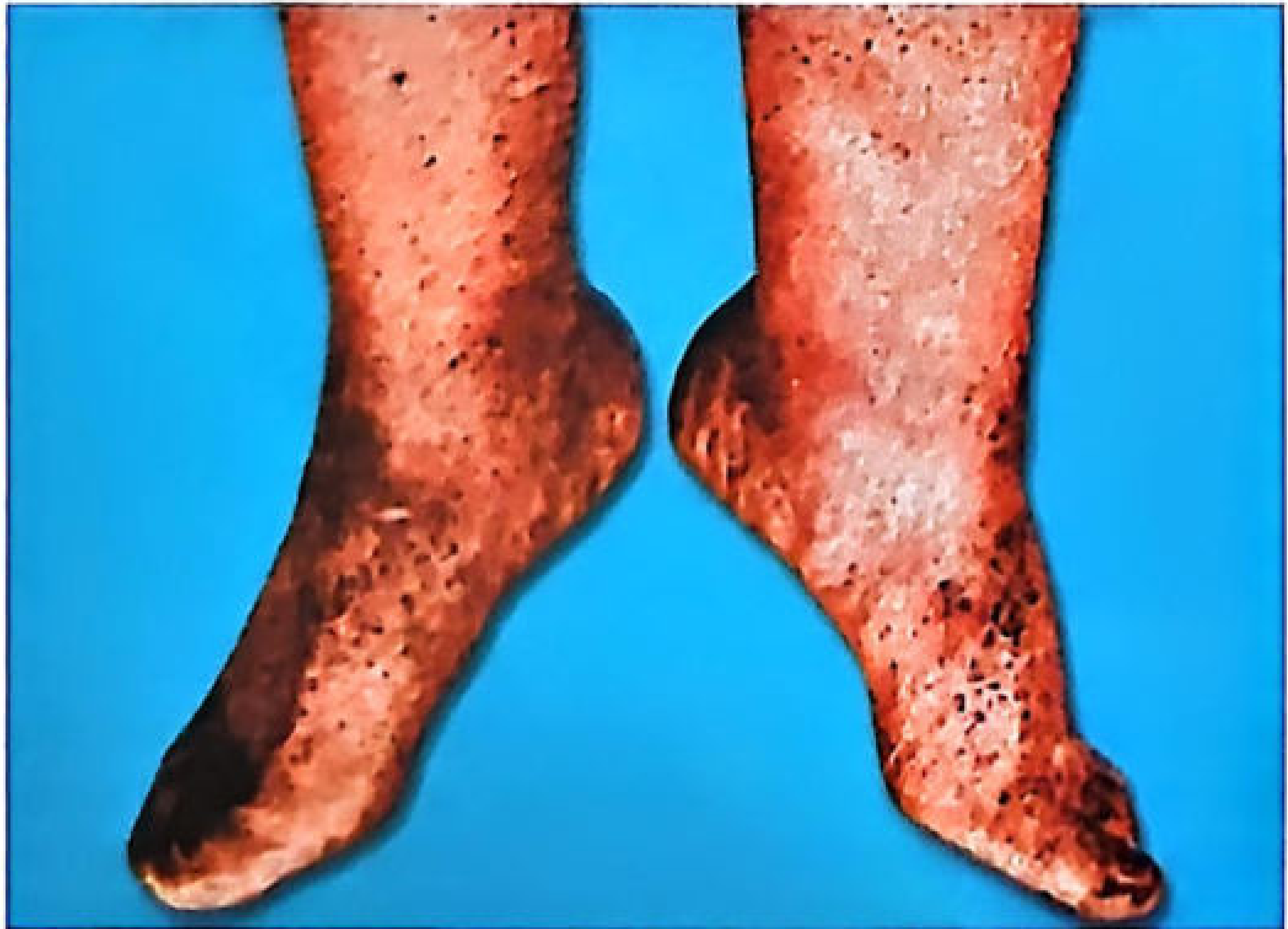
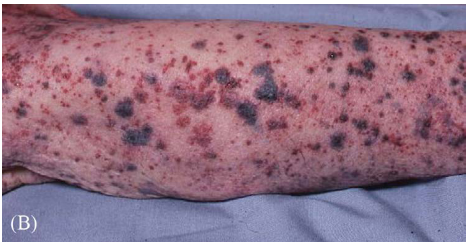
- Face, eyelids (outer and inner surface)
- Sclerae and conjunctivae (very characteristic - seen even in fair skin)
- Skin of face and neck (above level of compression)
- Behind the ears (best seen after blood drains out at completion of autopsy)
Internal (at autopsy):
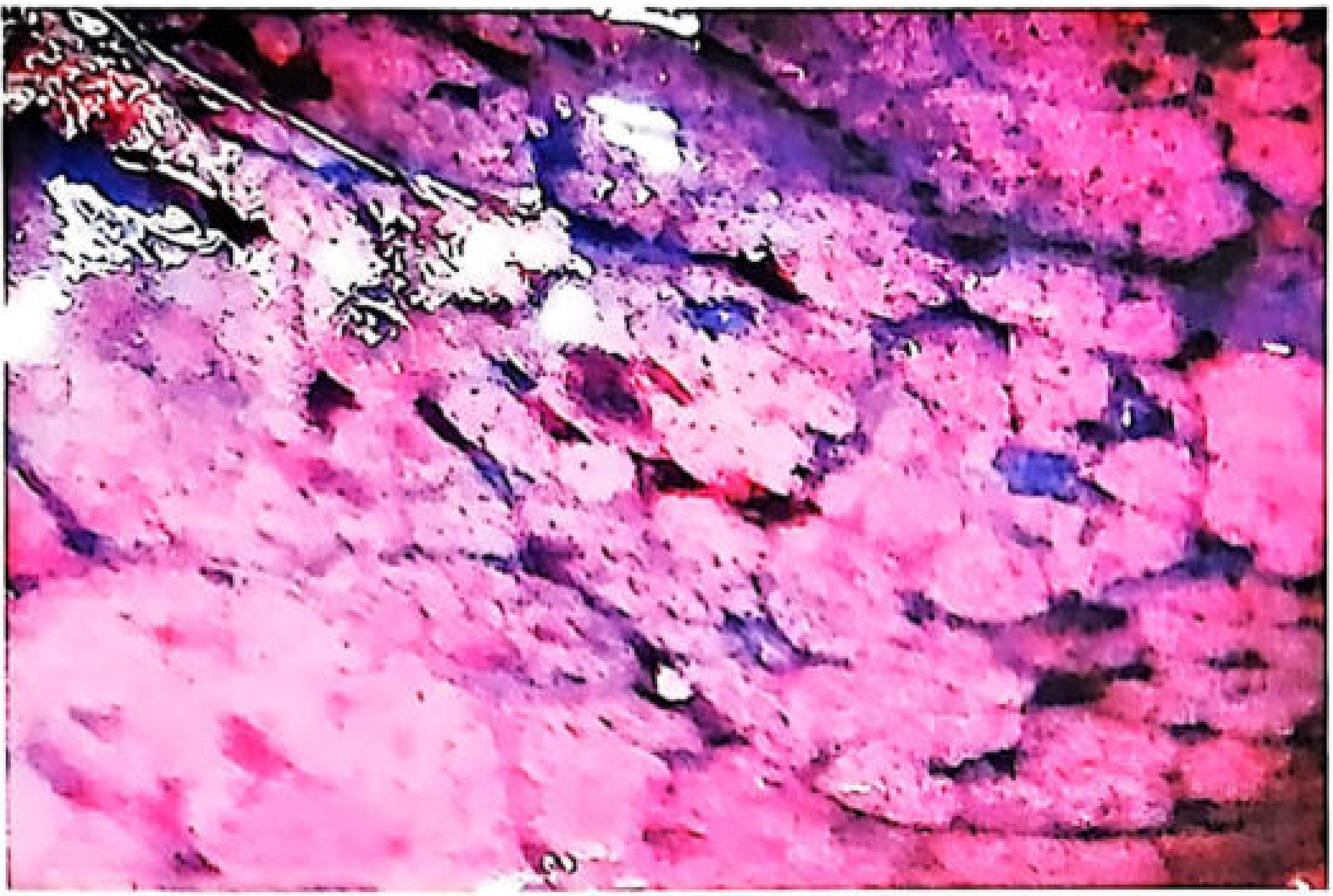
| Location | Notes |
|---|---|
| Visceral pleura (subpleural) | TRUE Tardieu spots - most characteristic location |
| Interlobar fissures and hilum | Subpleural petechiae |
| Pericardium / Epicardium | Very prominent |
| Thymus | Especially prominent in infants and children |
| Brain (white matter) | Petechiae in substance |
| Subarachnoid space | Larger patches from ruptured superficial vessels |
| Mucosal surfaces of mouth | Especially epiglottis |
| Meninges | Present |
Rule: They are most prominent where capillaries are least supported - face, conjunctivae, epiglottis, serous surfaces of heart and lungs, meninges, thymus.
Where They Are Most Pronounced (Diagnostic Significance)
| Cause of Death | Location of Prominent Spots |
|---|---|
| Strangulation / Hanging | Above the level of the ligature/constriction on neck |
| Traumatic asphyxia | On the chest |
| Hanging (suspended body) | Feet and legs (dependent areas) |
This distribution pattern can be DIAGNOSTIC of the cause of asphyxia!
When Are Tardieu Spots ABSENT?
- In drowning - Tardieu spots are rarely prominent (important exception)
- When carotid arteries (not veins) are obstructed - facial/orbital petechiae are less intense
- In sudden complete carotid obstruction - facial pallor occurs instead
Key exam rule: Absence of Tardieu spots does NOT exclude death from asphyxia.
Antemortem vs Postmortem Tardieu Spots
| Feature | True (Antemortem) Tardieu Spots | Postmortem Petechiae |
|---|---|---|
| Location | Serous surfaces (visceral pleura, pericardium, conjunctivae) | Skin of hands, feet, legs only (dependent areas) |
| Appearance | Small, round, well-defined, pinhead | Larger, less circumscribed |
| Cause | Raised venous pressure + anoxia during life | Gravitational settling of blood after death rupturing capillaries |
| Time to develop | 15-30 seconds | 18-24 hours (postmortem), or 2-4 hours in hanging limbs |
| Diagnostic value | Highly significant | Not significant for asphyxia |
Important: Postmortem petechiae on skin of hands and legs of a suspended body must NOT be mistakenly called Tardieu spots - they are due to gravity, not asphyxia.
Tardieu Spots vs Artificial Respiration Haemorrhages
- During CPR/artificial respiration: Haemorrhages may appear under pleura
- These can be confused with Tardieu spots
- Distinction: CPR haemorrhages are more extensive and bear the pattern of rib markings
- Tardieu spots are smaller and not rib-patterned
Differential Diagnosis of Petechial Haemorrhages
- Coronary deaths (cardiac failure)
- Terminal stages of any illness
- Blood diseases - in diseases, haemorrhages are smaller, more numerous, greater distribution; blood examination may reveal underlying cause
Summary Quick Revision
| Feature | Key Point |
|---|---|
| Named after | Dr. Auguste Tardieu, French Police Surgeon, 1866 |
| What they are | Petechial haemorrhages (pinpoint bleeds) |
| Size | 0.1-2 mm (pinhead) |
| Colour | Dark red / dark purple |
| Best (true) location | Visceral pleura (subpleural surface) |
| Also seen | Conjunctivae, sclerae, epiglottis, pericardium, thymus (in children) |
| Mechanism | Raised venous pressure + increased capillary permeability |
| Min. time to form | 15-30 seconds |
| Absent in | Drowning (rarely prominent) |
| Postmortem spots | On dependent skin - NOT true Tardieu spots |
| Medico-legal value | Distribution tells us how the person died |
One-line exam definition: Tardieu's spots are dark-red, pinhead-sized petechial haemorrhages seen on the visceral pleura, conjunctivae, and serous surfaces, caused by raised venous pressure and increased capillary permeability during asphyxia, first described by French Police Surgeon Auguste Tardieu in 1866.
Tardieus spots and petechia hemorrhages
🔴 Tardieu's Spots vs Petechial Haemorrhages
Are They the Same Thing?
Tardieu's spots ARE petechial haemorrhages - but not all petechial haemorrhages are Tardieu's spots.
| Term | Meaning |
|---|---|
| Petechial haemorrhage | Broad term - any tiny pinpoint bleed (0.1-2 mm) from a ruptured capillary or venule |
| Tardieu's spots | Specific petechial haemorrhages occurring in asphyxia, named after Dr. Auguste Tardieu (1866) |
1. Petechial Haemorrhages
Definition
Mechanism
NOT caused by hypoxia of vessel walls directly.
Minimum time to form
Locations in Asphyxia
| Site | Notes |
|---|---|
| Sclerae | Highly characteristic |
| Conjunctivae (bulbar and palpebral) | Very visible |
| Outer and inner surfaces of eyelids | Common |
| Skin of face and neck | Visible externally |
| Mucosal surfaces of mouth | Epiglottis especially |
| Skin of upper chest and back | Visible in traumatic asphyxia |
| Visceral pleura (Tardieu spots) | Most diagnostic internal site |
| Pericardium | Internal |
| Brain (white matter) | Internal |
| Meninges | Internal |
| Thymus | Especially in infants and children |
2. Tardieu's Spots (Specific Type of Petechial Haemorrhage)
Definition
Named After
Two Mechanisms
1. Raised venous stasis
→ Congestion → increased intracapillary pressure → rupture of vessels
2. Increased capillary permeability due to hypoxia
→ Pores between endothelial cells enlarge → blood transudes → rupture
Key Comparison Table
| Feature | Petechial Haemorrhages (General) | Tardieu's Spots (Specific) |
|---|---|---|
| Definition | Any pinpoint bleed from capillary rupture | Petechiae specifically on visceral pleura in asphyxia |
| Size | 0.1 - 2 mm | Pinhead (same) |
| Colour | Dark red | Dark red / purple |
| Shape | Round, well-defined | Round, well-defined |
| Location | Sclerae, conjunctivae, skin, serous surfaces | Visceral pleura primarily - also pericardium, thymus |
| Cause | Raised venous pressure / hypoxia | Raised venous pressure + increased capillary permeability |
| Significance | Cardinal sign of asphyxia | Most diagnostic internal sign of asphyxia |
| Named after | - | Dr. Auguste Tardieu, 1866 |
| Where most prominent | Above level of compression | Interlobar fissures, around hilum |
| In children | Present | Thymus shows numerous petechiae |
Antemortem vs Postmortem Petechiae
| Feature | Antemortem (True Tardieu / Asphyxia) | Postmortem (Artefact) |
|---|---|---|
| Location | Visceral pleura, pericardium, conjunctivae, sclerae | Skin of hands, legs, feet only (dependent areas) |
| Size | Small, pinhead, well-defined | Larger, less circumscribed |
| Cause | Raised venous pressure + anoxia during life | Gravity pulls blood to dependent parts → capillary over-distension after death |
| Time to form | 15-30 seconds | 18-24 hours (general) or 2-4 hours in hanging limbs |
| Medico-legal value | HIGH - indicates asphyxia | LOW - not significant |
| Diagnostic of asphyxia? | YES | NO |
Rule: Postmortem petechiae on the skin of the hands and legs of a suspended body must NOT be called Tardieu spots - they are due to gravity, not asphyxia.
Distribution = Diagnosis
| Pattern | Cause of Death |
|---|---|
| Above level of ligature mark on neck | Strangulation / Hanging |
| Prominent on chest | Traumatic asphyxia (compression of chest) |
| On feet and legs | Dependent areas in hanging |
| Prominent on face + sclerae | Throttling / Manual strangulation |
| Rarely seen | Drowning (Tardieu spots rare in drowning) |
| In carotid obstruction | Petechiae less intense (arterial block = pallor, not congestion) |
Important Exceptions and Rules
| Rule | Explanation |
|---|---|
| Absence of Tardieu spots does NOT exclude asphyxia | Especially true in drowning |
| Tardieu spots in drowning | Rarely prominent |
| In carotid artery obstruction | Facial petechiae are less prominent; may see pallor instead |
| In conjunctivae | Look behind the ears after blood drains - petechiae more visible there |
| CPR artefact petechiae | Appear under pleura, bear rib marking pattern - distinguish from Tardieu spots |
| Scalp reflection petechiae | Common autopsy artefact - no significance |
Diseases That Cause Petechiae (Differential Diagnosis)
| Condition | Feature |
|---|---|
| Coronary deaths | Petechiae present |
| Blood diseases (leukaemia, pancytopenia) | Smaller, more numerous, wider distribution |
| Terminal stages of any illness | Non-specific |
| Postmortem hypostasis | Seen in dependent areas |
In disease-related petechiae: blood examination (CBC, coagulation profile) may reveal the underlying cause.
Summary - One Line Each
-
Petechial haemorrhage = Pinpoint bleed (0.1-2 mm) from capillary/venule rupture due to raised venous pressure - a cardinal sign of asphyxia
-
Tardieu's spots = Petechiae specifically on the visceral pleura (and other serous surfaces) - named after Tardieu (1866) - the most specific internal finding of asphyxial death
-
Key rule: All Tardieu spots are petechial haemorrhages, but NOT all petechial haemorrhages are Tardieu spots.
-
Medico-legal importance: Location and distribution of petechiae helps determine the type and mechanism of asphyxial death.
Plz help me to learn it my forensic ppt
python3 -c " import pdfplumber pdf_path = '/tmp/workspace/attachments/7baef118-ebeb-463e-9aa5-9455fc0001cc/PM changes-Sowdi Sir.pdf.pdf' with pdfplumber.open(pdf_path) as pdf: print(f'Total pages: {len(pdf.pages)}') all_text = [] for i, page in enumerate(pdf.pages): text = page.extract_text() if text and text.strip(): all_text.append(f'=== PAGE {i+1} ===') all_text.append(text.strip()) print('\n'.join(all_text)) "
📚 POST MORTEM CHANGES
Signs of Death and Postmortem Changes
🗂️ OVERVIEW - Three Categories
POST MORTEM CHANGES
├── 1. IMMEDIATE CHANGES
├── 2. EARLY CHANGES
└── 3. LATE CHANGES
🔷 PART 1: IMMEDIATE CHANGES
| System | Changes |
|---|---|
| Brain | Loss of sensations, reflexes, EEG rhythm → Brain stem death |
| Circulation | Confirmed by 6 clinical tests + flat ECG for 5 minutes |
| Respiration | Confirmed by 3 simple bedside tests |
A. Brain Function Cessation
- Loss of sensations (touch, pain, temperature)
- Loss of ALL reflexes
- Total loss of EEG rhythm
- Confirmation of Brain stem death
Brain Stem Death Criteria:
- Patient in coma for at least 6 hours
- No abnormal decorticate or decerebrate postures
- All brain stem reflexes absent:
| Reflex | Sign of Death |
|---|---|
| Pupillary reflex | Pupils fixed and dilated |
| Corneal reflex | Absent |
| Oculo-cephalic reflex | Absent |
| Vestibulo-ocular reflex | Absent |
| Motor response | None |
| Gag reflex | Absent |
| Respiratory movements | None after disconnecting ventilator |
B. Circulatory Function Cessation
| Test | How It Works |
|---|---|
| Magnus's test | Ligature tied around finger - no colour change if circulation has stopped |
| Fingernail test | Press nail - no blanching/return of colour |
| Diaphanous test | Fingers held against light - no red glow (blood no longer circulating) |
| Icard's Fluorescein test | Fluorescein dye injected - does not spread if heart is stopped |
| Heat test | No response to heat stimulus |
| Artery incision test | No spurting of blood |
| Flat ECG | Continuous flat line for 5 minutes |
C. Respiratory Function Cessation
| Test | Method |
|---|---|
| Mirror test | Mirror held in front of mouth and nose - no condensation |
| Feather test | Feather placed near nostrils - no movement |
| Winslow's glass water test | Glass of water placed on chest - no ripple movement |
⚠️ Suspended Animation (Pitfall!)
Condition where vital functions are so low they cannot be detected by clinical examination - person appears dead but is ALIVE.
- Duration: Few seconds to minutes
- Voluntary: By yogis
- Involuntary: Drowning, electrocution, heat stroke, typhoid fever
🔷 PART 2: EARLY CHANGES
EARLY CHANGES
├── 1. Changes in the Eye
├── 2. Changes in the Skin
├── 3. Cooling of the Body (Algor Mortis)
├── 4. Post Mortem Lividity (Hypostasis)
└── 5. Rigor Mortis (Cadaveric Rigidity)
1. 👁️ Changes in the Eye
Corneal Changes:
- Loss of clear glistening appearance
- Becomes dry, cloudy and opaque
- Light reflex abolished
- Intraocular tension falls → eyeballs become flaccid and sink into the orbit
- Blood flow in retina becomes dotted and segmented (segmentation of blood column)
- Optic disc looks pale
- Pupils - fully dilated early, then constrict later due to rigor mortis
Scleral Changes:
- Brownish discolouration due to cellular debris and dust
- "Taches Noires" = triangular brownish patches on exposed sclera due to drying
2. 🧴 Changes in the Skin
- Loss of translucency
- Pale and ashy white appearance
- Loss of elasticity
- Wounds inflicted after death will NOT gape
- Wounds inflicted during life retain their characteristic features (gaping, bleeding, vital reaction)
3. 🌡️ Cooling of the Body (Algor Mortis)
- Rate of cooling is fast during first few hours, then slows
- Recorded using Thanotometer (chemical thermometer, 25 cm)
- Normal baseline = Rectal temperature
Post-Mortem Caloricity (Temperature RISES after death):
| Condition | Mechanism |
|---|---|
| Sun stroke / Pontine haemorrhage | Disturbed heat-regulating mechanism |
| Tetanus / Strychnine poisoning | Increased heat production in muscles |
| Acute bacterial/viral infections | Lobar pneumonia, typhoid, encephalitis |
| Intense asphyxia | Rise of temp by 2-3°C at death |
Factors Modifying Rate of Cooling:
| Factor | Effect |
|---|---|
| Age | Children and elderly cool faster |
| Condition of body | Obese = slower cooling |
| Mode of death | Asphyxia = higher initial temp |
| Surroundings | Clothing, bedding retain heat |
| Environmental temperature | Cold = faster cooling |
4. 🟣 Post-Mortem Lividity (Hypostasis)
Timeline:
| Time | Event |
|---|---|
| 1-3 hours | Starts (faint patches appear) |
| 6-8 hours | Fixes (becomes permanent) |
Key Points to Know:
- Site of distribution = lowest/dependent parts of body
- Tells the position of the body at time of death
- Colour of lividity suggests cause of death:
| Colour | Cause |
|---|---|
| Bright red / Cherry red | CO poisoning / Cold exposure |
| Dark blue/purple | Normal / Asphyxia |
| Pink | Cyanide poisoning / Refrigerated body |
| Brown | Nitrite/methaemoglobin poisoning |
Medico-Legal Importance:
- Reliable sign of death
- Gives information about position of body at time of death
- Helps estimate time since death
- Colour suggests cause of death
- Distribution gives information about manner of death
Lividity vs Bruise:
| Feature | Lividity | Bruise |
|---|---|---|
| Location | Dependent areas | Anywhere |
| Margins | Ill-defined | Defined |
| Cut surface | Blood flows away | Blood does NOT flow (clotted) |
| Colour | Uniform purple-red | Various stages (yellow, green) |
| Time | Appears after death | Before death (antemortem) |
5. 💪 Changes in Muscles / Rigor Mortis
Three Stages of Muscle Change:
PRIMARY RELAXATION → RIGOR MORTIS → SECONDARY RELAXATION
Stage 1 - Primary Relaxation (Immediately after death):
- Drop of lower jaw
- Eyeballs lose tension
- Pupils dilated
- Joints are flabby
- Incontinence of urine and faeces (smooth muscle relaxation)
Stage 2 - Rigor Mortis (Cadaveric Rigidity)
ATP (Adenosine Triphosphate) depleted after death
↓
Myosin + Actin cross-bridges CANNOT release
↓
Muscles become stiff and rigid
Time of Onset:
| Climate | Onset |
|---|---|
| Temperate | 3-6 hours |
| Tropical | 1-2 hours |
Duration:
| Climate | Duration |
|---|---|
| Temperate | 2-3 days |
| Tropical (winter) | 24-48 hours |
| Tropical (summer) | 18-36 hours |
The 12-12-12 Rule (General):
Sets on → 1-2 hours
Develops → 12 hours
Maintains → 12 hours
Passes off → after 12 hours
Order of Rigor:
- First appears in involuntary muscles (heart)
- Then small muscles of face, jaw, neck
- Then trunk and upper limbs
- Last to be affected = fingers and toes
- Follows Nysten's law (head → neck → trunk → limbs)
Facts:
- NOT related to nerve action
- Develops in paralysed limbs also
- Very rare in premature infants
- Slow in adolescents and healthy adults
Factors Modifying Rigor Mortis:
| Factor | Effect |
|---|---|
| Age (infants, elderly) | Onset early, disappears quickly |
| Healthy muscular person at rest | Slow onset, longer duration |
| Wasting diseases (cholera, TB, cancer, typhoid) | Onset early, disappears soon |
| Pneumonia, Asphyxia, nervous conditions | Onset delayed |
| Dry cold air | Slow onset, lasts long |
| Warm moist air | Rapid onset, short duration |
Stage 3 - Secondary Relaxation:
- Muscles become soft and flaccid
- Do NOT respond to mechanical or electrical stimulus
- Due to decomposition of muscles
⚠️ Conditions Simulating Rigor Mortis (Must Know!):
| Condition | Description |
|---|---|
| Heat Stiffening | Body exposed to extreme heat → proteins coagulate → stiffening (NOT true rigor) |
| Cold Stiffening | Body in freezing conditions → fat and tissues freeze → stiffens (thaws on warming) |
| Cadaveric Spasm | Instantaneous rigidity at time of death - no primary relaxation phase - seen in extreme emotion, fear, drowning |
Cadaveric Spasm is very important medico-legally - the hand may clutch objects (weapons, vegetation in drowning) at the instant of death, helping identify manner of death.
🔷 PART 3: LATE CHANGES
LATE CHANGES
├── 1. Decomposition / Putrefaction
├── 2. Adipocere Formation / Saponification
└── 3. Mummification
1. 🦠 Decomposition / Putrefaction
A. Autolysis (Self-digestion):
- Rise of enzyme levels in tissue cells after death
- Softening and liquefying of body tissue
- Starts 3-4 hours after death, continues for 2-3 days
B. Bacterial Action:
- Clostridium welchii, Streptococci, E. coli, B. Proteus
- Act on carbohydrates, fats, proteins
- Favoured by warmth and moisture
Characteristic Features of Putrefaction:
Colour Changes (Timeline):
| Time | Change |
|---|---|
| 18-24 hours | Greenish discolouration over caecum and flanks (first sign) |
| 24-36 hours | Spreads to whole body (abdomen → genitals → chest → neck → face → arms → legs) |
| Progressive | Greenish → black (sulphmethaemoglobin from H₂S) |
| Vessels | Marbling = arborescent (tree-like) pattern over shoulder and groin |
Appears early in summer, delayed in winter
Gases of Putrefaction:
- H₂S (hydrogen sulphide), ammonia, phosphated hydrogen, CO₂, methane
- Develop under skin and hollow organs in 18-36 hours
- In solid organs in 24-48 hours
- Cause pseudo-rigidity
- Excess gas → body floats in water
Pressure Effects of Putrefactive Gases:
- Diaphragm pushed upwards
- Froth from mouth and nose (fluid + gases)
- Bloating of face and features
- Shifting of hypostasis (lividity changes position)
- Changes in skin, hair and wounds
- Extrusion of fluid from mouth and nose
- Emptying of the heart
- Changes in appearance of genitals
Maggots (Forensic Entomology):
| Time | Event |
|---|---|
| 18-36 hours | Flies lay eggs (nose, mouth, vagina, anus) |
| 24-36 hours | Eggs hatch into larvae/maggots |
| 4-5 days | Develop into pupae |
| 7-8 days | Develop into adult fly |
Other Sequelae:
- Fall of teeth
- Separation of skull sutures
- Liquefied brain matter oozes out
- "Colliquative putrefaction" = complete liquefaction, occurs in 7-14 days
Internal Organ Changes:
| Organ | Change |
|---|---|
| Stomach | Dark red patches on walls, perforation due to autolysis |
| Liver | Softens, becomes spongy = "Foamy liver" |
Factors Modifying Putrefaction:
| Factor | Effect |
|---|---|
| Temperature | Begins at 10°C, rapid at 37°C; freezing = no putrefaction |
| Clothing | Initially hastens, then protects from flies |
| Moisture | Accelerates multiplication of organisms |
| Air | Casper's dictum: 1 week in air = 2 weeks in water = 8 weeks buried |
| Coffin | Airtight coffin = very little change for long periods |
| Deep graves | Very slow putrefaction |
| Lime burial | Retards decomposition |
| Sandy/porous soil | Conducive to mummification |
2. 🕯️ Adipocere Formation (Saponification)
Fat → Hydrolysis (by bacterial lecithinase + moisture)
→ Palmitic, stearic, hydroxystearic, oleic acids
→ Yellowish-white, greasy wax with RANCID smell
| Season | Time Required |
|---|---|
| Summer | 3 weeks |
| Tropics | 5-15 days |
- Establish identity of deceased
- Determine cause of death
- Estimate time since death
- Identify place of death
3. 🏺 Mummification
- Rusty brown colour, dry, leathery skin adherent to bones
- Internal organs become a thick brown mass
- Bodies buried in shallow graves in dry sandy soils
- Hot, dry, well-ventilated environments
- Identification of the deceased
- Determine cause of death
- Estimate time since death
- Identify place of death
🔷 PART 4: TIME SINCE DEATH (Post-Mortem Interval)
Methods to Estimate Time Since Death:
| Method | Clue |
|---|---|
| Cooling of body (Algor Mortis) | Temperature drop rate |
| Post-mortem lividity | Fixed vs unfixed; colour |
| Rigor mortis | Stage of development/passing off |
| Decomposition changes | Stage of putrefaction/adipocere/mummification |
| Stomach contents | Gastric emptying time (~4-6 hrs for full meal) |
| Bladder contents | Degree of filling |
| Biochemical changes | Potassium in vitreous humour (most reliable) |
| Circumstantial evidence | Last seen alive, mail, newspapers, etc. |
📋 MASTER QUICK REVISION TABLE
| Change | Timing | Key Point |
|---|---|---|
| Cessation of brain functions | Immediate | EEG flat, brain stem reflexes absent |
| Cessation of circulation | Immediate | Flat ECG for 5 min |
| Cessation of respiration | Immediate | Mirror/feather/Winslow test |
| Eye changes | Within hours | Taches Noires on sclera |
| Algor Mortis | Immediate onset | ~1°C/hour fall, rectal temp |
| Postmortem Caloricity | At death | +2-3°C in asphyxia |
| Lividity starts | 1-3 hours | Dependent areas |
| Lividity fixes | 6-8 hours | Cannot be shifted after this |
| Primary relaxation | Immediate | Jaw drops, incontinence |
| Rigor Mortis (tropical) | 1-2 hours | Lasts 18-36 hrs (summer) |
| Rigor Mortis (temperate) | 3-6 hours | Lasts 2-3 days |
| 12-12-12 Rule | - | Sets on / Develops / Maintains |
| Autolysis begins | 3-4 hours | |
| Greenish skin (first sign of putrefaction) | 18-24 hours | Over caecum and flanks |
| Flies lay eggs | 18-36 hours | |
| Gases under skin | 18-36 hours | Pseudo-rigidity |
| Maggots hatch | 24-36 hours | |
| Colliquative putrefaction | 7-14 days | |
| Adipocere (summer) | 3 weeks | Waxy, rancid smell |
| Mummification | 3 months - 2 years | Dry, leathery, brown |
| Casper's Dictum | - | 1 wk air = 2 wks water = 8 wks buried |
🎯 TOP EXAM QUESTIONS FROM THIS CHAPTER
- What are the three types of post-mortem changes?
- What are the tests for confirmation of brain stem death?
- What is Algor Mortis? What is Post-mortem Caloricity?
- What is Livor Mortis? State its medico-legal importance.
- Differentiate Lividity from Bruise.
- Define Rigor Mortis. Give the 12-12-12 rule.
- State Nysten's law.
- What is Cadaveric Spasm? How does it differ from Rigor Mortis?
- What is Cold Stiffening and Heat Stiffening?
- Describe the stages of putrefaction with timeline.
- What is Casper's Dictum?
- What is Adipocere? Give its MLI.
- What is Mummification? Where does it occur?
- How do you estimate Time Since Death?
- What is "Taches Noires"?
RIGER MORTIS ONSET AND DURATION WHAT FACTORS CAUSE UT
💪 RIGOR MORTIS - Onset, Duration & Modifying Factors
Definition
Biochemistry (Why Does It Happen?)
IN LIFE:
ATP produced → Actin + Myosin can interdigitate (contract) AND release
AFTER DEATH:
ATP production STOPS → consumption continues
↓
Glycogen used up → no more ATP synthesis
↓
Actin + Myosin filaments PERMANENTLY fused into stiff dehydrated gel
↓
RIGOR MORTIS
- Muscle reaction shifts from slightly alkaline → distinctly ACID (due to lactic acid formation)
- Autolysis of myosin and actin filaments during putrefaction → muscles soften → secondary relaxation
Order / Sequence of Rigor Mortis
Nysten's Law:
Rigor mortis passes from head downwards and passes off in the same order.
| Muscles | Time of Appearance |
|---|---|
| Involuntary muscles (heart) | Within 1 hour of death |
| Eyelid muscles | 3-4 hours |
| Face muscles | 4-5 hours |
| Neck and trunk | 5-7 hours |
| Upper extremities | 7-9 hours |
| Legs | 9-11 hours |
| Fingers and toes (LAST) | 11-12 hours |
Rigor mortis passes off in the SAME order it appeared (head → feet)
Onset and Duration
The 12-12-12 Rule (India / Tropical):
| Phase | Time |
|---|---|
| Sets on | 2-3 hours after death |
| Fully develops (head to foot) | 12 hours |
| Maintained / persists | 12 hours |
| Passes off | 12 hours |
| Total duration | ~36 hours |
Climate-Wise Comparison:
| Climate | Onset | Duration |
|---|---|---|
| Tropical (summer) | 1-2 hours | 18-36 hours |
| Tropical (winter) | 2-3 hours | 24-48 hours |
| Temperate | 3-6 hours | 2-3 days |
DiMaio (Western textbook):
- Appears: 2-4 hours after death
- Fully develops: 6-12 hours
- Can vary greatly depending on circumstances
How to TEST Rigor Mortis:
- Attempt to lift the eyelids
- Depress the jaw
- Gently bend the neck and joints of the body
Special Sign - Cutis Anserina ("Goose Skin"):
- Skin presents granular, puckered appearance = "Goose skin" (cutis anserina)
- Hair stands on end
- Mainly affects extremities
- Also seen in cold water drowning (due to spasm of erector pilae)
⭐ FACTORS MODIFYING ONSET AND DURATION OF RIGOR MORTIS
- HASTENS onset + SHORTENS duration, or
- DELAYS onset + PROLONGS duration
🔴 FACTORS THAT HASTEN ONSET / SHORTEN DURATION:
| Factor | Mechanism / Example |
|---|---|
| Hot/warm, moist atmosphere | Speeds bacterial action and ATP depletion |
| High body temperature | Accelerates ATP depletion; rigor can appear within minutes |
| Violent muscular exertion before death | Pre-depletes ATP; rigor sets in faster (even instantaneously = cadaveric spasm) |
| Severe convulsions before death | Depletes ATP rapidly (e.g., strychnine, tetanus) |
| Wasting/exhausting diseases | Cholera, typhoid, TB, cancer, plague - glycogen depleted before death |
| Infants and elderly | Small muscle mass, less glycogen = rapid rigor that passes off quickly |
| Electric shock | Sudden ATP depletion |
| Septicaemia / high fever | Metabolic exhaustion |
🔵 FACTORS THAT DELAY ONSET / PROLONG DURATION:
| Factor | Mechanism / Example |
|---|---|
| Cold/dry atmosphere | Slows bacterial growth, preserves ATP; cold or freezing delays AND prolongs rigor |
| Freezing | Rigor mortis does not appear until body thaws |
| Healthy, muscular, well-nourished adult at rest | More glycogen reserve → more ATP available → delayed onset, longer duration |
| Adolescents and healthy adults | Slow onset, longer duration |
| Pneumonia, Asphyxia, nervous conditions | Onset delayed |
| Air-conditioned / refrigerated environment | Cold slows all reactions |
📊 FACTOR-WISE SUMMARY TABLE:
| Factor | Effect on ONSET | Effect on DURATION |
|---|---|---|
| Hot moist air | ⬆️ RAPID | ⬇️ SHORT |
| Cold dry air | ⬇️ SLOW | ⬆️ LONG |
| Freezing | Very SLOW / absent | Very LONG (until thawed) |
| Healthy muscular adult at rest | ⬇️ SLOW | ⬆️ LONG |
| Infant / Premature infant | ⬆️ RAPID (or absent in premature) | ⬇️ SHORT |
| Old age | ⬆️ RAPID | ⬇️ SHORT |
| Wasting diseases (TB, cholera) | ⬆️ RAPID | ⬇️ SHORT |
| Exercise/convulsions before death | ⬆️ VERY RAPID | ⬇️ SHORT |
| High temperature at death | ⬆️ VERY RAPID (minutes!) | ⬇️ SHORT |
| Asphyxia / pneumonia | ⬇️ DELAYED | Variable |
⚠️ Special Points That Can Confuse You
1. Breaking of Rigor Mortis
- Rigor can be broken by mechanical force (forcibly flexing a stiff joint)
- After breaking → limb becomes flaccid and STAYS flaccid (will NOT return)
- Medico-legal danger: If body is moved from crime scene to mortuary, rigor can be partially broken → misleads the doctor in estimating time since death
- Always note the stage of rigor at the CRIME SCENE
2. Rigor Mortis is NOT nerve-dependent
- Develops in paralysed limbs too
- Purely a chemical/biochemical process
3. Premature Infants
- Rigor mortis is very rare in premature infants (very little muscle mass and glycogen)
4. Heart (Involuntary Muscle)
- Rigor in heart appears within 1 hour of death
- Left chambers appear more affected than right (thicker walls)
Conditions Simulating Rigor Mortis (Must Not Confuse!)
| Condition | Description | How to Distinguish |
|---|---|---|
| Heat Stiffening | Body exposed to extreme heat → muscle proteins coagulate → stiff | Muscles are shortened + cooked appearance; NOT preceded by relaxation |
| Cold Stiffening | Extreme cold → fat and tissues freeze → stiff | Thaws on warming and becomes flaccid again |
| Cadaveric Spasm | Instantaneous rigidity at moment of death - NO primary relaxation phase | Follows intense emotion, fear, violent death, drowning; hand clutches object |
Cadaveric Spasm - Key Facts:
- Occurs at the instant of death (no relaxation period)
- Caused when: violent exertion / extreme emotion / fear depletes ATP completely before death
- Medico-legal importance: Hand may firmly grip a weapon, vegetation (in drowning), rope - proving it was held at the time of death - important to distinguish suicide vs homicide
Time Since Death - Using Rigor Mortis
| Rigor Status | Time Since Death |
|---|---|
| No rigor present | Within 2 hours |
| Starting in face/neck only | 2-6 hours |
| Fully developed head to toe | 12-24 hours |
| Beginning to pass off | 24-36 hours |
| Completely passed off | More than 36-48 hours |
COMPLETE SUMMARY CARD
RIGOR MORTIS AT A GLANCE
─────────────────────────────────────────
Cause: Depletion of ATP → actin-myosin permanent fusion
Order: Involuntary → Eyelids → Face → Neck →
Trunk → Upper limbs → Legs → Fingers/Toes
Passes off: SAME order (Nysten's Law)
TIMING (India/Tropical):
Onset → 1-2 hrs (summer) / 2-3 hrs (winter)
Full dev → 12 hours
Persists → 12 hours
Passes → 12 hours
TOTAL → ~36 hours
HASTENED BY: DELAYED BY:
Heat, moisture Cold/dry air
Exercise before death Healthy muscular body
Wasting diseases Cold/temperate climate
Infants/elderly Adolescents
High fever Asphyxia/pneumonia
Convulsions
SIMULATED BY:
Heat stiffening / Cold stiffening / Cadaveric spasm
─────────────────────────────────────────