
Give major dermatologixal cases in canines with pictures
canine sarcoptic mange scabies dog skin
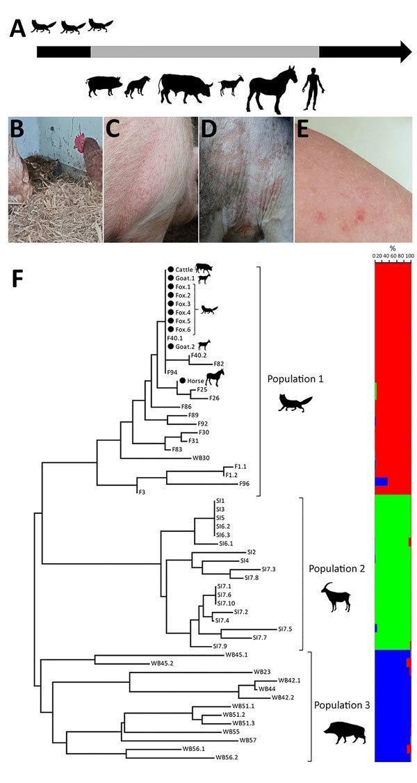
This composite educational resource illustrates a zoonotic outbreak of sarcoptic mange (Sarcoptes scabiei). The figure includes a timeline (A) showing cross-species transmission from foxes to livestock (pigs, oxen, goats, horses) and humans. Clinical photographs (B-E) demonstrate varied manifestations of the infestation: severe hyperkeratosis and lethargy in a red fox (B); erythematous papules on the shoulder and thorax of a pig (C); localized alopecia and erythema in the perineal region of an ox (D); and pruritic erythematous papules on the forearm of a teenage girl (E), characteristic of human scabies acquired from animal contact. A genetic component (F) features a neighbor-joining tree and multilocus microsatellite bar plot, providing molecular evidence of mite population clustering. The content highlights the clinical signs of sarcoptic mange across different hosts and the use of molecular epidemiology to trace transmission pathways in a shared farm environment. It serves as a study aid for dermatology, infectious disease, and public health veterinary medicine.
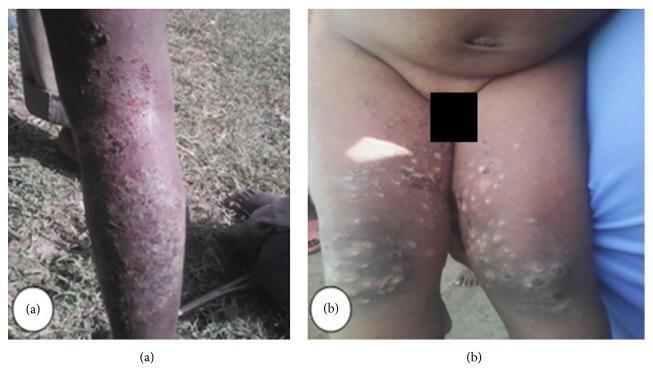
Two clinical photographs (a and b) demonstrating dermatological manifestations of a scabies infestation in human patients. Image (a) presents the posterior view of a lower leg, showing a widespread, dense distribution of small, erythematous papules and patches. The rash covers the calf and ankle regions with evidence of excoriations and crusting, suggesting secondary bacterial infection or intense pruritus. Image (b) shows the intergluteal region and posterior thighs of a child. This view exhibits a polymorphous eruption consisting of scattered papules, larger nodules, and pustular lesions. Some lesions show signs of inflammation and hyperpigmentation. The distribution is characteristic of scabies, frequently involving skin folds and areas of contact. These images illustrate the typical clinical presentation of sarcoptic mange (scabies) in a community outbreak setting, highlighting common complications such as secondary impetiginization and extensive cutaneous involvement.
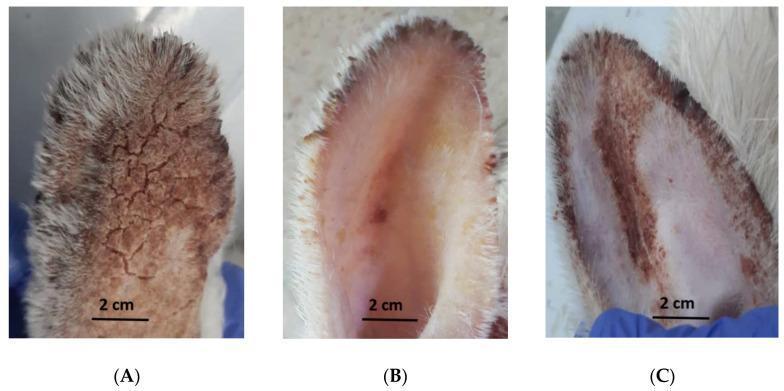
This clinical photograph displays a comparative macroscopic examination of rabbit ears affected by sarcoptic mange (Sarcoptes scabiei) across three experimental groups. Panel A (Control) demonstrates severe clinical manifestations of mange, characterized by extensive brownish discoloration, thick hyperkeratotic crusts, and a 'cracked-mud' skin texture indicating heavy scale formation and chronic inflammation. Panel B (Coconut Seed Extract group) shows a significant therapeutic response, featuring smooth, pinkish skin with a near-complete absence of scabs and scales, suggesting effective resolution of the parasitic infestation and tissue healing. Panel C (Ivermectin group) depicts an intermediate clinical state; while there is an improvement compared to the control, residual erythematous patches and localized scabbing remain visible, particularly along the ear margins. The image illustrates the efficacy of different scabicidal treatments on skin integrity, inflammatory response, and epithelial recovery in a zoonotic dermatological model. Key educational concepts include the morphology of parasitic dermatitis, hyperkeratosis, and macroscopic evaluation of wound healing.
canine demodectic mange demodicosis dog
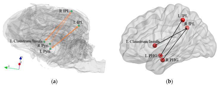
Comparative neuroanatomical diagram illustrating functional connectivity networks in the canine (a) and human (b) brains, specifically highlighting regions associated with familiarity and recollection judgments. Both panels use semi-transparent 3D brain renderings to display spatial relationships. In the dog brain (a), the network features connections between the bilateral inferior parietal regions (IPL), the bilateral pyriform cortex (Pyri), and the left claustrum/insula, represented by orange vectors. In the human brain (b), the homologous network connects the bilateral IPL, the bilateral parahippocampal gyrus (PHG), and the left claustrum/insula, represented by black vectors. The diagram illustrates how these specific regions of interest (ROIs) form functional circuits. The differences in anatomical distribution—such as the prominence of the pyriform cortex in the canine model versus the parahippocampal gyrus in the human model—highlight species-specific neurofunctional specializations. These diagrams are used in neuroimaging research to map behavioral changes to connectivity shifts within the fronto-parietal and limbic systems.
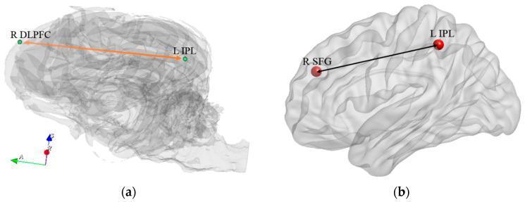
Anatomical comparison of functional connectivity within the fronto-parietal network (FPN) in the canine and human brain. (a) A 3D wireframe reconstruction of a dog brain illustrating the connectivity between the Right Dorsolateral Prefrontal Cortex (R DLPFC) and the Left Inferior Parietal Region (L IPL), indicated by an orange arrow. (b) A 3D cortical rendering of a human brain showing homologous connectivity between the Right Superior Frontal Region (R SFG) and the Left Inferior Parietal Region (L IPL), indicated by a black line. These visualizations highlight the evolutionary preservation of the FPN, a critical system involved in cognitive control, attention, and memory retrieval. The diagram serves to demonstrate how specific functional connections in these regions correlate with behavioral performance and neuroplasticity during training. Labels use standard neuroanatomical nomenclature, and R/L indicate right and left hemispheres respectively.
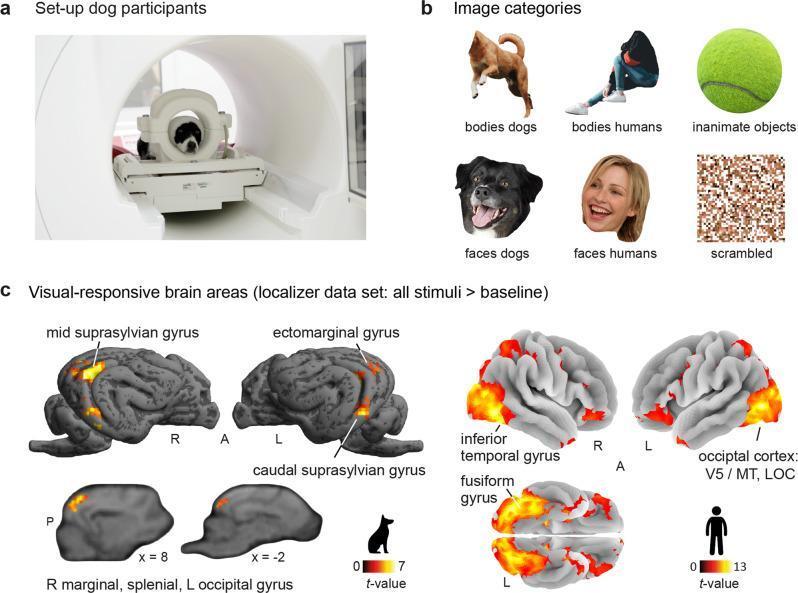
An educational infographic summarizing a comparative functional magnetic resonance imaging (fMRI) study on visual processing in dogs and humans. Panel A shows the experimental setup with a dog positioned inside a 3T MRI scanner using a custom-made head coil. Panel B illustrates the six stimulus categories: canine bodies, human bodies, inanimate objects (tennis ball), canine faces, human faces, and scrambled control images. Panel C displays statistical parametric maps of visual-responsive brain areas (all stimuli > baseline) projected onto species-specific cortical surfaces. In dogs, activation is localized to the mid suprasylvian gyrus, ectomarginal gyrus, caudal suprasylvian gyrus, and marginal/splenial regions, with t-values ranging from 0 to 7. In humans, activation is prominent in the inferior temporal gyrus, fusiform gyrus, and occipital cortex (including V5/MT and the lateral occipital cortex), with t-values ranging from 0 to 13. The image demonstrates the comparative neuroanatomy of the ventral visual stream and the specialization of higher-order sensory cortices across species for animate and inanimate stimulus perception.
canine pyoderma bacterial skin infection dog pustules
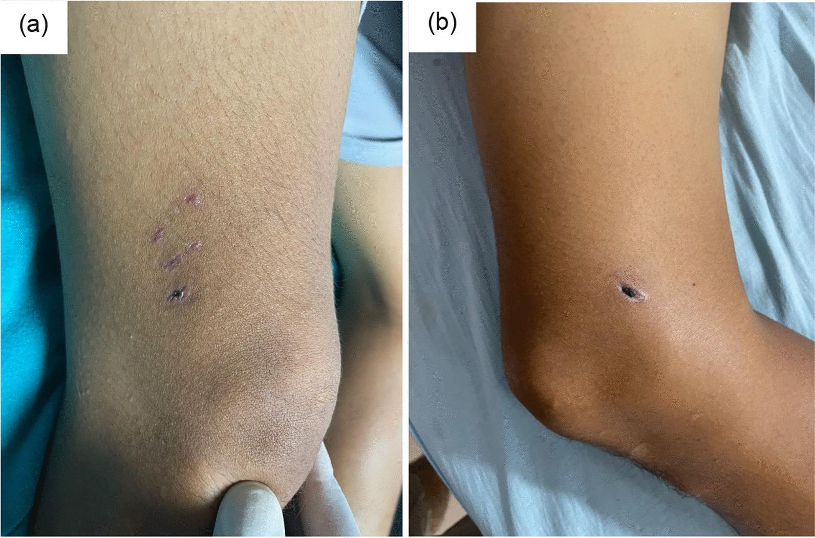
Clinical photograph of the right lower extremity in a 35-year-old patient demonstrating healing skin lesions from a dog bite occurring 25 days prior. (a) Lateral view of the distal thigh shows three well-defined, linear healing lacerations arranged in a pattern consistent with canine dentition. The wounds exhibit pink-to-purple post-inflammatory hyperpigmentation with closed edges and no signs of active discharge or perilesional cellulitis. (b) Medial view of the same limb reveals a single, 1 cm deep puncture wound characterized by a prominent, dry, black necrotic scab (eschar). The surrounding skin shows mild induration but lacks significant erythema or fluctuance, suggesting a dry healing state without secondary bacterial infection. These images represent characteristic cutaneous manifestations of animal bite trauma and are contextually linked to a clinical case of rabies encephalomyelitis, emphasizing the importance of identifying primary inoculation sites in zoonotic disease workups.
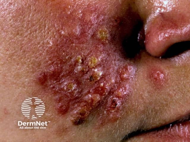
Clinical photography of the mid-face demonstrates a severe inflammatory rosacean eruption consistent with pyoderma faciale (rosacea fulminans). The image shows an asymmetric, left-sided distribution involving the malar cheek and perioral skin with confluent erythema, edematous plaques, and numerous yellow-brown crusts overlying fragile erosions. Superficial pustules and papules coalesce into broad plaques, with surrounding telangiectatic vasculature and subtle induration. The skin appears tender to palpation in the affected zones; the nose, nasolabial folds, and surrounding cheek show intense inflammation, while adjacent non-lesional skin remains relatively unf acted. There is no obvious sign of systemic infection; the presentation is acute in onset with abrupt inflammatory burden, suggesting rosacea spectrum rather than acne vulgaris or impetiginized dermatitis. The imaging modality, a high-resolution clinical photograph, provides rapid documentation of morphology, distribution, and severity, assisting differential diagnosis, treatment planning, and longitudinal monitoring. Clinically, this entity requires prompt, targeted therapy to reduce inflammation, prevent scarring, and manage potential triggers; management commonly involves systemic antibiotics (e.g., doxycycline) and anti-inflammatory regimens, with cautious corticosteroid use in select cases. Recognizing pyoderma faciale within the rosacea family is essential to differentiate from acne fulminans and infectious etiologies, guiding dermatologic evaluation and patient counseling. Accurate labeling supports diagnostics, research, and education globally.
canine allergic dermatitis atopic dog skin rash
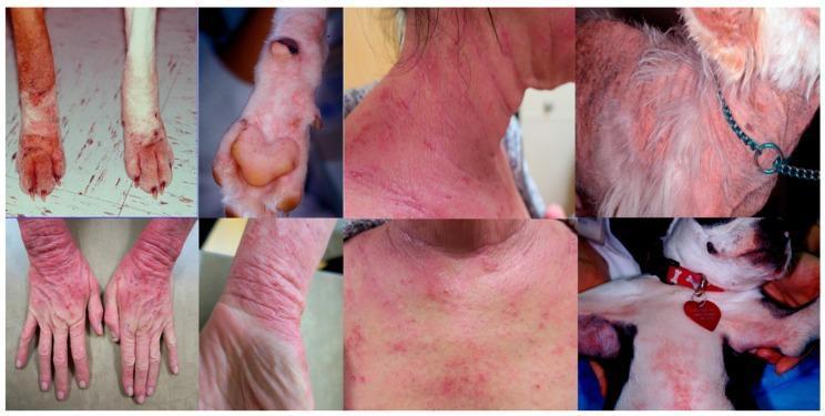
This clinical comparison chart displays eight photographs illustrating the manifestations of atopic dermatitis (AD) in both human and canine subjects. The images highlight a shared distribution pattern of inflammatory skin lesions across both species. In humans, clinical signs include diffuse erythema, lichenification, and scaling on the hands, wrists, neck, and upper chest. In canines, similar presentations of erythematous dermatitis are shown on the paws (pododermatitis), neck, and ventral chest (axillary region). Morphological features common to both species include intense redness and evidence of chronic irritation. The canine images specifically show secondary changes like alopecia, hyperpigmentation, and potential ulceration on the paws. The comparison serves to demonstrate the similarities in the clinical phenotype of AD across species, particularly targeting areas with high skin permeability and flexural surfaces. Educational focus is placed on recognizing these shared anatomical predilections—the face, neck, chest, and extremities—which are central to the diagnosis of allergic skin disease in a One Health clinical context.
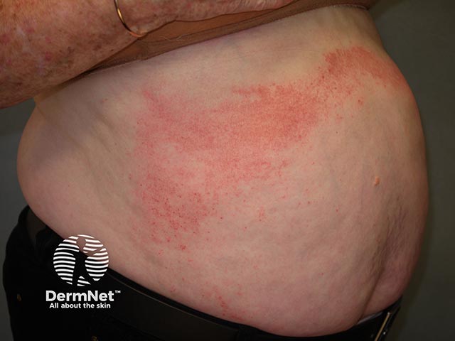
Clinical photograph of the anterior abdominal skin illustrating allergic contact dermatitis. Modality: Dermatologic photography; technique: standard color image, high resolution. Primary diagnosis: contact dermatitis due to butylphenol formaldehyde resin, with exposure commonly encountered in adhesives, resins, or medical-grade cements used in prosthetics or cosmetics. On the skin, there are diffuse erythematous patches across the ventral abdomen, with ill-defined margins and slight surface scaling and mild edema. The rash appears as coalescent red macules and papules, arranged in an irregular, patchy distribution along the exposed skin area. The image captures a localized inflammatory reaction with surface warmth and possible excoriations from scratching. Differential considerations include irritant contact dermatitis, atopic dermatitis, and less likely urticaria or drug eruption, but the temporal association with exposure to resin supports an allergic mechanism. Clinically, patch testing can confirm sensitization to butylphenol formaldehyde resin; avoidance is key, along with topical corticosteroids or calcineurin inhibitors to reduce inflammation; emollients to restore barrier. This image is relevant for dermatology education, allergology, occupational skin disease, and clinical decision support, illustrating the characteristic pattern of contact dermatitis on the abdomen, the importance of exposure history, and the role of diagnostic testing in suspected resin allergy. Useful for case-based learning and comparison with other eczematous eruptions.
ringworm dermatophytosis dog fungal skin lesion
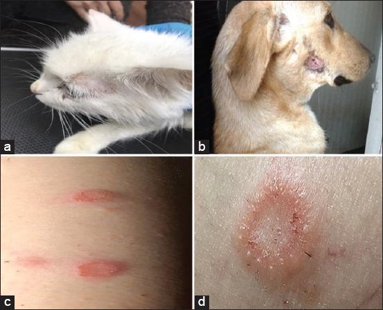
This composite clinical photograph illustrates dermatophytosis (ringworm) across multiple species, highlighting zoonotic transmission and comparative morphology. Panel (a) shows a white cat with a periocular lesion characterized by focal alopecia and mild erythema. Panel (b) depicts a dog with a well-defined, circular erythematous plaque on the lateral muzzle, exhibiting crusting and central hair loss. Panels (c) and (d) focus on human cutaneous manifestations. Panel (c) shows multiple small, oval, erythematous maculopapular lesions on the trunk. Panel (d) provides a close-up of a classic tinea corporis lesion, featuring a distinctive annular (ring-shaped) morphology with a raised, erythematous, scaly border and relative central clearing. The educational focus of the image is to demonstrate the clinical presentation of Microsporum or Trichophyton infections, emphasizing the varied presentation of scale, alopecia, and circular erythema across feline, canine, and human hosts, which is essential for diagnosing fungal skin infections in clinical and veterinary practice.
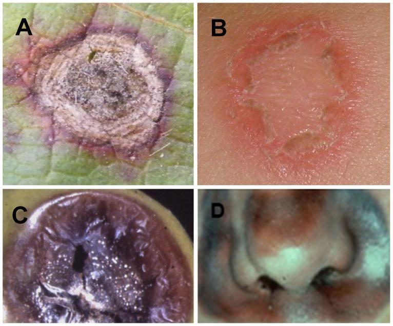
A comparison chart consisting of four panels (A-D) illustrating morphological similarities between plant and human fungal infections. Panel B shows a clinical photograph of dermatophytosis (ringworm) on human skin, characterized by a classic annular (ring-shaped) lesion with a raised, erythematous, and slightly scaly border and central clearing. Panel D presents a clinical photograph of invasive mucormycosis affecting the human nasal region, demonstrating extensive tissue necrosis with dark brown to black discoloration and ulceration around the nostrils and nasal bridge. For educational comparison, Panels A and C show botanical fungal infections: panel A depicts a Cercospora shot hole lesion on a leaf with necrotic centers and pigmented margins, while panel C shows Botrytis fruit rot with soft, dark, necrotic tissue and fungal spores. This visual resource is designed for medical students and dermatologists to understand fungal pathogenesis and the cross-kingdom similarities in host tissue responses such as necrosis and inflammation.
canine hot spot acute moist dermatitis dog
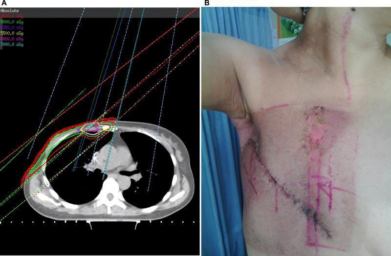
This composite educational graphic illustrates the correlation between radiation therapy planning and acute skin toxicity in a post-mastectomy patient. Image A is a diagnostic axial CT scan of the chest showing a conventional radiotherapy dosage plan for breast cancer. Colored isodose curves (45 Gy to 70 Gy) are superimposed on the anterior chest wall, revealing 'hot spots' where radiation fields overlap between the medial tangent and the internal mammary node (IMN) field. Image B is a clinical photograph of the same patient’s right chest and axilla demonstrating acute radiation dermatitis. Key findings include extensive moist desquamation characterized by denuded, erythematous skin and serous exudate in the axillary fold and the parasternal region. The distribution of the skin lesions corresponds directly to the high-dose 'hot spots' identified in the radiation plan. Clinical markers and surgical scars are also visible, highlighting the spatial relationship between the treatment fields and the resulting cutaneous damage. This visual demonstrates the pathophysiology of radiation-induced skin injury and the clinical significance of dose distribution in oncological treatment.
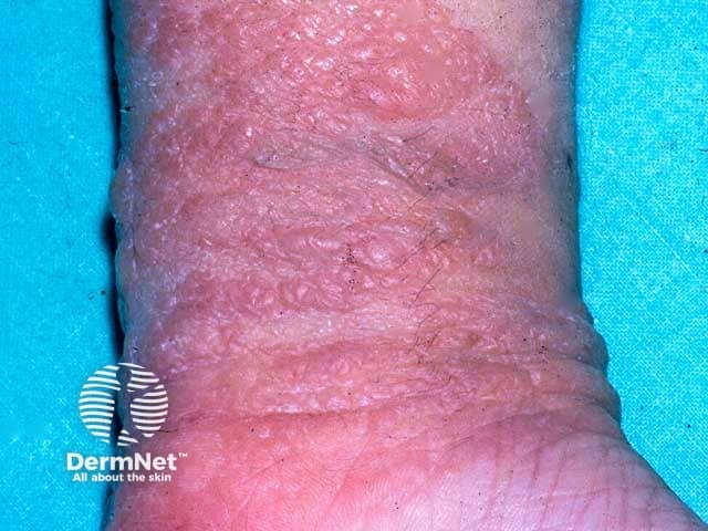
Clinical photography of the dorsal left wrist demonstrates an acute, moist dermatitis confined to the adhesive contact zone of a sticking plaster. High-resolution color image shows erythematous, inflamed skin with pronounced edema, surface crinkling, and serous oozing. Superficial vesiculation is present at the margins of the lesion, with excoriations and crust formation indicating ongoing excoriation from pruritus and rubbing. The distribution is linear to irregular along the plaster edge, reflecting contact with the adhesive and backing material. The surrounding skin is mildly inflamed but without deep ulceration or panniculitis. The appearance is compatible with allergic contact dermatitis, a type IV hypersensitivity reaction to components of plaster adhesives such as acrylates or natural resins (colophony) in susceptible individuals. Differential diagnoses include irritant contact dermatitis from adhesive exposure, irritant dermatitis, or to a lesser extent eczema/atopic dermatitis restricted to the site. Clinical significance lies in identifying a contact allergen and guiding avoidance; patch testing to confirm sensitization to plaster constituents is recommended. Management implications include allergen avoidance, topical corticosteroids to reduce inflammation, and emollients to restore barrier function. This image is relevant for dermatology education, allergology, patch-testing literature, and clinical practice guidelines on contact dermatitis and plaster allergies. Useful for clinical education.
canine mast cell tumor dog skin neoplasm
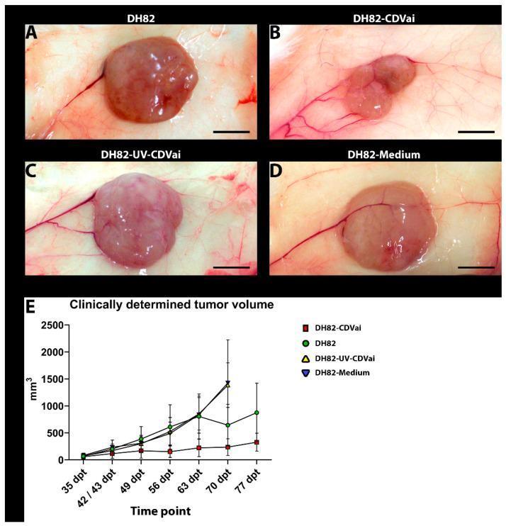
This composite educational resource presents clinical images and a line graph illustrating tumor growth in a DH82 cell line xenograft model, evaluating the effects of Canine Distemper Virus (CDV) infection. Figures A-D show representative gross photographs of subcutaneous neoplasms at 63 days post-transplantation (dpt) across four groups: non-infected controls (DH82), CDV-infected (DH82-CDVai), UV-inactivated CDV-injected (DH82-UV-CDVai), and medium-injected controls. The DH82-CDVai neoplasm (B) is visually smaller and exhibits more lobulated surface characteristics compared to the larger, smoother, and more spherical neoplasms in the control groups (A, C, D). Prominent peritumoral vasculature is visible in all specimens. A scale bar representing 0.5 cm is included in each image. Figure E displays a line graph of clinically determined tumor volume (mm³) from 35 to 77 dpt. The graph demonstrates that CDV-infected xenografts maintain a relatively stable, significantly lower volume over time compared to the continuous progression and rapid growth observed in the non-infected and medium-treated control groups.
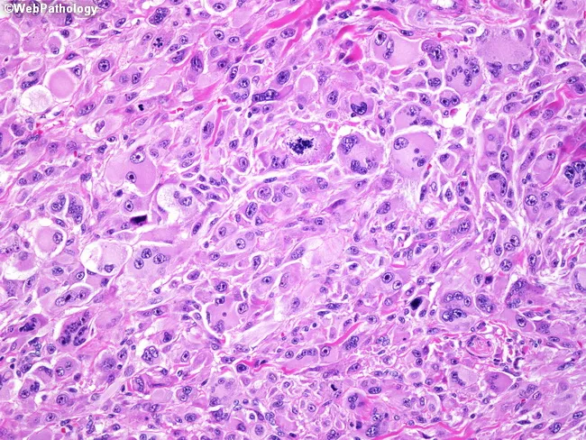
High-magnification hematoxylin and eosin stained skin biopsy reveals a highly atypical spindle cell neoplasm with storiform and fascicular growth patterns. Tumor cells are pleomorphic, with elongated nuclei, hyperchromasia, conspicuous nucleoli, and abundant mitotic figures including atypical mitoses, consistent with a high-grade sarcomatoid phenotype. The lesion displays prominent cellularity in a dermal-to-subcutaneous setting, with occasional multinucleated cells and hemorrhagic/inflammatory background. Immunophenotype can mimic melanoma or spindle cell squamous cell carcinoma, necessitating ancillary testing. Atypical fibroxanthoma (AFX) typically shows positivity for vimentin, HAM56, and alpha1-antitrypsin, while lacking lineage-specific markers. In this case, tumor cells are negative for S-100, Melan A (MART-1), desmin, and cytokeratins, supporting non-epithelial and non-melanocytic differentiation. The differential diagnosis includes spindle cell melanoma, spindle cell squamous cell carcinoma, and other cutaneous sarcomas; IHC panel is critical to exclude melanocytic and epithelial origins. Clinically, AFX arises in sun-exposed, chronically damaged skin of elderly patients and is treated by complete surgical excision with clear margins; prognostication depends on complete removal and absence of metastasis. This image is educational for pathology education, differential diagnosis, and tailored immunohistochemical workup; it demonstrates characteristic spindle morphology, storiform architecture, high mitotic rate, and immunoprofile supporting AFX.
canine seborrhea sebaceous skin disorder dog scales
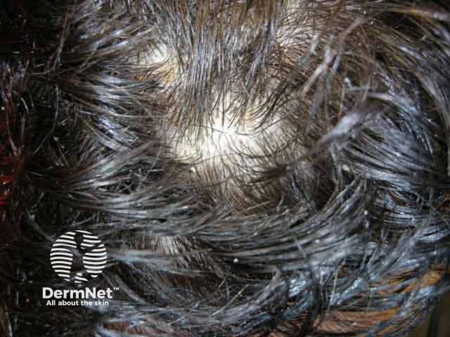
Clinical photograph of the scalp depicting seborrheic dermatitis with visible scales and dandruff. Modality: clinical photography; technique: standard bright-light macro close-up; magnification approximately 1.5–2x. Anatomic location: hair-bearing skin of the scalp (vertex and frontal regions), with erythematous, inflamed surrounding skin and greasy yellowish-white scales adherent to hair shafts and interfollicular zones. Visual features include diffuse flaking, oily sheen, crusting at hairline, and punctate erythema beneath flaking areas. The scale is fine to moderately thick, loosely adherent to microscopic hair shafts, often described as dandruff in milder cases. The image captures typical seborrheic dermatitis morphology: erythematous plaque-like patches with greasy scales, preferentially in sebaceous-rich regions, sometimes involving eyebrows and the glabella. Pathological correlation: clinically arcuate to patchy inflammation with hyperseborrhea; Malassezia yeast and lipid-rich microenvironment contribute to flaking. Diagnostic significance: supports diagnosis of seborrheic dermatitis; helps differentiate from dry scalp, psoriasis, or tinea capitis; guides therapeutic decisions. Potential clinical use cases include documentation of disease severity, patient education about chronic relapsing course, and evaluation of treatment response to antifungal shampoos (ketoconazole, zinc pyrithione) and anti-inflammatory agents (low-potency steroids, calcineurin inhibitors). When in doubt, correlate with symptoms such as pruritus, distribution, and presence of associated conditions (facial seborrhea, dandruff).
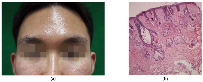
This composite image illustrates the clinical and histological features of facial seborrhea. Panel (a) is a clinical photograph of a young male's face, demonstrating characteristic greasy skin and prominent surface shine on the forehead and nose, indicative of excessive sebum production. Enlarged facial pores (ostia) are clearly visible on the cheeks. Panel (b) provides a correlating histological view of facial skin (nasal region) stained with hematoxylin and eosin (H&E) at x50 magnification. Key microscopic findings include multiple lobules of mature, hypertrophic sebaceous glands located in the dermis. A significantly dilated follicular infundibulum is observed, filled with lamellated keratinous material and sebum. This educational material highlights the relationship between macroscopic 'oily skin' and the underlying hyperactive pilosebaceous unit, a common factor in the pathogenesis of acne vulgaris and seborrheic dermatitis.
major dermatological conditions canines clinical signs treatment 2024
flea allergy dermatitis dog skin lesion alopecia
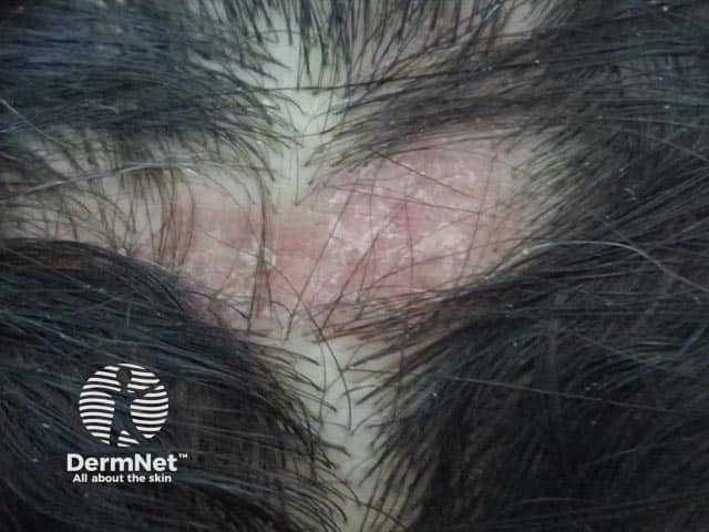
This is a dermatology clinical photograph of a scalp lesion in a patient with discoid lupus erythematosus (DLE). Modality and technique: color digital clinical photography, high-resolution close-up scalp image, frontal orientation. Anatomic localization: scalp skin over the vertex with involvement of the parietal regions. Visual characteristics include red-to-violaceous erythematous plaques with thick adherent scale, focal crusting, and patchy, scarring alopecia with hair shaft destruction; perifollicular hyperkeratosis and follicular plugging may be evident. The lesion shows surface inflammation with atrophic scarring on the affected areas, consistent with active discoid lupus and potential cicatricial alopecia. If biopsy were performed, histology would typically reveal interface dermatitis with a lymphocytic infiltrate and dermal mucin; clinical context remains essential. This image is diagnostic for a scarring alopecic disorder and is clinically significant for differentiating DLE from psoriasis, seborrheic dermatitis, tinea capitis, and lichen planopilaris. Diagnostic significance includes recognition of scarring alopecia risk, need for serologic evaluation, and consideration of antimalarials and topical therapies. Clinical correlation includes sun protection and monitoring for systemic lupus erythematosus. Educational use encompasses dermatology training, case-based learning, and AI-assisted image analysis of scalp inflammatory dermatoses. The image also serves as a reference for documenting progression and treatment response in both pediatric and adult patients.
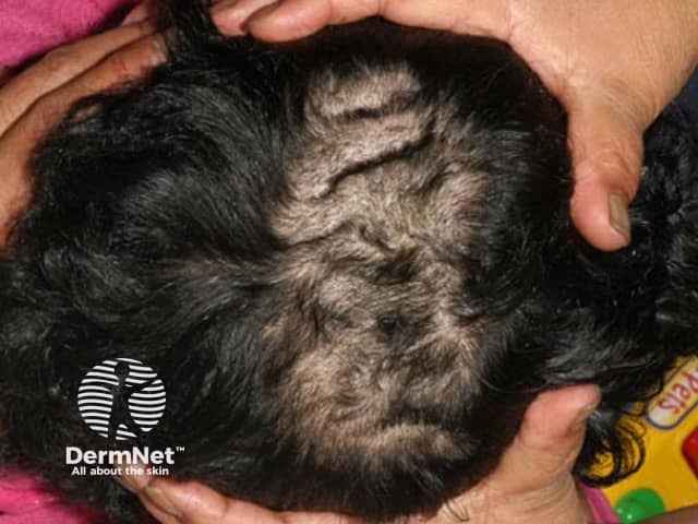
This clinical dermatology photograph captures a posterior view of the scalp illustrating patchy non-scarring alopecia with conspicuous scaling and crusting along several discrete patches in the occipital region. Hair within affected patches is sparse, with visible hair shaft breakage and reduced density. Perifollicular desquamation and mild erythema are evident at lesion margins, while surrounding scalp skin remains largely uninvolved. The distribution is focal and localized to the occipital scalp, with no obvious regrowth in the affected zones. The appearance is most consistent with tinea capitis (scalp ringworm) or another dermatophyte infection, though alopecia areata and seborrheic dermatitis are important differential considerations. Clinical significance lies in distinguishing infectious from inflammatory alopecias to direct appropriate therapy and infection control measures. The image demonstrates classic features: patchy alopecia with scale, crust, and fragile hairs; reduced pigment variation; discrete borders; and preserved epidermal integrity outside patches. Imaging modality: standard high‑resolution clinical photography, color-accurate to facilitate visual diagnosis, documentation, and educational use. Anatomical context: scalp skin over the occipital region, hair-bearing epithelium with involved follicles. Potential diagnostic workflow includes fungal microscopy (KOH prep) and culture, followed by systemic antifungal treatment (griseofulvin, terbinafine) and topical antifungals. This image supports training in pediatric dermatology and differential diagnosis of patchy scalp alopecia with scaling.
Major Dermatological Conditions in Canines
1. Flea Allergy Dermatitis (FAD) - Most Common Allergic Skin Disease
- Intense pruritus, especially over the dorso-lumbar region (rump/base of tail)
- Papular and crusting eruptions, alopecia
- Self-trauma, secondary bacterial infection
- Even a single flea bite can trigger a severe reaction in sensitized dogs
2. Canine Atopic Dermatitis (CAD)
- Chronic, relapsing pruritus
- Erythema and alopecia of paws (pododermatitis), face, ears, ventral abdomen, axillae, groin
- Lichenification and hyperpigmentation in chronic cases
- Secondary Malassezia and bacterial infections
3. Sarcoptic Mange (Canine Scabies)
- Intense, non-seasonal pruritus (often worse at night)
- Papular crusting eruptions starting on ear margins, elbows, ventral abdomen
- Alopecia, excoriations, thick yellow-gray crusts in severe cases
- Zoonotic - can transiently infect humans
4. Demodicosis (Demodectic Mange)
- Localized - patchy alopecia, usually young dogs, often self-limiting
- Generalized - widespread, severe; associated with immunosuppression (steroids, cancer, hypothyroidism)
- Non-pruritic (unless secondary infection present) patchy to diffuse alopecia
- Comedones, follicular papules and pustules
- "Moth-eaten" appearance
- Secondary bacterial pyoderma (furunculosis)
5. Dermatophytosis (Ringworm)
- Circular areas of alopecia with erythema, scaling, and crusting
- Classic "ring-shaped" lesion with central clearing
- Most common on face, paws, and trunk
- Usually NOT severely pruritic
- Zoonotic - transmissible to humans and cats
6. Pyoderma (Bacterial Skin Infection)
- Surface - hot spots (acute moist dermatitis), intertrigo (skin fold dermatitis)
- Superficial - impetigo (puppies), superficial folliculitis (papules, pustules, epidermal collarettes)
- Deep - furunculosis, cellulitis; often secondary to demodicosis, hypothyroidism, or atopy
- Pustules, papules, epidermal collarettes (ring-shaped scale)
- Alopecia, crusts, and hyperpigmentation
- Deep pyoderma: pain, draining tracts, hemorrhagic bullae
7. Malassezia Dermatitis
- Erythema, greasy scales, and rancid/musty odor
- Intense pruritus
- Predilection sites: ventral neck, ear canals, axillae, interdigital spaces, skin folds, perianal region
- Reddish-brown staining of fur (especially paws in allergic dogs)
- Chronic cases: lichenification and hyperpigmentation ("elephant skin")
8. Canine Seborrhea
- Seborrhea sicca - dry, flaky dandruff
- Seborrhea oleosa - greasy, oily coat with rancid odor
- Seborrheic dermatitis - combination with inflammation
9. Canine Hypothyroid-Related Dermatosis
- Symmetric, non-pruritic alopecia (trunk, flank, neck, tail)
- Dry, dull coat; skin thickening ("tragic face" from myxedema)
- Hyperpigmentation
- Secondary infections and poor wound healing
10. Skin Neoplasia
Mast Cell Tumor (MCT) - Most Common Canine Skin Tumor
- Classically "the great imitator" - can look like any mass
- Variable appearance: soft to firm, often hairless, sometimes ulcerated
- Releases histamine - can cause erythema and urticaria when manipulated (Darier's sign)
- Predisposed breeds: Boxers, Bulldogs, Pugs, Boston Terriers, Golden Retrievers
- Graded I-III; treatment: surgical excision +/- chemotherapy, targeted therapy (toceranib)
Histiocytoma
- Round, button-like, erythematous nodule
- Young dogs (under 3 years), typically on head or limbs
- Usually self-regressing within 3 months
Squamous Cell Carcinoma (SCC)
- Ulcerated, non-healing lesions on poorly pigmented or sun-exposed skin (nasal planum, ear tips)
- Predisposed breeds: Light-coated dogs (Dalmatians, Bull Terriers)
- Treatment: surgery, radiation, photodynamic therapy
Summary Table
| Condition | Primary Cause | Key Feature | Zoonotic? |
|---|---|---|---|
| Flea Allergy Dermatitis | Flea hypersensitivity | Dorso-lumbar pruritus | No |
| Atopic Dermatitis | Environmental allergens | Paw/ear/ventral involvement | No |
| Sarcoptic Mange | Sarcoptes mite | Intense pruritus, ear margins | Yes |
| Demodicosis | Demodex mite | Follicular alopecia, non-pruritic | No |
| Dermatophytosis | Dermatophyte fungi | Circular alopecic lesions | Yes |
| Pyoderma | Staphylococcus | Pustules, epidermal collarettes | Rare |
| Malassezia Dermatitis | Yeast overgrowth | Greasy, malodorous skin | No |
| Seborrhea | Multifactorial | Scales, greasy coat | No |
| Hypothyroid Dermatosis | Low thyroid hormone | Symmetric alopecia | No |
| Mast Cell Tumor | Neoplasia | Any skin mass in Boxer-type breeds | No |