

Explain the slides in simple mbbs language
🫀 Slide 1: Introduction & Hemodynamics
What is Eisenmenger Syndrome?
- It starts as a left-to-right shunt (e.g., VSD, ASD, PDA) - blood flows from the high-pressure left side to the low-pressure right side
- Over time, this extra blood flow damages the pulmonary vessels → causes Pulmonary Arterial Hypertension (PAH)
- Eventually, pulmonary pressure becomes so high that the shunt reverses → now blood flows right to left (deoxygenated blood enters systemic circulation → cyanosis)
- Eisenmenger Complex = specifically when VSD causes this reversal
Key Hemodynamics (Easy to Remember):
| Defect | What Happens |
|---|---|
| VSD/PDA | RV only gets concentric hypertrophy (no dilation) because R-to-L shunt decompresses the RV |
| PDA | Deoxygenated blood enters the descending aorta → lower limbs turn blue, upper limbs stay pink = DIFFERENTIAL CYANOSIS (classic!) |
| ASD/VSD | Deoxygenated blood enters the ascending aorta → fingers AND toes both cyanosed = EQUAL CYANOSIS |
| ASD (atrial level) | RV also dilates + cardiac enlargement because RV pressure can exceed systemic pressure |
🩺 Slide 2: Clinical Features
Symptoms (what patient complains of):
- Cyanosis (bluish discoloration)
- Fatigue and dyspnea (breathlessness on effort)
- History of repeated chest infections in childhood (because the original L-to-R shunt causes excess blood in lungs)
Examination findings:
- Cyanosis + Clubbing (chronic hypoxia causes finger clubbing)
- Differential cyanosis (lower limb blue, upper pink) = PDA ; equal cyanosis = VSD/ASD
- Parasternal impulse = RV heave felt at left sternal border (sign of RV hypertrophy)
- Palpable S2 at pulmonary area (because pulmonary pressure is very high)
- P2 louder than A2 - the pulmonary component of the second heart sound becomes loud (normally A2 > P2)
🔊 Slide 3: Auscultation + Investigations
S2 Splitting - High-Yield Table:
| Defect | S2 Character |
|---|---|
| VSD | SINGLE S2 (A2 and P2 superimposed - both ventricles equalize) |
| PDA | Normal splitting of S2 |
| ASD | Wide and FIXED split (classic!) - fixed because ASD equalizes pressures throughout respiration |
Other Heart Sounds:
- Constant pulmonary ejection click in ASD (heard in both inspiration AND expiration - distinguishes it from other clicks)
- Graham-Steell murmur = harsh, high-pitched early diastolic murmur at left sternal border due to pulmonary regurgitation (from high pulmonary pressure)
- Tricuspid regurgitation can also develop in ASD patients
Investigations:
- ECG: Right axis deviation + RVH + P-pulmonale (tall peaked P waves in lead II)
- CXR:
- Prominent pulmonary arterial segment (big pulmonary artery shadow)
- Large right and left main pulmonary arteries (central vessels dilated)
- Peripheral lung fields look empty = "pruning" (classic CXR finding - vessels disappear at periphery)
💊 Slide 4: Treatment (partially visible)
- Repair the defect early (before pulmonary hypertension develops) - surgical correction of VSD/ASD/PDA in childhood
- Once Eisenmenger develops, surgical correction is contraindicated (closing the defect would remove the "safety valve" and cause acute RV failure)
- Management then becomes: Pulmonary vasodilators (sildenafil, bosentan), avoid dehydration/iron deficiency, and ultimately heart-lung transplant
Quick Memory Tricks:
- PDA = Differential cyanosis (lower blue, upper pink) - think "P for Peripheral/lower limb"
- ASD = Fixed wide split S2 - classic exam answer
- VSD in Eisenmenger = Single S2
- Pruning on CXR = Eisenmenger (central vessels dilate, peripheral vessels disappear)
- Graham-Steell murmur = PR due to PAH (not a structural pulmonary valve problem)
Hemodynamics in Epstein anomaly
Ebstein anomaly hemodynamics diagram tricuspid valve
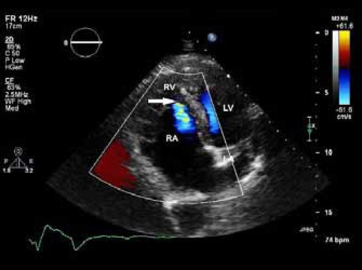
This diagnostic image is a transthoracic echocardiogram in the apical four-chamber view, utilizing color flow Doppler to evaluate cardiac hemodynamics. The visual demonstrates characteristic features of Ebstein’s anomaly, most notably the apical displacement of the septal leaflet of the tricuspid valve, which results in the 'atrialization' of the proximal portion of the right ventricle. There is significant dilation of both the right atrium (RA) and right ventricle (RV), with the RA appearing markedly enlarged compared to the left ventricle (LV). The color Doppler overlay reveals a blue-tinted regurgitant jet (indicated by a white arrow) moving from the right ventricle back into the right atrium, consistent with mild-to-moderate tricuspid regurgitation. A red zone at the periphery of the sector indicates flow towards the transducer. This image serves as an educational example of congenital right-sided heart disease and valvular pathology, illustrating the anatomical remodeling and functional consequences associated with tricuspid valve malformation.
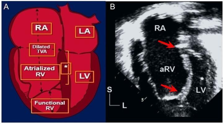
Educational panel illustrating Ebstein anomaly through a comparison of anatomical schematic (A) and diagnostic ultrasound (B). Figure A is a pathophysiology diagram showing apical displacement of the septal tricuspid valve leaflet, creating a tripartite right heart structure: the true right atrium (RA), an atrialized portion of the right ventricle (aRV), and a small functional right ventricle (RV). It highlights the dilated tricuspid valve annulus (TVA) and the characteristic large, sail-like anterior leaflet. Figure B is an apical 4-chamber echocardiogram during systole, showing the clinical manifestation of the anomaly. Red arrows delineate the displacement index—the distance between the mitral valve annulus and the apically displaced septal tricuspid leaflet. This measurement, when indexed to body surface area (>8 mm/m²), is a critical diagnostic criterion to distinguish Ebstein anomaly from other causes of right ventricular enlargement. Visible chambers include the massively dilated RA, the aRV, and the compressed left ventricle (LV).
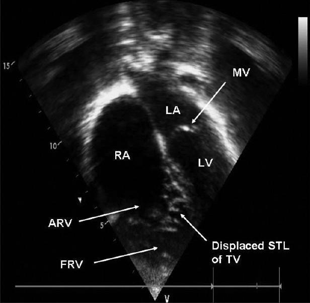
This diagnostic image is a transthoracic echocardiogram in an apical four-chamber view, demonstrating Ebstein’s anomaly of the tricuspid valve. The visual highlights a significant apical displacement of the septal tricuspid leaflet (STL) of the tricuspid valve (TV) relative to the mitral valve (MV). This displacement results in the 'atrialization' of the superior portion of the right ventricle (labeled as ARV - Atrialized Right Ventricle), which is functionally continuous with the right atrium (RA). The image also depicts the remaining functional right ventricle (FRV), which appears reduced in size due to the inferior displacement of the valve. Key anatomical landmarks labeled include the left atrium (LA), left ventricle (LV), and right atrium (RA). This imaging modality is essential for visualizing the structural and spatial rearrangement characteristic of congenital tricuspid valve malformations. The clinical significance of this finding relates to the assessment of right heart hemodynamics and the degree of functional impairment in pediatric and adult congenital heart disease.
Ebstein's Anomaly - Hemodynamics (MBBS Level)
🔑 The Core Defect (Understand This First)
Diagnostic criterion: septal leaflet displaced >8 mm/m² more than the mitral valve insertion, or >20 mm in adults.
🫀 The "Two-Part RV" Concept (Most Important!)
| Part | What It Is | What It Does |
|---|---|---|
| Atrialized RV (aRV) | The portion of RV above the displaced valve - now "belongs" to the RA functionally | Thin, dilated, noncontractile - behaves like RA, not RV |
| Functional RV (fRV) | The portion below the displaced valve - the only true RV left | Small, does the actual pumping |
🔄 Step-by-Step Hemodynamics
Step 1: Tricuspid Regurgitation (TR)
- The displaced, dysplastic valve cannot close properly
- Blood regurgitates back from RV → RA during systole
- This is the primary hemodynamic problem - it's the most common congenital cause of TR
Step 2: Right-Sided Volume Overload
- The RA receives normal venous return from IVC/SVC PLUS the regurgitant blood from RV
- RA becomes massively dilated
- The atrialized RV also dilates (it's a thin-walled, noncontractile dead zone)
Step 3: Reduced Pulmonary Blood Flow
- The functional RV is small → it can't generate enough forward stroke volume
- Less blood flows forward into the pulmonary artery
- Result: Reduced pulmonary blood flow (oligemic lung fields on CXR)
Step 4: Raised RA Pressure
- RA is overloaded with blood and dilated
- RA pressure rises significantly
Step 5: Right-to-Left Shunt through ASD/PFO
- ~95% of Ebstein's patients have an ASD or PFO (patent foramen ovale)
- When RA pressure exceeds LA pressure, blood shunts right-to-left (RA → LA)
- Deoxygenated blood enters systemic circulation → CYANOSIS
- This shunt also "decompresses" the overloaded RV/RA (a partial relief valve)
Step 6: LV Dysfunction (Ventricular Interdependence)
- The massively dilated RA and aRV physically compress the LV
- The dilated RV shifts the interventricular septum to the left (paradoxical septal motion)
- This impairs LV filling and function (ventricular interdependence)
- In severe cases, this can cause LVOT obstruction or mitral valve prolapse
📊 Hemodynamics Summary Flowchart
Displaced TV leaflets
        ↓
   TR (valve leaks)
        ↓
RA + aRV overloaded and dilated
        ↓
  ↗ RA pressure rises
  ↓
R-to-L shunt via ASD/PFO → CYANOSIS
        ↓
Small fRV → Reduced pulmonary blood flow
        ↓
LV compression by huge RA/aRV → LV dysfunction
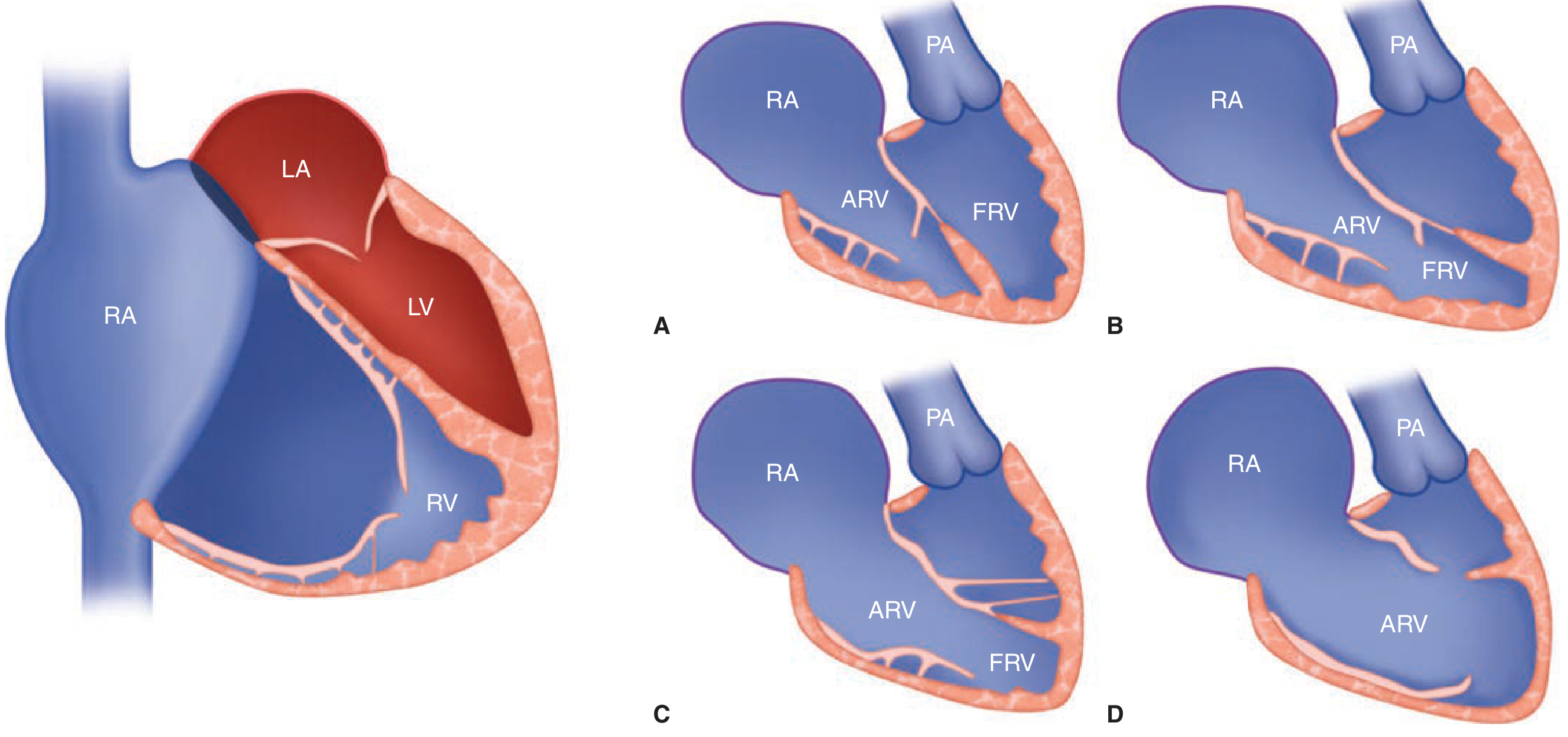
🧠 Carpentier Classification (Severity)
| Type | Atrialized RV | Functional RV | Notes |
|---|---|---|---|
| A | Small, contractile | Adequate | Mildest - valve repair possible |
| B | Large, noncontractile | Small | Marked displacement |
| C | Large, noncontractile | Very small | Restricted anterior leaflet |
| D | Almost entire RV atrialized | Only infundibulum left | Worst - near-complete atrialization |
⚡ Special Hemodynamic Points (High Yield for Exams)
| Feature | Explanation |
|---|---|
| Anterior leaflet is sail-like | It is elongated, redundant, tethered - can cause intracavitary RV obstruction |
| Neonatal Ebstein's | High pulmonary vascular resistance at birth → even more resistance to forward flow → severe cyanosis ("functional pulmonary atresia") |
| Accessory pathways (WPW) | The dilated AV junction has direct muscular connections between RA and RV → substrate for accessory pathways → arrhythmias (AVRT, AF, AFL) |
| R-to-L shunt as safety valve | The ASD shunt offloads the RV, but causes cyanosis and paradoxical embolism as a trade-off |
| "Wall-to-wall heart" | Severe fetal Ebstein's → massive cardiomegaly compresses lungs → pulmonary hypoplasia → intrauterine death |
💊 Clinical Consequences of These Hemodynamics
| Hemodynamic Problem | Clinical Result |
|---|---|
| TR + RA dilation | Jugular venous distension, hepatomegaly, peripheral edema |
| R-to-L shunt via ASD | Cyanosis, clubbing, polycythemia, paradoxical embolism (stroke!) |
| Low cardiac output | Fatigue, exercise intolerance, dyspnea |
| Accessory pathways | Palpitations, WPW, sudden cardiac death |
| Cardiomegaly | "Box-shaped heart" on CXR |
Echo Image of Ebstein's
Quick Memory Hook
"EBSTEIN = Eaten RV" - The RV gets partially "eaten" by the RA because the valve slid down. What's left (fRV) is too small to pump efficiently → TR → RA overload → R-to-L shunt → Cyanosis.
- Source: Fuster and Hurst's The Heart, 15th Ed. and Braunwald's Heart Disease