
CVA:ic bleed vs infarct treatment protocol
"ischemic stroke" AND "hemorrhagic stroke" AND "treatment guidelines"
ischemic stroke vs hemorrhagic stroke CT scan comparison
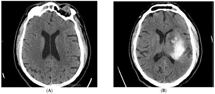
This figure presents a side-by-side comparison of non-contrast axial cerebral CT scans from the same patient, illustrating the progression of an acute stroke. (A) Initial CT scan at admission shows brain parenchyma with normal density and preserved grey-white matter differentiation. Symmetrical lateral ventricles and midline alignment are maintained, with no visible established ischemic lesions or hemorrhage at this early stage. (B) Follow-up CT scan performed 24 hours after thrombolytic therapy reveals a significant hemorrhagic transformation. A large, irregularly shaped hyperdense (bright) area is visible within the left hemisphere, specifically involving the left middle cerebral artery (MCA) territory. This hyperdensity indicates an intraparenchymal hematoma. There is evidence of mild mass effect on the adjacent left lateral ventricle. This comparison serves as a clinical example of a common complication following reperfusion therapy in acute ischemic stroke, emphasizing the importance of follow-up neuroimaging to detect secondary intracranial hemorrhage.
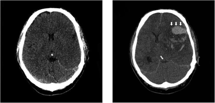
Side-by-side axial non-contrast CT head scans demonstrating the progression of an acute ischemic stroke. The initial scan (left) shows extensive loss of gray-white matter differentiation and sulcal effacement throughout the left cerebral hemisphere, consistent with a large acute middle cerebral artery (MCA) territory infarction. The follow-up scan (right) obtained two days later reveals hemorrhagic transformation within the infarcted region, visualized as a focal area of high-density parenchymal hematoma (indicated by white arrows). Associated mass effect is evident by the effacement of the ipsilateral lateral ventricle and midline shift toward the right. This comparison illustrates a severe complication of large-vessel ischemic stroke, highlighting the transition from cytotoxic edema to secondary intracranial hemorrhage and subsequent increased intracranial pressure.
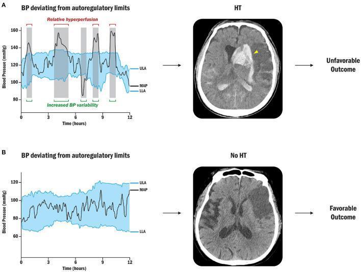
Educational infographic and diagnostic comparison illustrating the relationship between blood pressure (BP) autoregulation and hemorrhagic transformation (HT) following ischemic stroke. The visual is divided into two clinical scenarios (A and B). Row A (Unfavorable Outcome): A line graph shows Mean Arterial Pressure (MAP) frequently deviating above the Upper Limit of Autoregulation (ULA), indicated by shaded gray vertical bars representing relative hyperperfusion. The corresponding non-contrast axial CT scan of the brain reveals a large area of hyperdensity (indicated by a yellow arrowhead) within a hypodense ischemic territory in the left hemisphere, characteristic of parenchymal hematoma or HT. There is associated mass effect and ventricular compression. Row B (Favorable Outcome): The graph depicts MAP maintained strictly within the personalized limits of autoregulation (between ULA and LLA). The corresponding axial CT scan shows a stable, well-demarcated area of hypodensity in the left middle cerebral artery territory, representing an established infarct without evidence of hyperdense blood products (No HT). This comparison demonstrates how maintaining cerebral perfusion pressure within autoregulatory limits can prevent secondary brain injury and improve clinical outcomes by mitigating the risk of post-stroke hemorrhage.
CVA: Intracerebral Hemorrhage vs. Ischemic Infarct - Treatment Protocol
Quick Differentiation First
| Feature | Hemorrhagic Stroke (ICH) | Ischemic Infarct |
|---|---|---|
| CT appearance | Hyperdense (bright) lesion | Hypodense (dark) lesion (may be normal early) |
| Onset characteristics | Sudden; often with severe headache, vomiting, markedly elevated BP | Sudden focal deficits; headache less common |
| Prevalence | 10-15% of all strokes | ~85% of all strokes |
| Management philosophy | Supportive + hemostasis + BP control | Restore perfusion (tPA / thrombectomy) |
ISCHEMIC STROKE - Treatment Protocol
1. IV Thrombolysis (alteplase / tPA)
| Time Window | Recommendation |
|---|---|
| 0-3 hours | Strongly recommended for eligible patients ≥18 yrs |
| 3-4.5 hours | Recommended for ≤80 yrs, no diabetes + prior stroke, NIHSS ≤25, not on oral anticoagulants, <1/3 MCA territory involved |
| BP prerequisite | Must be <185/110 mmHg before giving tPA (or safely lowered to this) |
| Glucose | Must be >50 mg/dL |
- Any intracranial hemorrhage on CT
- Active internal bleeding
- Recent (3 months) intracranial/spinal surgery or head trauma
- Intracranial neoplasm, AVM, aneurysm
- Current use of direct thrombin inhibitors or factor Xa inhibitors (unless labs normal or last dose >48h)
- INR >1.7 or elevated aPTT if on heparin
- Platelet count <100,000/mm³
- BP >185/110 that cannot be reduced safely
2. Mechanical Thrombectomy
- Indicated for large vessel occlusion (LVO) - proximal intracranial arteries
- Time window extended up to 24 hours in selected patients with salvageable penumbra on imaging (DAWN/DEFUSE-3 criteria)
- Stent retriever-assisted thrombectomy is standard of care at stroke centers
- Can be done even if tPA was already given
- Considered for basilar artery, carotid terminus, MCA occlusions
3. Acute BP Management in Ischemic Stroke
- If eligible for tPA: Lower BP to <185/110 before administration; maintain <180/105 for 24h after
- If NOT eligible for tPA: Only treat if BP >220/120 (permissive hypertension - maintains perfusion to ischemic penumbra); lower by ~15% in first 24h
- Preferred agents: labetalol, nicardipine
4. Antiplatelet Therapy
- Aspirin 160-325 mg within 24-48 hours of onset (not within 24h of tPA)
- Dual antiplatelet (aspirin + clopidogrel) for 21 days in minor stroke / high-risk TIA (POINT/CHANCE trials)
5. General Supportive Care
- Oxygen if SpO₂ <94%; avoid hyperoxia
- Treat hypoglycemia; manage hyperglycemia (target 140-180 mg/dL)
- Avoid fever; treat with antipyretics if temp >38°C
- DVT prophylaxis
- Dysphagia screen before oral intake
- Statin therapy (high-intensity, e.g. atorvastatin 80 mg)
- Early rehabilitation
INTRACEREBRAL HEMORRHAGE (ICH) - Treatment Protocol
1. Blood Pressure Control
- Target SBP: Lower to 140 mmHg in mild-moderate ICH (volume 6-19 mL) with SBP 150-220 mmHg - improves 3-month functional outcomes
- Do NOT lower to <110 mmHg - increases renal adverse events without added benefit
- Use agents with rapid onset and short duration: nicardipine, labetalol, clevidipine
- Avoid nitroprusside (reflex vasodilation can increase ICP)
2. Reversal of Anticoagulation
| Anticoagulant | Reversal Agent | Notes |
|---|---|---|
| Warfarin | Vitamin K 5-10 mg IV + PCC 50 IU/kg IV + FFP 2 units | IV Vit K onset 2h; PCC onset ~30 min |
| Unfractionated heparin | Protamine 1 mg per 100 units UFH (max 50 mg) | Rate ≤5 mg/min; caution fish allergy |
| LMWH (within 8h) | Protamine 1 mg per 1 mg LMWH (max 50 mg) | Reduce dose if last dose 8-12h ago |
| Dabigatran | Idarucizumab 5 g IV | Specific reversal agent |
| Factor Xa inhibitors | Andexanet alfa (rivaroxaban, apixaban) | Per institutional protocol |
| Antiplatelet agents | Platelet transfusion - consult first | CAUTION: may worsen outcome; risk of arterial thrombosis |
3. Surgical Management
- Cerebellar hemorrhage: Surgical evacuation recommended if diameter >3 cm, deteriorating neurologically, or brainstem compression
- Supratentorial ICH: Surgical benefit uncertain; consider if accessible lobar hematoma, clinical deterioration, and accessible location
- Intraventricular extension: External ventricular drain (EVD) for obstructive hydrocephalus
- Minimally invasive approaches (e.g., MISTIE III) under ongoing investigation
4. ICP Management
- Head of bed elevation 30°
- Avoid hyponatremia; maintain euvolemia
- Mannitol or hypertonic saline for acute herniation
- EVD if hydrocephalus develops
- Avoid aggressive hyperventilation except as bridge to definitive treatment
5. Seizure Management
- Treat clinical seizures with antiepileptics (levetiracetam preferred; phenytoin/fosphenytoin alternative)
- NO prophylactic antiepileptics - not proven beneficial and may worsen outcomes
- Continuous EEG monitoring in comatose patients to detect subclinical seizures
6. General Supportive Care
- Strict glycemic control (140-180 mg/dL)
- Fever control (antipyretics; external cooling if needed)
- Pneumonia and DVT prophylaxis
- Stool softeners (avoid Valsalva)
- ICU monitoring (at minimum 24-48h)
- NPO until dysphagia screen completed
7. Secondary Prevention After ICH
- If ICH related to vitamin K antagonist: switch to a DOAC (lower recurrence risk)
- If on antiplatelets only: benefit-risk assessment individually
- Control of BP long-term is the single most important modifiable risk factor
- Anticoagulation for atrial fibrillation after ICH: reassess at 4-8 weeks; if high thromboembolic risk and not eligible, consider left atrial appendage closure
ICH Severity Scoring - ICH Score
| Parameter | Points |
|---|---|
| GCS 3-4 | 2 |
| GCS 5-12 | 1 |
| GCS 13-15 | 0 |
| ICH volume ≥30 mL | 1 |
| IVH present | 1 |
| Infratentorial origin | 1 |
| Age ≥80 | 1 |
Critical "Never Do" Reminders
| In ICH | In Ischemic Stroke |
|---|---|
| Never give tPA | Never delay CT - it changes everything |
| Never give anticoagulants (except CVST) | Never give tPA if BP >185/110 without treatment first |
| Never give prophylactic antiepileptics | Do not aggressively lower BP if not eligible for tPA |
| Avoid aggressive BP lowering below SBP 110 | Do not give aspirin within 24h of tPA |
- Rosen's Emergency Medicine, Table 87.5 - 2019 AHA/ASA Alteplase Guidelines
- Goldman-Cecil Medicine, Chapter 377 - Hemorrhagic Cerebrovascular Disease
- Tintinalli's Emergency Medicine, Chapter 166/167 - Hemorrhagic/Ischemic Stroke
- Frameworks for Internal Medicine, Chapter 43 - Hemorrhagic Stroke