
feature of neuronal cells of medulla of adrenal gland
Reading File
Now let me search the medical library for information on adrenal medulla neuronal cells.
Finding Sources
Finding Sources
Good results. Let me now read the key sections from histology and pathology textbooks for detailed features.
Reading File
Reading File
Reading File
Excellent content. Let me now fetch the histology image and also get the physiology content.
Loading Image
Reading File
I now have comprehensive content from multiple authoritative textbooks. Here is the full answer:
Neuronal (Chromaffin) Cells of the Adrenal Medulla
The cells of the adrenal medulla are called chromaffin cells (also called medullary cells). They are best understood as modified neurons. Here is a systematic breakdown of their features:
1. Embryological Origin
- Derived from neural crest cells (neuroectoderm), the same precursors that give rise to postganglionic neurons of sympathetic and parasympathetic ganglia.
- They migrate to colonize the adrenal medulla during development.
- Langman's Medical Embryology confirms the suprarenal medulla originates from neural crest ectoderm.
2. Nature as Modified Neurons
- Chromaffin cells are fundamentally modified sympathetic postganglionic neurons - they have lost their axons and dendrites and are instead specialized as secretory (endocrine) cells.
- They are considered equivalent to postsynaptic neurons, but lack axonal processes in vivo.
- In culture, they can extend axon-like processes, but in the intact gland, glucocorticoids from the adrenal cortex suppress axonal growth, maintaining the endocrine phenotype.
- Because their secretory products enter the bloodstream via fenestrated capillaries rather than a synapse, they function as endocrine cells.
(Histology: A Text and Atlas, Ross & Pawlina)
3. Histological Features
- Large, pale-staining, polyhedral (epithelioid) cells arranged in cords or clumps.
- Supported by a reticular fiber network.
- Profuse sinusoidal (fenestrated) capillaries run between the cords, enabling rapid hormone delivery to the bloodstream.
- A few parasympathetic ganglion cells are scattered in the medulla.
- On H&E: faintly stained cytoplasmic granules visible in most cells.
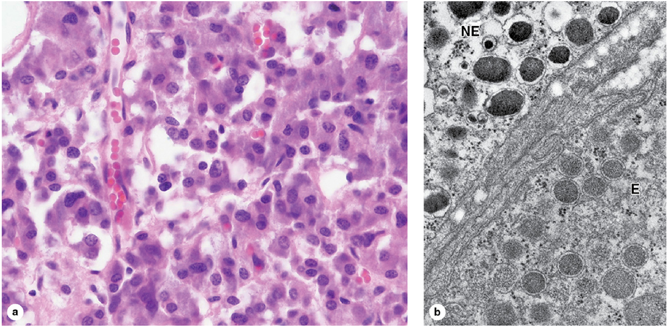
4. Ultrastructure (Electron Microscopy)
- Contain many electron-dense secretory granules, 150-350 nm in diameter, for catecholamine storage and secretion.
- Two cell types based on granule appearance:
- Epinephrine (E) cells: granules are less electron-dense and generally smaller.
- Norepinephrine (NE) cells: granules are more electron-dense (due to chromogranins binding catecholamines).
- Granule contents: catecholamines (epinephrine or norepinephrine) + Ca²⁺ + ATP + chromogranins (49-kDa proteins) - stored as granular complexes.
(Junqueira's Basic Histology, 17e)
5. The Chromaffin Reaction (Naming Origin)
- Named because they react with chromate salts (chromic acid or potassium dichromate) - the chromaffin reaction.
- This reaction involves oxidation and polymerization of the catecholamines within the secretory vesicles.
- Chromaffin cells are part of the APUD system (Amine Precursor Uptake and Decarboxylation).
(Histology: A Text and Atlas, Ross & Pawlina)
6. Innervation and Secretory Mechanism
- Innervated by myelinated preganglionic sympathetic nerve fibers (via the greater splanchnic nerve, with cell bodies in the thoracic spinal cord).
- Preganglionic fibers release ACh, which activates nicotinic receptors on chromaffin cells.
- Upon activation, chromaffin cells release catecholamines directly into the circulation - acting like a "gang-lion in the blood."
(Costanzo Physiology, 7th Ed.)
7. Secretory Products and Proportions
| Product | % Secreted | Where Else Made |
|---|---|---|
| Epinephrine (adrenaline) | ~80% | Only in adrenal medulla |
| Norepinephrine | ~20% | Also sympathetic nerve endings |
- The conversion of norepinephrine → epinephrine requires the enzyme PNMT (phenylethanolamine-N-methyltransferase), which is present only in chromaffin cells of the adrenal medulla.
- PNMT activity depends on cortisol supplied via the portal venous drainage from the adrenal cortex - this is why cortisol from the cortex controls the medullary phenotype in two ways: inhibits axon growth AND enables epinephrine synthesis.
(Costanzo Physiology, 7th Ed.)
8. Supporting Cells
- Sustentacular cells support the chromaffin cells (analogous to glial/satellite cells in ganglia).
(Robbins, Cotran & Kumar - Pathologic Basis of Disease)
9. Paraganglion System
- Chromaffin cells are not exclusive to the adrenal medulla. Scattered clusters exist in paravertebral and prevertebral sympathetic ganglia and viscera - collectively called paraganglia.
- Extra-adrenal chromaffin cells typically secrete only norepinephrine (lack PNMT because they are distant from cortisol supply).
10. Clinical Relevance - Pheochromocytoma
A tumor of chromaffin cells. Key features follow the "rule of 10s":
- 10% extra-adrenal (paragangliomas)
- 10% in children
- 10% bilateral/multiple
- 10% not hypertensive
- 10% malignant
- 10% familial
- 10% recur after surgery
- Pheochromocytoma secretes mainly norepinephrine (tumor is too far from cortical cortisol to express PNMT effectively)
Summary in one sentence: Chromaffin cells are neural crest-derived, modified sympathetic postganglionic neurons that have traded their axons for secretory granules, are innervated by preganglionic cholinergic fibers, and secrete predominantly epinephrine (80%) into the blood under stress.
This is a shared conversation. Sign in to Orris to start your own chat.