

How to treat this under eye 45 yr old female Diabetic 7.6 hba1c
periorbital hyperpigmentation dark circles under eye treatment
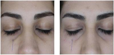
This clinical photograph is a side-by-side comparison chart showing the 'before' and 'after' results of carboxytherapy for dark under-eye circles (DUC) in a patient with Fitzpatrick skin type 3. The images provide a close-up view of the periorbital and infraorbital regions with eyes closed. The 'before' image (left) demonstrates significant infraorbital hyperpigmentation characterized by a dark, shadowed appearance in the tear trough area, along with fine lines and visible micro-vessels beneath the lower eyelid. The 'after' image (right) shows a visible reduction in pigmentation, resulting in a more even skin tone and decreased shadowing. Additionally, there is a subtle improvement in skin texture with a softened appearance of fine lines and a reduction in the prominence of superficial vascularity. This comparison illustrates the clinical effectiveness of carboxytherapy in improving microcirculation and reducing localized hyperpigmentation in the dermatological management of aesthetic periorbital concerns.
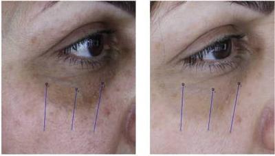
This clinical photograph presents a side-by-side 'before' and 'after' comparison of the periorbital and malar regions in a patient with pigmented dark under-eye circles (DUC). The 'before' image (left) shows significant infraorbital hyperpigmentation with a dark, mottled brown appearance extending from the medial canthus to the lateral periorbital area. There is visible skin texture irregularity and prominent fine lines in the lower eyelid and upper cheek regions. The 'after' image (right) demonstrates the clinical outcome following carboxytherapy, showing a marked reduction in pigmentation intensity and a more uniform skin tone. The treated area exhibits a smoother texture with diminished visibility of pores and fine wrinkles. Blue vertical lines are overlaid for measurement reference, likely marking the lateral, middle, and medial zones for quantitative ΔE (color change) analysis. This comparison illustrates the efficacy of carboxytherapy in managing periorbital hyperpigmentation and improving overall skin quality in Fitzpatrick skin type III individuals.
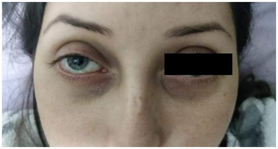
This clinical photograph displays the facial features and periorbital region of a 28-year-old patient. Key diagnostic observations include translucent, fair skin and a narrow nasal bridge. Both eyes exhibit significant infraorbital hyperpigmentation and hollows, often referred to as periorbital dark circles or 'raccoon eyes' appearance, which in this clinical context may relate to underlying vascular pathology or connective tissue disorders like Ehlers-Danlos Syndrome (EDS). The patient's right eye shows mild conjunctival injection at the inner canthus. The patient also demonstrates thin vermilion lips, a characteristic phenotypic feature associated with certain vascular-type connective tissue conditions. This image serves to illustrate the extraocular physical manifestations that can accompany internal vascular emergencies such as carotid-cavernous fistulas or central retinal artery occlusion (CRAO).
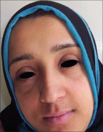
Clinical photograph of a patient's face, specifically highlighting the periocular region. The image illustrates mild-to-moderate periocular hyperpigmentation, commonly referred to as dark circles or infraorbital hyperchromia. The discoloration is most prominent along the inferior orbital rim and lower eyelids, with a higher intensity of faint brownish pigmentation noted at the medial canthi. The skin texture appears relatively smooth, with the primary finding being the focal increase in melanin or vascular congestion in the periorbital tissues. This image serves as a post-treatment documentation frame, showing a satisfactory clinical response where the transverse and vertical dimensions of the hyperpigmentation have reduced. In an educational context, this visual demonstrates the clinical manifestation of periocular darkening which can be associated with external factors like kohl application or systemic issues like lead toxicity, and is used to evaluate the efficacy of dermatological or chelation interventions.
periorbital hyperpigmentation treatment
PMID: 41615388
PMID: 41362125
Assessment: Periorbital Hyperpigmentation (Dark Circles / Infraorbital Hyperchromia)
Clinical Classification (Important for Treatment Choice)
| Type | Appearance | Key Feature |
|---|---|---|
| Pigmented (melanin) | Brown, darker in day | Improves with wood's lamp |
| Vascular | Bluish/purple, worse on stretching skin | Prominent vessels |
| Structural/Shadow | Gray-brown, worse with fat loss | Tear trough hollowing |
| Mixed | Brown + purple | Most common (likely here) |
Considerations Specific to This Patient
- Age 45, diabetic (HbA1c 7.6%) - moderately controlled diabetes. This matters because:
- Skin healing is slower; avoid aggressive procedures until sugars are optimized
- Diabetic microangiopathy can worsen the vascular component of POH
- Risk of post-inflammatory hyperpigmentation (PIH) is higher in darker skin types (Fitzpatrick IV-V, as seen here)
- Chemical peels and lasers carry higher risk of PIH; caution + lower concentrations advised
Treatment Algorithm (Stepwise)
Step 1: Photoprotection (Non-negotiable, First Line)
- Broad-spectrum SPF 50+ sunscreen daily (tinted sunscreens preferred - they block visible light which also triggers melanogenesis in darker skin)
- This is the foundation for ALL other treatments - 2026 global consensus (PMID 41362125) emphasizes this as mandatory before any other intervention
Step 2: Topical Agents (First-line adjunct)
| Agent | Dose/Use | Notes for Diabetic patient |
|---|---|---|
| Hydroquinone 2-4% | Apply to dark area at night x 3 months | Avoid prolonged use; risk of exogenous ochronosis in dark skin |
| Kojic acid 1-2% + glycolic acid | Combined cream, nightly | Well-tolerated, gentler option |
| Azelaic acid 15-20% | BD application | Safe, anti-inflammatory, good for Fitzpatrick IV-V |
| Retinol/tretinoin 0.025-0.05% | Every other night | Start low - periorbital skin is thin; avoid if there are wound healing issues |
| Vitamin C (L-ascorbic acid 10-20%) | Morning, under sunscreen | Antioxidant + pigment suppression |
| Niacinamide 4-5% | BD | Inhibits melanosome transfer, excellent tolerability, no concerns in diabetics |
Step 3: Procedures (Second-line, once glycemia is better controlled - target HbA1c <7%)
- Chemical peels (lactic acid 15-30%, glycolic acid 20-35%, Jessner's) - effective for superficial melanin-type POH. Use lower concentrations in darker skin to avoid rebound PIH
- Carboxytherapy (CO₂ injections) - particularly effective for vascular component; good safety profile, improves microcirculation
- PRP (Platelet-rich plasma) injections - promotes collagen and skin quality; systematic review 2025 (PMID 41190633) supports use for periorbital rejuvenation
- Microneedling - targets skin texture and collagen; can be combined with PRP or topicals via transdermal delivery
- Q-switched Nd:YAG laser or fractional laser - for melanin type; caution in Fitzpatrick IV-V (test patch mandatory); systematic review of light devices (PMID 37661041) supports efficacy
- Hyaluronic acid filler (tear trough) - for structural/shadow component; must be done by trained clinician; 0.5-1 mL HA filler can dramatically improve the tear trough shadow
Step 4: Address the Diabetes
- Encourage better glycemic control (HbA1c 7.6% is borderline - target <7%)
- Poorly controlled diabetes worsens vascular POH via microangiopathy and delays treatment response
What to Avoid in This Patient
- High-strength chemical peels (TCA >20%) until sugars improve
- Aggressive laser sessions without test patch
- Hydroquinone >3 months continuously (risk of ochronosis in dark skin)
- Any procedures under the eye without proper ophthalmologic screening if there is any suggestion of diabetic retinopathy
Suggested Initial Prescription (Conservative, Safe to Start Now)
- Sunscreen SPF 50+ (tinted) - every morning, reapply midday
- Niacinamide 5% + kojic acid 2% cream - apply to under-eye at night
- Vitamin C serum 10% - morning under sunscreen
- Azelaic acid 15% cream - alternate nights (or add to nightly routine after 4 weeks)
- Review at 8-12 weeks; if inadequate response, plan superficial chemical peel or carboxytherapy
- Global consensus on melanin hyperpigmentation 2026 (PMID 41362125) - Passeron T et al.
- Under Eye Treatment Systematic Review 2026 (PMID 41615388) - Beer J et al.
- PRP vs PRF periorbital rejuvenation 2025 (PMID 41190633)