
Free radical injury
free radical reactive oxygen species cell injury mechanism diagram
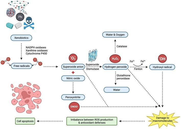
This pathophysiology diagram illustrates the biochemical pathways of reactive oxygen species (ROS) production, detoxification, and their roles in oxidative stress and cell death. The process initiates with 'Xenobiotics' (e.g., heavy metals like Pb, pharmaceuticals, and environmental toxins), which are converted into free radicals via enzymes such as NADPH oxidases, xanthine oxidases, and Cytochrome P450. Key intermediates include the Superoxide anion (O₂⁻), which can react with Nitric oxide to form Peroxynitrite (ONOO⁻) or be converted by Superoxide Dismutase (SOD) into Hydrogen peroxide (H₂O₂). The diagram details two detoxification routes for H₂O₂: conversion to water and oxygen by Catalase, or to water by Glutathione peroxidase. Alternatively, H₂O₂ can undergo the Fenton reaction (involving Fe²⁺ to Fe³⁺ transition) to generate the highly reactive Hydroxyl radical (•OH). Downstream effects focus on the 'Imbalance between ROS production & antioxidant defenses,' leading to 'Damage to macromolecules' (proteins, lipids, and nucleic acids). The final clinical outcome depicted is 'Cell apoptosis,' represented by a morphological illustration of a fragmenting cell. This flowchart serves as an educational tool for understanding oxidative stress mechanisms and cellular pathology.
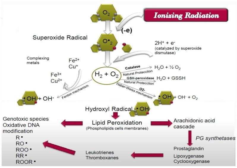
This pathophysiology diagram illustrates the biochemical mechanisms of cellular injury induced by ionizing radiation, specifically focusing on reactive oxygen species (ROS) and lipid peroxidation. The flowchart begins with ionizing radiation triggering the reduction of molecular oxygen (O2) into the superoxide radical (O2•-). This radical is converted via superoxide dismutase into hydrogen peroxide (H2O2), which can be neutralized by natural protective enzymes like catalase and glutathione peroxidase. However, in the presence of complexing metals (Fe, Cu), the Fenton and Haber-Weiss mechanisms facilitate the conversion of ROS into the highly reactive hydroxyl radical (•OH). The lower section details the pathological consequences of hydroxyl radicals, primarily lipid peroxidation of cell membranes. This process branches into two significant pathways: the generation of genotoxic species leading to oxidative DNA modification (represented by R•, RO•, and ROO• radicals) and the activation of the arachidonic acid cascade. The latter involves PG synthetases, lipoxygenase, and cyclooxygenase, ultimately producing inflammatory mediators such as prostaglandins, leukotrienes, and thromboxanes. The diagram serves as an educational tool for understanding radiation-induced oxidative stress and systemic inflammatory responses.
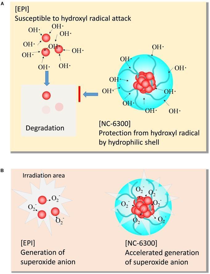
This pathophysiology diagram illustrates the comparative mechanism of action and stability of Epirubicin (EPI) and NC-6300 (an epirubicin-incorporating micelle) during sonodynamic therapy (SDT). Panel A depicts chemical stability against reactive oxygen species. It shows that free EPI is susceptible to hydroxyl radical (OH•) attack, leading to drug degradation, visually represented by fading red spheres. In contrast, NC-6300 features a protective hydrophilic shell (PEG-polyamino acid polymer) that shields the drug core from hydroxyl radicals, maintaining stability. Panel B demonstrates the sonosensitizing activity within an irradiation area. While both substances generate superoxide anions (O2•−) upon irradiation, NC-6300 shows accelerated generation compared to free EPI. This is graphically indicated by a higher density of O2•− symbols and larger radial sparks around the micellar structure. The diagram highlights two key pharmaceutical advantages of the NC-6300 nanocarrier: protection of the active moiety from premature degradation and enhanced therapeutic reactive oxygen species production for antitumor effects.
Free Radical Injury
Definition
- Robbins, Cotran & Kumar Pathologic Basis of Disease, p. 64
Reactive Oxygen Species (ROS) - The Main Players
| Species | Nature | Key Feature |
|---|---|---|
| Superoxide (O2-) | Radical | Generated in mitochondria, by phagocyte oxidase; dismutates to H2O2 |
| Hydrogen peroxide (H2O2) | Non-radical | Weak oxidizer; lipid-soluble - can diffuse across membranes; precursor to OH• |
| Hydroxyl radical (OH•) | Radical | Most reactive ROS; principal species damaging lipids, proteins, and DNA; no enzymatic inactivation |
| Peroxynitrite (ONOO-) | Radical | Formed by interaction of O2- and nitric oxide (NO); damages lipids, proteins, DNA |
- Basic Medical Biochemistry - A Clinical Approach, p. 897-898
Generation of Free Radicals
1. Normal Metabolic Processes
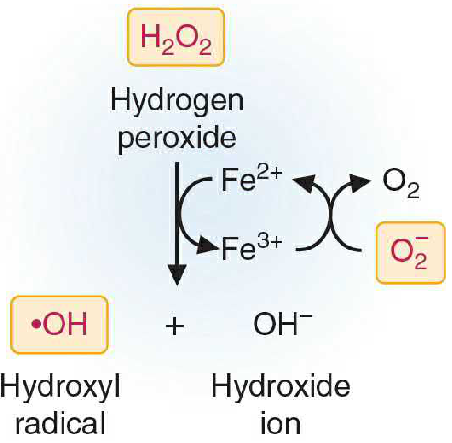
2. Fenton Reaction and Haber-Weiss Reaction
3. Ionizing Radiation
4. Enzymatic Reactions
- NADPH oxidase in leukocytes (deliberate production during phagocytosis - "oxidative burst")
- Cytochrome P450, xanthine oxidase, and other metal-containing enzymes produce superoxide as accidental by-products.
5. Nitric Oxide
- Robbins, p. 64-65; Basic Medical Biochemistry, p. 898-904
Pathologic Conditions Caused by Free Radicals
| Category | Examples |
|---|---|
| Cardiovascular | Atherogenesis, ischemia-reperfusion injury, stroke |
| Neurological | Alzheimer disease, Parkinson disease, ALS, multiple sclerosis |
| Metabolic | Diabetes, alcohol-induced liver disease |
| Pulmonary | COPD |
| Other | Aging, cervical cancer, retrolental fibroplasia, Down syndrome, OXPHOS diseases |
- Basic Medical Biochemistry, Table 25.1, p. 897
Mechanisms of Cell Injury by Free Radicals
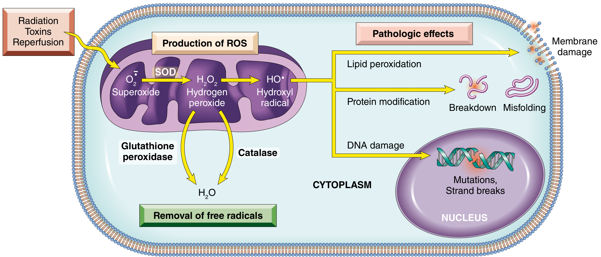
1. Lipid Peroxidation (Membrane Damage)
- Initiation: OH• + LH → L• (lipid radical)
- Propagation: L• + O2 → LOO• → LOOH (lipid peroxide) → more radicals
- Termination: Vitamin E donates a single electron to LOO•, forming a stable oxidized product
2. Protein Modification
- Oxidation of amino acid side chains
- Formation of protein-protein cross-links (fragmentation of polypeptides)
- Protein misfolding
3. DNA Damage
- Single- and double-strand breaks
- Mutations (especially oxidation of guanine to 8-hydroxyguanosine)
- Cross-linking of DNA strands
- Robbins, p. 64-66; Basic Medical Biochemistry, p. 908-912
Cellular Defense Against Free Radicals (Antioxidant Systems)
Enzymatic Scavengers
| Enzyme | Reaction | Location |
|---|---|---|
| Superoxide dismutase (SOD) | 2O2- + 2H+ → H2O2 + O2 | Mitochondria (Mn-SOD), cytosol (Cu/Zn-SOD) |
| Catalase | 2H2O2 → 2H2O + O2 | Peroxisomes |
| Glutathione peroxidase | H2O2 + 2GSH → 2H2O + GSSG | Cytosol, mitochondria |
| Peroxiredoxins | ONOO- → HNO2 | Cytosol, mitochondria |
Non-enzymatic Antioxidants
-
Vitamin E (tocopherol): Lipid-soluble; terminates lipid peroxidation chain reactions in membranes
-
Vitamin C (ascorbate): Water-soluble scavenger; also regenerates vitamin E
-
Glutathione (GSH): Major intracellular thiol antioxidant
-
Beta-carotene: Quenches singlet oxygen in lipid environments
-
Ceruloplasmin / transferrin / ferritin: Sequester iron and copper, preventing Fenton reactions
-
Robbins, p. 65-66; Basic Medical Biochemistry, p. 912-915
Oxidative Stress: The Imbalance
- Chemical and radiation injury
- Ischemia-reperfusion injury (restoration of blood flow paradoxically generates a burst of ROS)
- Cellular aging
- Microbial killing by phagocytes (intentional)
- Cancer, neurodegeneration, atherosclerosis
Ischemia-Reperfusion Injury: A Clinically Important Example
- Basic Medical Biochemistry, p. 911 (RNOS and Inflammation section)
Summary: Key Concepts
- Free radicals (especially ROS) have a single unpaired electron and are highly reactive
- Sources: mitochondrial respiration, NADPH oxidase (leukocytes), Fenton reaction (iron/copper), radiation, xanthine oxidase
- OH• is the most damaging species; no enzymatic defense exists against it
- Three main targets: membrane lipids (peroxidation), proteins (oxidation/cross-linking), DNA (strand breaks/mutations)
- Defense: SOD, catalase, glutathione peroxidase, vitamins E and C, glutathione, metal-binding proteins
- Clinical significance: ischemia-reperfusion, atherosclerosis, aging, neurodegeneration, cancer