
Mgd in ophtha
meibomian gland dysfunction MGD eyelid
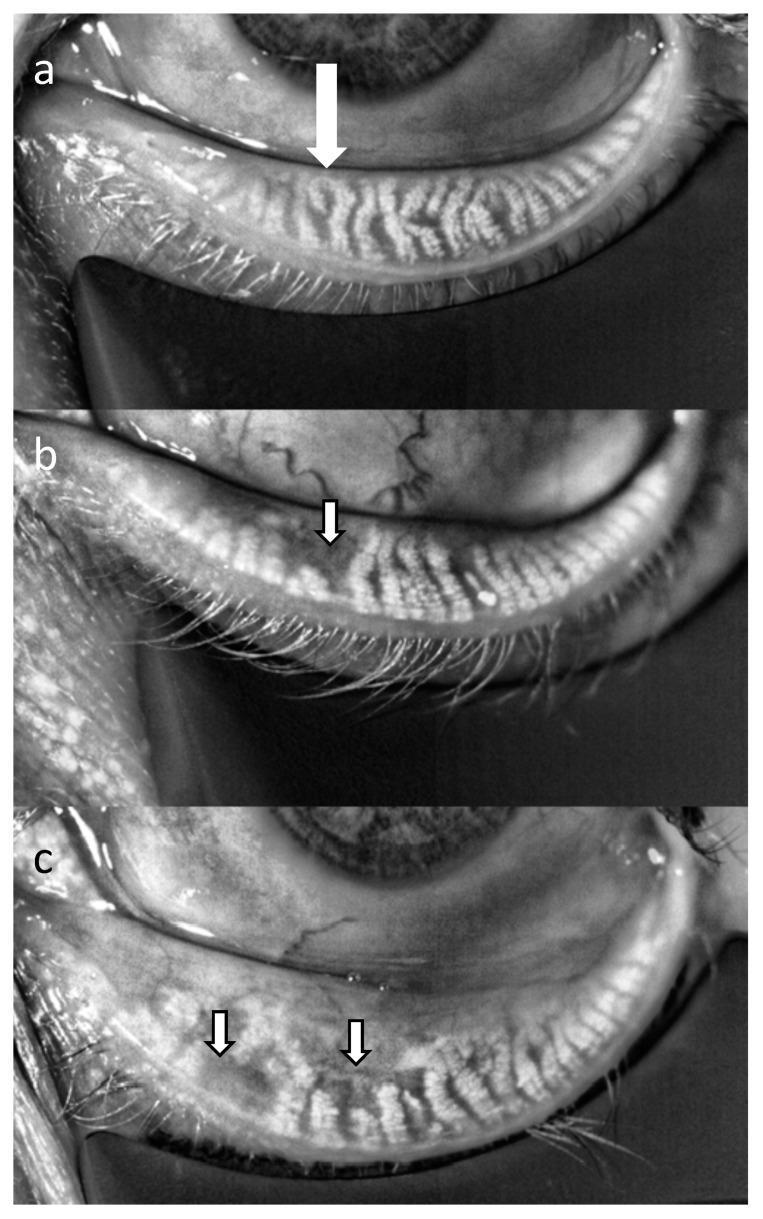
This diagnostic image displays three infrared meibography panels (a, b, and c) of the lower eyelid, illustrating the progression of Meibomian gland dysfunction (MGD). Panel (a) shows healthy Meibomian glands characterized by a dense, parallel, and organized array of hyperreflective elongated structures extending vertically toward the eyelid margin. In panel (b), representing mild MGD, there is a visible disruption in gland continuity and density, with subtle areas of glandular thinning indicated by an arrow. Panel (c) demonstrates moderate MGD, where significant pathology is evident through widespread gland dropout and architectural distortion. The arrows in (c) highlight dark, hyporeflective regions representing the loss of functional glandular tissue. This comparison serves as an educational tool for ophthalmology to identify different stages of Meibomian gland atrophy and calculate meiboscores. The images were captured using infrared interferometry, highlighting the autofluorescence of healthy meibum against the atrophied areas.
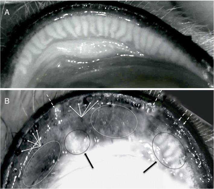
This diagnostic image displays non-contact infrared meibography of the upper eyelid, comparing a healthy state (A) with Meibomian Gland Dysfunction (MGD) (B). Image A demonstrates a normal anatomical distribution of meibomian glands, appearing as regular, vertical, hyperreflective linear structures. Image B illustrates several pathological features of MGD: diffused ductal occlusion and plugging of the gland orifices (indicated by dotted white arrows) and posterior migration of the mucocutaneous junction with retroplacement of ductal openings (indicated by multiple thin white arrows). Morphological changes within the glands are highlighted, including cystic dilatation of the ducts containing dense, opaque secretions (black arrows and black ovals). These areas alternate with regions of significant hyporeflectivity (white ovals), representing acinar atrophy and gland dropout. This comparison serves as a clinical educational tool for identifying terminal duct obstruction and glandular degeneration during ocular surface evaluations.
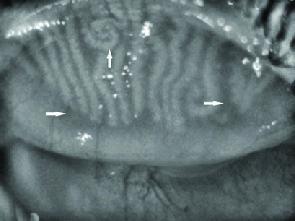
This diagnostic clinical image, likely obtained via meibography, displays the meibomian glands within a human eyelid. The glands are visible as vertically oriented, light-colored parallel structures. Pathological changes associated with meibomian gland dysfunction (MGD) are highlighted by white arrows. A vertical arrow in the upper-left quadrant points to a region of increased ductal tortuosity, where the gland exhibits a spiraling or twisting morphology rather than a linear path. Horizontal arrows on the left and right peripheries indicate areas of focal gland atrophy or dropout, where the normal vertical stripes are significantly diminished or absent against the tarsal background. The image serves as an educational tool for ophthalmology and optometry to demonstrate the structural degradation and morphological remodeling of oil-producing glands in chronic ocular surface disease.

Clinical slit-lamp photographs displaying the classification of Meibomian Gland Dysfunction (MGD) based on lid margin appearance and meibum characteristics. (A) Hypersecretory MGD: Demonstrates significant lid margin hyperemia (redness) and a large volume of expressible meibum (lipid) accumulating along the posterior lid margin, often associated with inflammation. (B) Hyposecretory MGD: Shows a relatively clean, less inflamed eyelid margin with minimal meibum production, characteristic of a low-delivery state. (C) Obstructive MGD: Features an irregular, thickened lid margin with visible plugging of the meibomian gland orifices. Small yellowish mounds or 'capping' may be present at the gland openings, indicating physical obstruction of lipid flow. All three images highlight the relationship between the eyelashes, meibomian gland orifices, and the mucocutaneous junction. These images are educational tools for ophthalmologists and optometrists to differentiate between high-delivery and low-delivery MGD subtypes during physical examination.
meibomian gland dysfunction treatment
Meibomian Gland Dysfunction (MGD)
Definition & Overview
Pathophysiology
- Gland obstruction - Keratinization and hyperproliferation of ductal epithelium blocks meibum flow
- Altered meibum - Bacterial lipases (especially S. aureus) convert triglycerides into free fatty acids, raising the melting point of meibum, making it thick and inspissated
- Lipid layer deficiency - Loss of the tear film phospholipid surfactants increases tear evaporation and osmolarity → unstable tear film
- Inflammation - Chronic inflammation of the lid margin and ocular surface
- Glandular atrophy - Progressive acinar atrophy and "gland dropout" with disease progression
Classification
| Type | Secretion | Characteristics |
|---|---|---|
| Hyposecretory (Obstructive) | Low delivery | Capped orifices, minimal expressible meibum - most common |
| Hypersecretory | High delivery | Turbid, frothy secretions expressed easily; lid margin hyperemia |
Clinical Features
Symptoms
- Burning, grittiness, stinging (often worse on waking)
- Mild photophobia, intermittent blurring
- Red-rimmed eyes, crusting
- Contact lens intolerance
- Symptoms worsen with dry/windy environments or prolonged screen use (reduced blink rate)
Lid Margin Signs
- Posterior lid margin changes: thickening, irregular margin, telangiectasia
- Meibomian orifice plugging ("capping") - toothpaste-like secretions
- Turbid, inspissated meibum - thick, opaque material on expression
- Foaming of tear meniscus at lid margin
- Associated chalazion and hordeolum (internal stye)
- Notching of lid margin in severe disease
Tear Film Changes
- Reduced tear break-up time (TBUT) < 10 seconds
- Abnormal lipid layer (interferometry)
- Increased tear osmolarity
Corneal Signs (in severe/longstanding MGD)
- Inferior punctate epithelial erosions
- Inferior peripheral vascularization
- Marginal corneal infiltrates
Diagnosis
Clinical
- Slit-lamp examination - inspect lid margins, express meibomian glands, assess meibum quality
- Schirmer test - to assess aqueous component (may be normal or reduced)
- TBUT - usually short (< 10 s)
- Fluorescein/rose bengal/lissamine green staining - inferior corneal and conjunctival staining
Imaging
- Meibography (infrared) - non-contact infrared transillumination of the tarsal plate to visualize gland morphology and detect dropout
Management
1. Eyelid Hygiene (First-line, cornerstone)
- Warm compresses - 10 minutes, twice daily; softens inspissated meibum (must be ~40°C)
- Lid massage - manual expression of glands after warming
- Lid scrubs - diluted baby shampoo or commercial wipes (removes debris, biofilm, Demodex)
2. Topical Therapy
- Lubricating drops - lipid-containing artificial tears preferred (e.g., sodium hyaluronate + lipid emulsion)
- Topical antibiotics - chloramphenicol or azithromycin eye drops/ointment
- Low-potency topical steroids (short course) - fluorometholone 0.1% or loteprednol 4x/day × 1 week for active inflammation or papillary conjunctivitis
- Topical ciclosporin - for severe dry eye with chronic inflammation
3. Systemic Antibiotics (for moderate-severe disease)
- Doxycycline 50-100 mg twice daily × 1 week, then daily for 6-24 weeks (most commonly used)
- Lymecycline 408 mg capsule daily × 3 months
- Azithromycin 500 mg/day × 3 days for 3 cycles at 1-week intervals
- Erythromycin 250 mg once or twice daily (if tetracyclines contraindicated)
- Tetracyclines are contraindicated in children < 12 yrs, pregnancy, breastfeeding
4. Omega-3 Fatty Acid Supplements
- Fish/plant oil supplements improve meibum quality and reduce symptoms
5. Demodex-Targeted Therapy (if collarettes present)
- Tea tree oil 50% scrub + 5% ointment to lid margins
- Lotilaner 0.25% (TP-03) ophthalmic solution - well tolerated, reduces Demodex within 4 weeks
- Topical/oral ivermectin in refractory cases
6. Physical/Device-Based Therapies
| Modality | Mechanism |
|---|---|
| LipiFlow (thermal pulsation) | Vectored thermal pulsation - heats and expresses meibomian glands simultaneously |
| Intense Pulsed Light (IPL) | Reduces telangiectasia, targets Demodex, reduces inflammation; effective for evaporative dry eye from MGD |
| Meibomian gland probing | Mechanical opening of obstructed orifices |
| Low-level light therapy | Relatively new; photobiomodulation of eyelid tissues |
Complications
| Complication | Notes |
|---|---|
| Chalazion | Retained inspissated meibum → lipogranuloma in tarsus |
| Hordeolum (internal stye) | Acute infection of meibomian gland |
| Evaporative dry eye | Most common cause |
| Punctate epitheliopathy | Inferior corneal staining |
| Marginal keratitis | Peripheral corneal infiltrates/thinning |
| Corneal vascularization | Inferior pannus in severe longstanding disease |
Key Associations
- Acne rosacea - strongest systemic association; often requires systemic doxycycline
- Seborrhoeic dermatitis
- Demodex blepharitis (esp. D. brevis in posterior blepharitis)
- Sjögren syndrome (dry eye + MGD overlap)
- Isotretinoin therapy (causes iatrogenic MGD)