
Function of promoter area primary motor area and supplementary motor area
Reading File
Now let me search the medical library for these motor cortex areas simultaneously.
Finding Sources
Finding Sources
Finding Sources
Reading File
I have the Guyton content. Let me read the supplementary motor area section and also get the Adams & Victor content in parallel.
Reading File
Reading File
I now have excellent content from both Guyton and Adams & Victor. Let me also fetch the motor cortex diagram image to include in the answer.
Loading Image
Loading Image
I now have comprehensive content from Guyton and Adams & Victor, along with the diagrams. Let me also get the Guyton premotor area content that was truncated, and fetch the motor homunculus from Adams & Victor.
Reading File
Loading Image
I now have all the information needed for a comprehensive answer. Here is the complete breakdown:
Motor Cortex Areas: Premotor, Primary Motor, and Supplementary Motor
The motor cortex occupies the precentral gyrus and the adjacent frontal cortex. It is divided into three functionally distinct areas, all working together to produce coordinated voluntary movement.
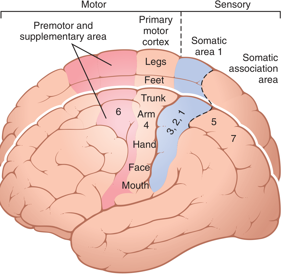
1. Primary Motor Cortex (Area 4)
Location: First convolution of the frontal lobe, immediately anterior to the central sulcus (precentral gyrus). Extends from the sylvian fissure laterally to the longitudinal fissure medially. Corresponds to Brodmann area 4.
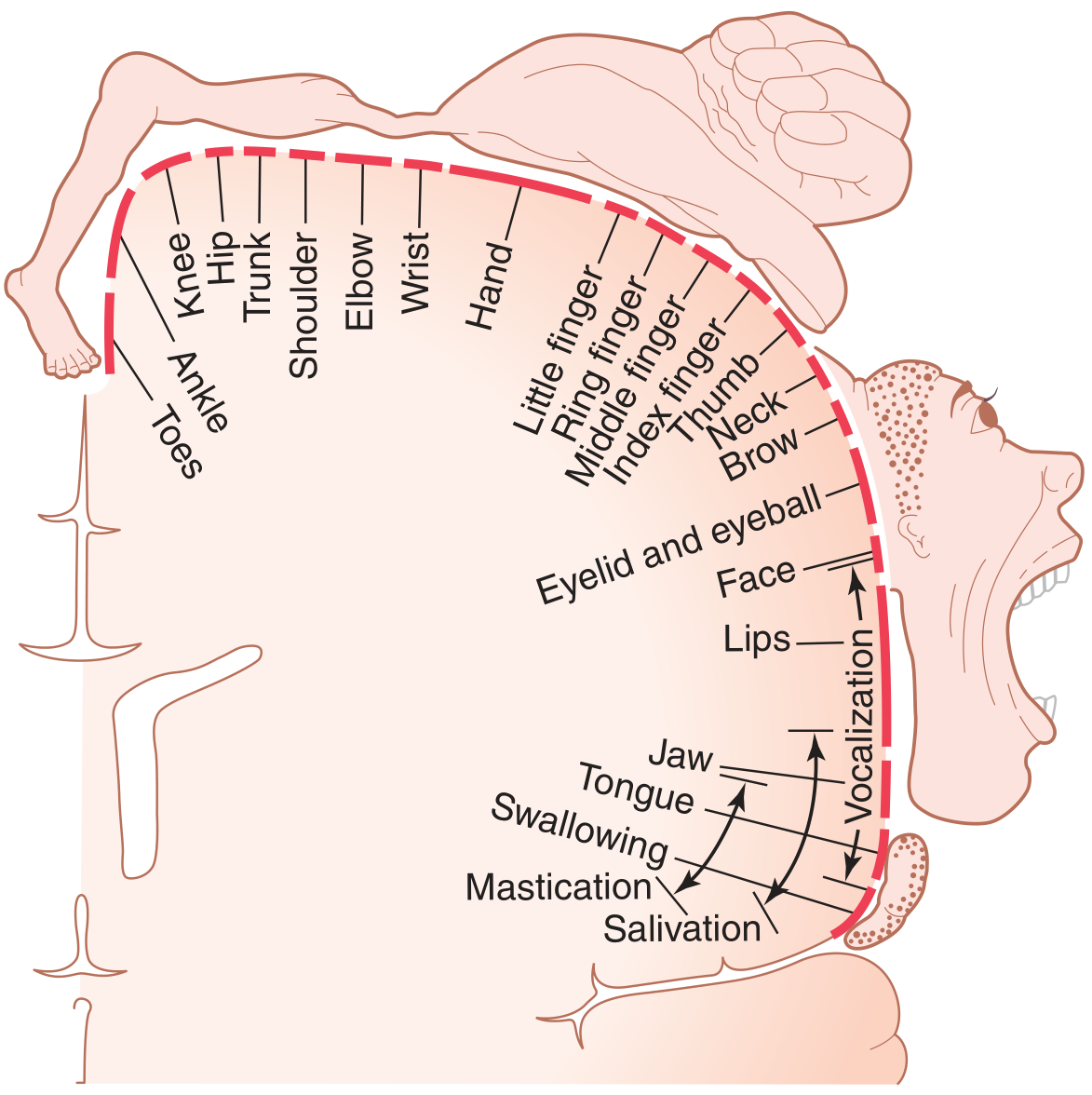
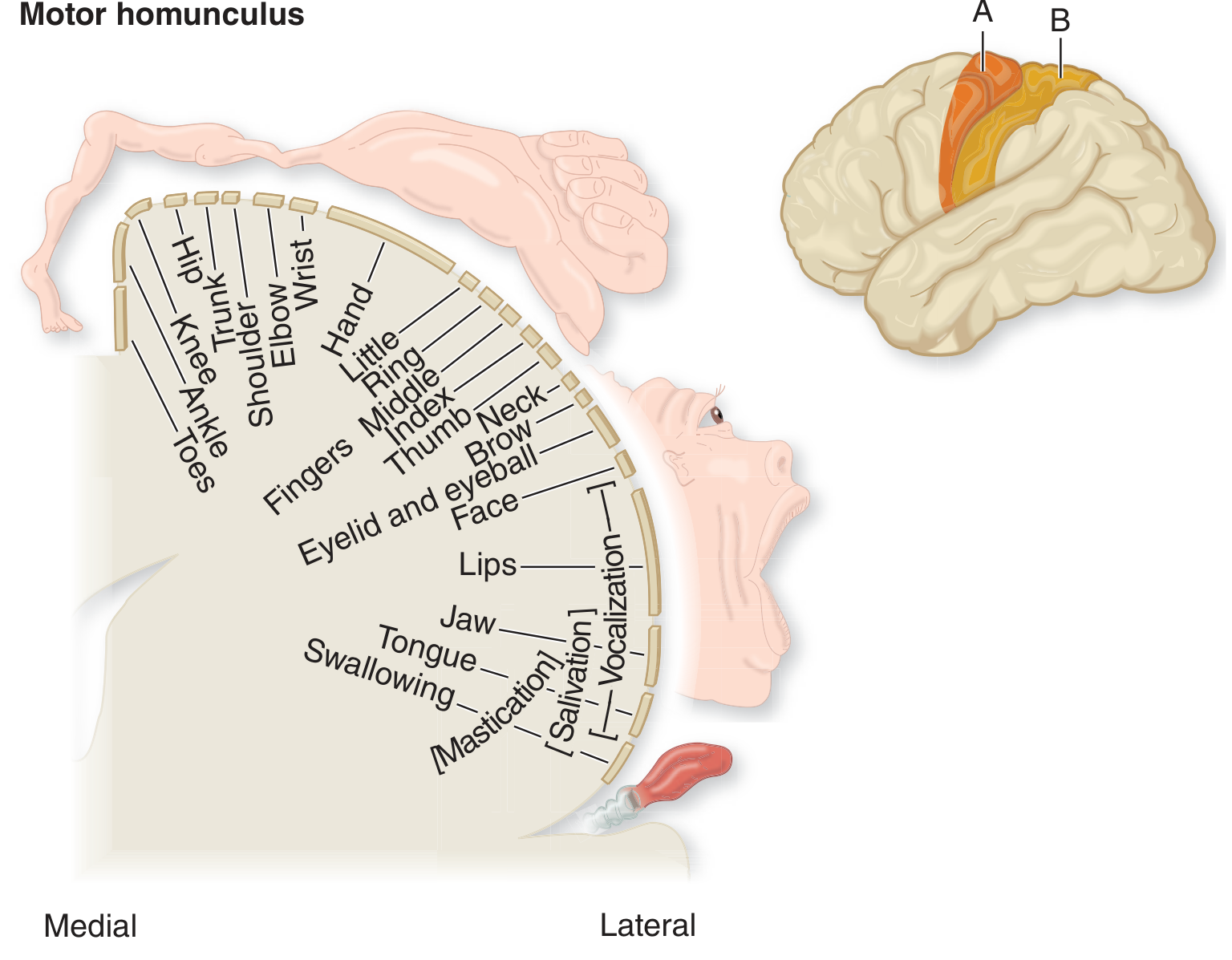
Topographic organization (Somatotopy):
- Face and mouth - most lateral (near sylvian fissure)
- Hand and arm - midportion
- Trunk - near apex
- Leg and foot - dip into the longitudinal fissure (paracentral lobule on medial surface)
This somatotopic map is the famous motor homunculus (first described by Wilder Penfield).
Key functions:
- Direct execution of voluntary movement - sends signals via the corticospinal and corticobulbar tracts to spinal motor neurons
- Stimulation at low threshold evokes contralateral discrete muscle contractions
- Controls fine, skilled movements, especially of the distal limbs (hands, fingers, face, speech muscles)
- More than half the primary motor cortex is devoted to the hands and speech muscles, reflecting the importance of manual dexterity and communication
- Each neuron typically excites a pattern of muscles producing a specific movement, not a single isolated muscle (concept of movement representation, not muscle representation - as articulated by Hughlings Jackson and Sherrington)
- Parts of the body capable of the most delicate movements have the largest cortical representation
2. Premotor Area (Area 6 - lateral portion)
Location: Lies 1-3 cm anterior to the primary motor cortex (Brodmann area 6). Extends from the sylvian fissure inferiorly to the longitudinal fissure superiorly. Topographic organization mirrors that of area 4 (face most lateral, leg most medial/superior).
Key functions:
- Generates more complex patterns of movement than area 4 - for example, positioning the shoulder and arm so the hand can perform a specific task
- The anterior premotor cortex develops a "motor image" of the total movement to be performed
- The posterior premotor cortex then translates this image into sequential muscle activation patterns, sending signals either directly to area 4 or via the basal ganglia-thalamus loop back to area 4
- Stimulation of the caudal premotor area produces responses similar to area 4 stimulation (via direct propagation to area 4); stimulation of the rostral premotor area elicits more general movement patterns of proximal limb musculature via pathways independent of area 4
- Contains mirror neurons - these fire both when performing an action and when observing someone else perform the same action. They are thought to be important for understanding others' actions and for learning by imitation
- Together with basal ganglia, thalamus, and area 4, it forms the circuit for complex coordinated muscle activity
Specialized premotor regions:
| Subregion | Function |
|---|---|
| Broca's area (inferior premotor) | Motor speech - word formation; damage causes expressive/Broca's aphasia |
| Frontal eye field (above Broca's) | Voluntary eye movements; damage causes eyes to "lock" onto objects |
| Head rotation area | Directs head toward objects (works with eye field) |
| Hand skills area | Coordinates purposeful hand movements; damage causes motor apraxia |
3. Supplementary Motor Area (SMA, area 6β / medial area 6)
Location: The most anterior portion of area 6 on the medial surface of the frontal lobe, lying mainly in the longitudinal fissure but extending a few centimeters onto the superior frontal cortex. It abuts the premotor area superiorly.
Key functions:
- Controls body-wide attitudinal and postural movements - provides background postural support for fine movements performed by the arms/hands
- Stimulation often produces bilateral responses (unlike area 4 which is predominantly contralateral) - e.g., bilateral grasping movements of both hands simultaneously (thought to be remnants of climbing behavior)
- Can also induce:
- Gross ipsilateral or contralateral limb movements
- Bilateral tonic contractions of the limbs
- Contraversive movements of head and eyes with contralateral arm tonic contraction
- Inhibition of voluntary motor activity and vocal arrest
- Provides fixation movements of body segments and positional movements of the head and eyes as background for finer movements
- Functions in concert with the premotor area to coordinate proximal/axial movements
Summary Comparison
| Feature | Primary Motor (Area 4) | Premotor (Area 6 lateral) | Supplementary Motor (Area 6 medial) |
|---|---|---|---|
| Brodmann area | 4 | 6 (lateral) | 6 (medial/SMA) |
| Location | Precentral gyrus | 1-3 cm anterior to area 4 | Medial frontal lobe / longitudinal fissure |
| Stimulation threshold | Low | Higher than area 4 | Higher |
| Type of movement | Discrete, fine, contralateral | Complex patterns, proximal | Bilateral, postural, axial |
| Main output | Corticospinal tract (direct) | Via area 4 or BG-thalamus | Via area 4 and premotor |
| Key role | Movement execution | Movement planning & sequencing | Postural setting, bilateral coordination |
| Lesion effect | Contralateral weakness/paresis | Apraxia, loss of complex patterns | Loss of postural control, vocal arrest |
Sources: Guyton and Hall Textbook of Medical Physiology, pp. 687-688; Adams and Victor's Principles of Neurology 12th Ed., pp. 70-71
This is a shared conversation. Sign in to Orris to start your own chat.