
SPINAL TRACTS IN SPINAL CORED TRANSVERSE SECTION
transverse section spinal cord ascending and descending tracts
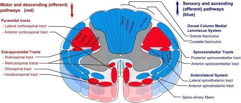
This medical anatomical diagram illustrates a transverse section of the human spinal cord, specifically detailing the organization of white matter tracts. The visual uses a color-coded schema to differentiate between motor and sensory pathways: descending (efferent) motor tracts are highlighted in red, while ascending (afferent) sensory tracts are shown in blue. Key anatomical structures identified include the Pyramidal tracts (lateral and anterior corticospinal tracts), Extrapyramidal tracts (rubrospinal, reticulospinal, olivospinal, and vestibulospinal), the Dorsal Column-Medial Lemniscus system (gracile and cuneate fasciculi), Spinocerebellar tracts, and the Anterolateral system (lateral and anterior spinothalamic tracts). A significant educational feature of the diagram is the depiction of somatotopic organization, showing the concentric arrangement of sacral, lumbar, thoracic, and cervical fibers within the lateral corticospinal and dorsal column tracts. The image provides a comprehensive overview of spinal cord neuroanatomy, useful for understanding clinical localization of spinal cord lesions and the functional topography of the central nervous system.
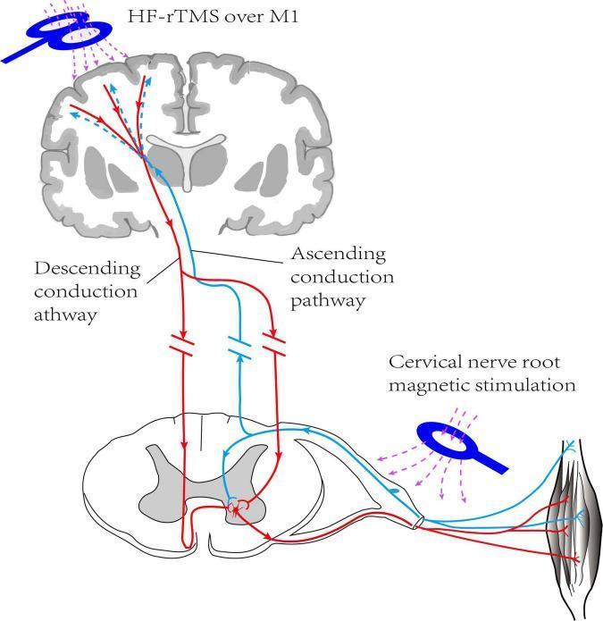
This medical schematic diagram illustrates a neuromodulation intervention combining central and peripheral magnetic stimulation. The visual depicts a coronal section of the human brain and a transverse cross-section of the cervical spinal cord. A high-frequency repetitive transcranial magnetic stimulation (HF-rTMS) coil is positioned over the primary motor cortex (M1), targeting the descending conduction pathway (indicated by red solid lines). Concurrently, a magnetic stimulation coil is applied to the cervical nerve root, targeting the ascending conduction pathway (indicated by light blue lines). The diagram highlights the neural circuit between the motor cortex, spinal interneurons, and effector muscles, demonstrating the dual-target approach for neurorehabilitation, likely in the context of post-stroke hemiplegia. Key anatomical landmarks include the cortical grey matter, subcortical white matter tracts, spinal dorsal and ventral horns, and a peripheral skeletal muscle. Arrows indicate the directional flow of neural signaling within the corticospinal and spinothalamic tracts, emphasizing the integration of central motor output and peripheral sensory input.
"spinal cord"[MeSH Terms] AND "corticospinal tract"
Spinal tracts in a transverse section of the spinal cord
Ascending (sensory) tracts
| Tract | Position in section | Modality | Decussation |
|---|---|---|---|
| Fasciculus gracilis | Posterior funiculus, medial | Fine touch, vibration, conscious proprioception from the lower limb and lower trunk (below T6) | Crosses in the lower medulla (internal arcuate fibres) |
| Fasciculus cuneatus | Posterior funiculus, lateral to gracilis (T6 and above only) | The same modalities from the upper limb and neck | Crosses in the lower medulla |
| Lateral spinothalamic | Anterolateral funiculus | Pain and temperature | Crosses in the anterior white commissure, 1-2 segments above entry |
| Anterior spinothalamic | Anterior funiculus | Crude touch and pressure | Crosses in the anterior white commissure |
| Dorsal (posterior) spinocerebellar | Lateral funiculus, posterolateral periphery | Unconscious proprioception from the lower limb | Uncrossed |
| Ventral (anterior) spinocerebellar | Lateral funiculus, anterolateral periphery | Unconscious proprioception from the lower limb | Double crossing |
| Spinoreticular and spinomesencephalic | Anterolateral system | Emotional and arousal aspects of pain; central pain modulation | Mostly crossed |
- Anterolateral pathway. Gray's describes it as three tracts: spinothalamic, spinoreticular, and spinomesencephalic. First-order neurons are in the dorsal root ganglion. Second-order axons cross in the anterior commissure and ascend to the VPL nucleus of the thalamus. Third-order neurons project through the posterior limb of the internal capsule to the somatosensory cortex. The spinoreticular tract carries the emotional and arousal aspects of pain, and the spinomesencephalic tract goes to the periaqueductal grey and superior colliculi (Gray's Anatomy for Students, Ascending Tracts in the Spinal Cord).
- Posterior column-medial lemniscal pathway. The gracile fasciculus carries input from the lower limb and trunk. The cuneate fasciculus carries input from the upper limb and neck. Both ascend ipsilaterally to the nucleus gracilis and nucleus cuneatus in the caudal medulla (Gray's Anatomy for Students, Ascending Tracts in the Spinal Cord).
- Lamination in the spinothalamic tract. Cervical fibres lie dorsomedially and sacral fibres lie ventrolaterally. Temperature fibres lie dorsolateral to pain fibres (Localization in Clinical Neurology, 8e, Ascending Tracts).
Descending (motor) tracts
| Tract | Position in section | Function | Decussation |
|---|---|---|---|
| Lateral corticospinal | Lateral funiculus, deep and posterior | Voluntary movement of the limbs, especially skilled distal movements | About 85-90% cross at the pyramidal decussation in the medulla |
| Anterior corticospinal | Anterior funiculus, next to the anterior median fissure | Axial and trunk muscles | Crosses at the segmental level |
| Rubrospinal | Lateral funiculus, anterior to the lateral corticospinal tract | Flexor tone of the upper limb | Crosses in the midbrain |
| Lateral vestibulospinal | Anterior funiculus | Extensor tone and posture, balance | Uncrossed |
| Medial vestibulospinal | Anterior funiculus, cervical and upper thoracic | Head position | Uncrossed (bilateral) |
| Reticulospinal (pontine and medullary) | Anterior and lateral funiculi | Posture and locomotion, modulation of tone | Mostly uncrossed |
| Tectospinal | Anterior funiculus | Reflex head and neck turning to visual and auditory stimuli | Crosses in the midbrain |
| Olivospinal | Anterolateral, cervical | Minor |
- Gray's groups the descending tracts into a lateral motor system and a medial motor system. Each tract has an upper motor neuron in the cortex or brainstem and a lower motor neuron in the spinal grey matter (Gray's Anatomy for Students, Descending Tracts in the Spinal Cord).
Quick map of one half of the cord
- Posterior funiculus: gracilis (medial) and cuneatus (lateral).
- Lateral funiculus: lateral corticospinal, rubrospinal, dorsal and ventral spinocerebellar, and lateral spinothalamic.
- Anterior funiculus: anterior corticospinal, vestibulospinal, reticulospinal, tectospinal, and anterior spinothalamic.
Somatotopy (high-yield)
- Lateral corticospinal: from medial to lateral, the order is cervical, thoracic, lumbar, sacral. Sacral fibres are the most lateral.
- Spinothalamic: cervical fibres are medial and sacral fibres are lateral. Sacral fibres are therefore the first to be affected by an extrinsic cord compression.
- Posterior columns: sacral and lumbar fibres are medial, and thoracic and cervical fibres are lateral.
Clinical correlation
- Posterior column lesion: loss of vibration and position sense on the same side, with a positive Romberg sign.
- Lateral corticospinal lesion: upper motor neuron signs on the same side below the lesion (spasticity, hyperreflexia, Babinski sign).
- Spinothalamic lesion: loss of pain and temperature on the opposite side, starting 1-2 segments below the lesion.
- Brown-Séquard syndrome (hemisection): ipsilateral loss of motor function and of vibration and proprioception below the lesion, and contralateral loss of pain and temperature.