
Explanation of cerebellum anatomy in detailed information
Reading File
Now let me search the medical library for cerebellum anatomy content in parallel.
Finding Sources
Finding Sources
Searching the Web
cerebellum anatomy detailed structure lobes nuclei cortex
Excellent! Found great sources. Let me now read the dedicated anatomy sections in parallel from the two best textbook sources.
Reading File
Reading File
Finding Sources
Finding Sources
Excellent content retrieved! Now let me get more detailed content on deep nuclei, peduncles, and fetch the anatomical images.
Reading File
Reading File
Loading Image
Loading Image
Now let me get the cerebellar peduncles and cortex layer details from the Neuroanatomy textbook.
Reading File
Reading File
Loading Image
Loading Image
I now have comprehensive textbook content and excellent anatomical images. Let me fetch the cortical layers diagram and the cerebellar cortex cell diagram.
Loading Image
Loading Image
I now have comprehensive, well-illustrated content from multiple authoritative textbooks. Let me compile the full detailed answer.
Cerebellum - Detailed Anatomy
The cerebellum ("little brain" in Latin) sits in the posterior cranial fossa, dorsal to the pons and medulla oblongata, forming the roof of the fourth ventricle. It is separated from the overlying occipital and temporal lobes by the tentorium cerebelli. Despite comprising only about 10% of total brain volume, it contains roughly half of all neurons in the entire central nervous system - the vast majority being tiny granule cells.
1. Gross Anatomy and Surface Features
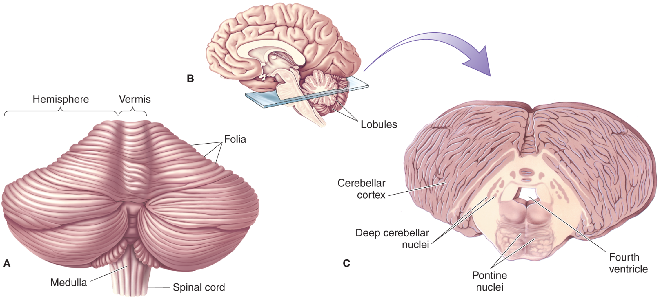
The outer surface resembles a piece of cauliflower. The visible dorsal surface is covered by a series of shallow parallel ridges called folia (singular: folium), which run transversely from side to side. Deeper transverse fissures, revealed on a sagittal section, divide the cerebellum into 10 lobules (numbered I-X, the Larsell classification). The white matter branching pattern within the cortical gray matter is called the arbor vitae ("tree of life").
Together, folia and lobules greatly increase the surface area of the cerebellar cortex - despite being thinner than the cerebral neocortex, the human cerebellar cortex has a surface area nearly equal to that of the entire neocortex.
2. Major Divisions
A. Vermis and Cerebellar Hemispheres
At the midline, a ridge courses like a backbone down the length of the cerebellum - this is the vermis (Latin for "worm"). The vermis separates the two lateral cerebellar hemispheres. Each hemisphere is further divided into an intermediate part and a lateral part.
- Vermis - sends output to brainstem structures controlling the ventromedial descending spinal pathways, which govern axial (head, neck, trunk) musculature
- Intermediate hemisphere - mainly involved in control of distal appendicular muscles in the arms and legs
- Lateral hemisphere - involved in planning motor programs for the extremities
B. Three Lobes (Anatomical Divisions)
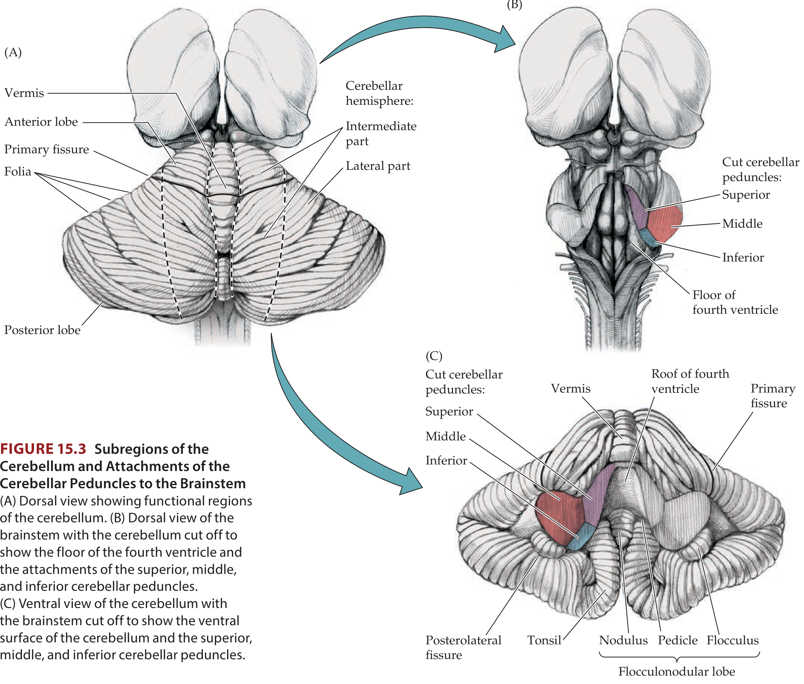
| Lobe | Fissure Boundary | Larsell Lobules |
|---|---|---|
| Anterior lobe | Separated from posterior lobe by the primary fissure | Lobules I - V |
| Posterior lobe | Separated from flocculonodular lobe by the posterolateral fissure | Lobules VI - IX |
| Flocculonodular lobe | Most inferior/anterior; connected by pedicles to the nodulus | Lobule X |
The cerebellar tonsils are important landmarks on the inferior surface of the posterior lobe. Mass lesions or severe elevated intracranial pressure can cause tonsillar herniation through the foramen magnum, compressing medullary respiratory centers and causing death.
3. Functional (Longitudinal) Subdivisions
| Functional Zone | Anatomical Basis | Function |
|---|---|---|
| Vestibulocerebellum | Flocculonodular lobe | Modulation of body equilibrium and eye movements; rich connections with vestibular system |
| Spinocerebellum | Vermis of anterior lobe + pyramis + uvula + paraflocculus | Modulation of muscle tone; axial and limb movements; receives input from spinal cord |
| Cerebrocerebellum | Middle vermis + most cerebellar hemispheres | Planning and initiation of movements; coordination of fine ipsilateral limb movements; receives corticopontocerebellar input |
The somatotopic organization of the cerebellum is well established:
- Hand: represented in the ipsilateral anterior lobe (lobules IV-V)
- Foot: represented in the ipsilateral central lobule (lobules II-III)
- Tongue: mapped posterior and lateral to the hand area, extending into lobules VI-VII
4. Cerebellar Cortex - Layers and Cell Types
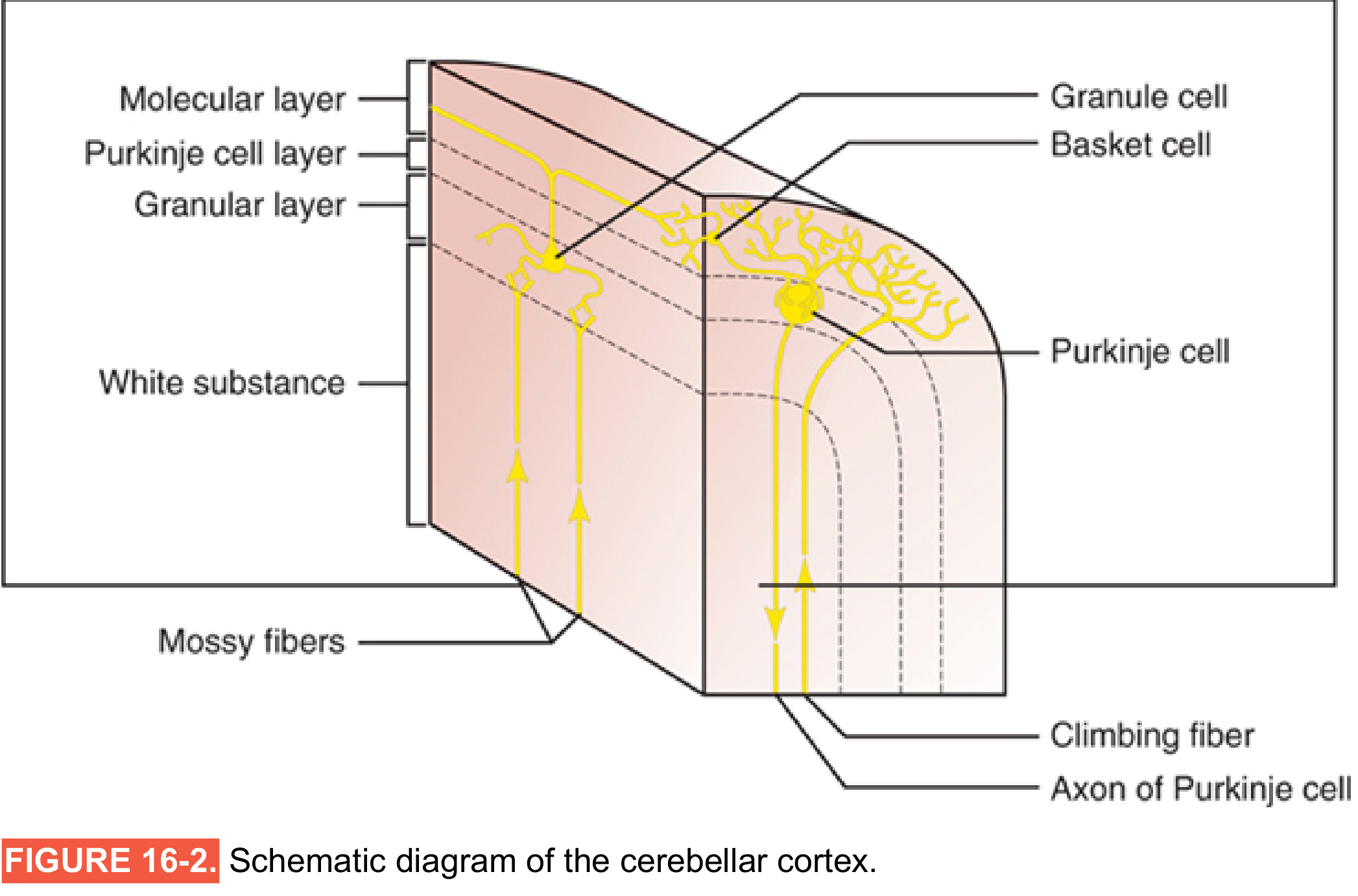
The cerebellar cortex has three distinct layers (mnemonic: Mother Please Go - Molecular, Purkinje, Granular):
Outer - Molecular Layer
- Contains the dendrites of Purkinje cells (extending upward like a flat fan)
- Contains parallel fibers (axons of granule cells that ascend from the granular layer and bifurcate in a T-shape, running parallel to the folia)
- Contains two types of inhibitory interneurons: stellate cells (outer portion) and basket cells (inner portion, which form basket-like axonal networks around Purkinje cell bodies)
Middle - Purkinje Cell Layer
- A single row of large flask-shaped Purkinje cells - the SOLE OUTPUT neurons of the cerebellar cortex
- Their axons project outside the cortex to synapse on the deep cerebellar nuclei (and some directly to vestibular nuclei)
- They are GABAergic (inhibitory)
- Their elaborate dendritic trees fan out into the molecular layer in one plane, perpendicular to the folia
Inner - Granular Layer
- Contains enormous numbers of tiny excitatory granule cells - their total number (~50 billion) equals roughly half of all neurons in the entire CNS
- Also contains Golgi cells (inhibitory interneurons that modulate granule cell activity)
- Granule cells receive mossy fiber input at specialized synaptic complexes called cerebellar glomeruli
All cell types except Purkinje cells are intrinsic neurons - only Purkinje cell axons project outside the cerebellum.
5. Cerebellar White Matter and Fiber Systems
The cerebellar white matter contains three groups of fibers:
- Intrinsic fibers - do not leave the cerebellum
- Afferent (input) fibers - enter mainly via the inferior and middle cerebellar peduncles, organized into three systems:
- Climbing fibers: terminal fibers of the olivocerebellar tract (from the inferior olive); each climbing fiber forms multiple synapses with a SINGLE Purkinje cell, providing powerful, precise excitation
- Mossy fibers: all other cerebellar afferent tracts; diffuse system where a single mossy fiber stimulates thousands of Purkinje cells via granule cell intermediaries
- Multilayered fibers: from hypothalamus, raphe nuclei, and locus ceruleus
- Efferent (output) fibers - the axons of Purkinje cells
6. Deep Cerebellar Nuclei
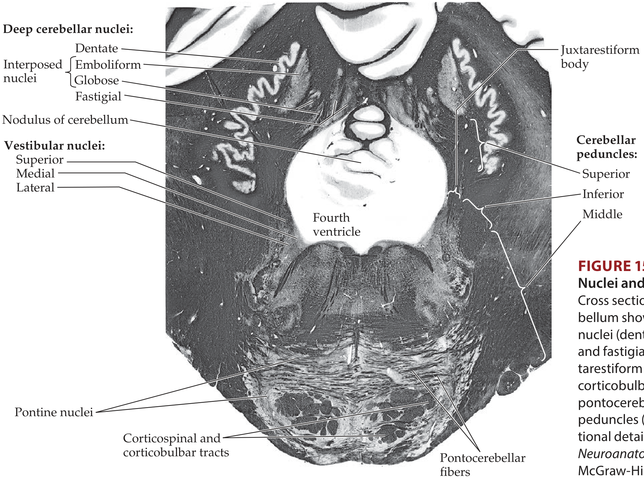
Embedded in the white matter core are three pairs of deep nuclei (also called roof nuclei), arranged from lateral to medial:
| Nucleus | Location | Connection | Function |
|---|---|---|---|
| Dentate nucleus | Most lateral; largest | Receives from lateral cerebellar hemispheres | Planning motor programs for extremities; largest deep nucleus |
| Emboliform nucleus | Next to dentate | Receives from intermediate hemisphere | Segmental reflexes; stability |
| Globose nucleus | Next to fastigial | Receives from intermediate hemisphere | Segmental reflexes; stability |
| Fastigial nucleus | Most medial | Receives from vermis | Stance, gait, sitting, standing, walking |
(Mnemonic: Don't Eat Greasy Foods - Dentate, Emboliform, Globose, Fastigial)
The emboliform and globose nuclei together form the nucleus interpositus.
Each deep nucleus has a somatotopic arrangement: caudal body regions mapped anteriorly, rostral body regions posteriorly, trunk laterally, and limbs medially. All efferent projections from these nuclei are excitatory, except those to the inferior olive (which are inhibitory).
7. Cerebellar Peduncles
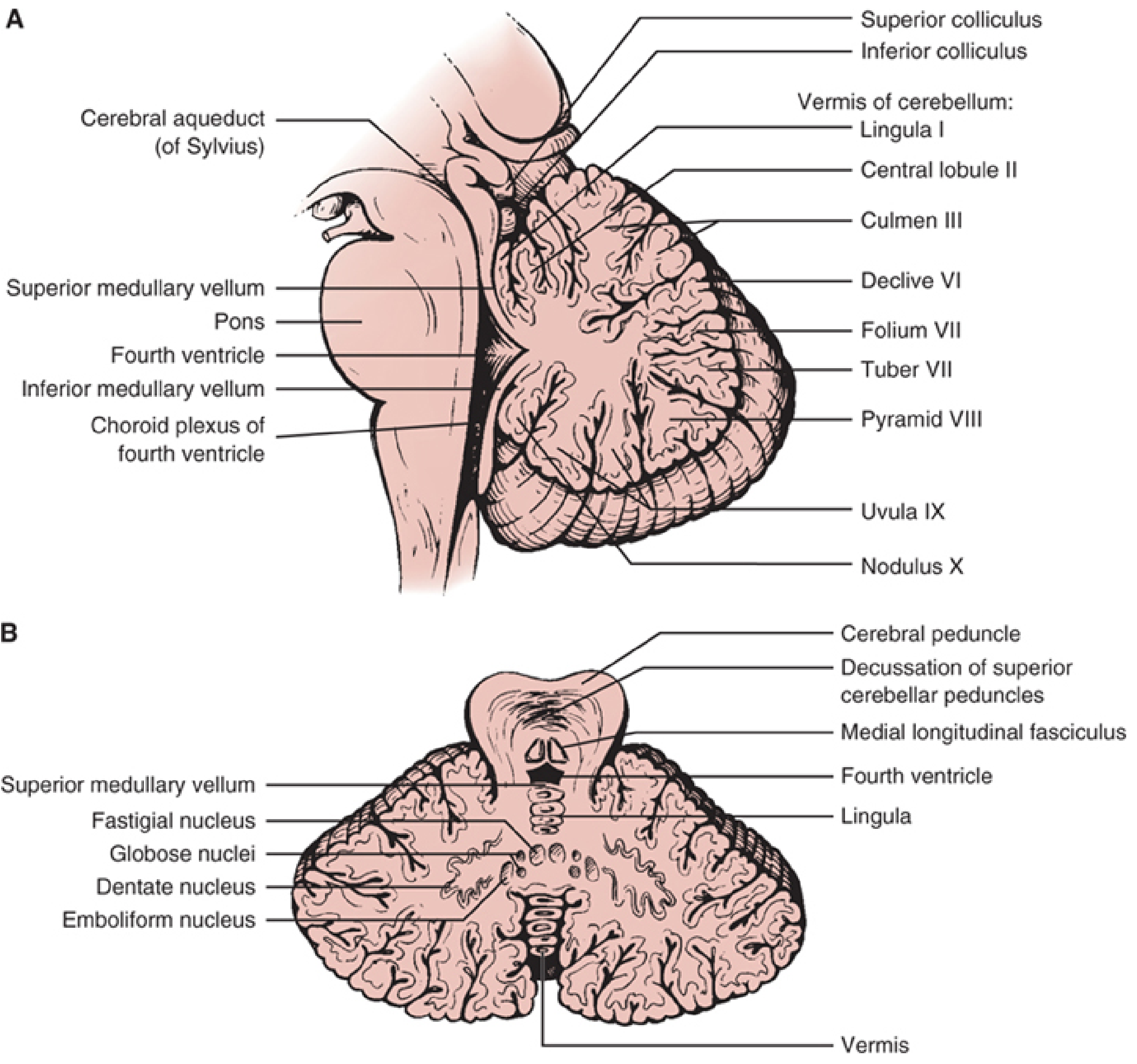
The cerebellum is attached to the brainstem by three paired white matter stalk-like bundles:
Inferior Cerebellar Peduncle (ICP / Restiform Body)
- Connects cerebellum to the medulla
- Afferent fibers: posterior spinocerebellar tract (proprioception from lower body), olivocerebellar tract (climbing fibers from inferior olive), vestibulocerebellar tract, cuneocerebellar tract
- Efferent fibers: cerebellovestibular and cerebelloreticular projections (via juxtarestiform body component)
Middle Cerebellar Peduncle (MCP / Brachium Pontis)
- Largest peduncle; connects cerebellum to the pons
- Contains almost exclusively afferent pontocerebellar fibers from the contralateral pontine nuclei
- Main route for corticopontocerebellar input (cerebral cortex → pontine nuclei → cerebellum)
Superior Cerebellar Peduncle (SCP / Brachium Conjunctivum)
- Connects cerebellum to the midbrain
- Primarily efferent: carries output from deep cerebellar nuclei
- Dentatorubral tract: to contralateral red nucleus
- Dentatothalamic tract: to contralateral ventrolateral thalamic nucleus (then to cerebral cortex)
- Uncinate bundle of Russell: to vestibular nuclei and reticular formation
- Also carries some afferent fibers: anterior spinocerebellar tract, tectocerebellar tract (auditory/visual from colliculi), trigeminocerebellar tract
The superior cerebellar peduncle decussates (crosses) in the caudal midbrain tegmentum before reaching the red nucleus and thalamus.
8. Vascular Supply
The cerebellum receives blood from three paired arterial branches:
| Artery | Origin | Territory |
|---|---|---|
| PICA (Posterior Inferior Cerebellar Artery) | Intracranial vertebral artery | Inferior cerebellar hemisphere, inferior vermis, lateral medullary tegmentum, inferior cerebellar peduncle |
| AICA (Anterior Inferior Cerebellar Artery) | Basilar artery (~1 cm from origin) | Anterior petrosal cerebellar surface, flocculus, lower middle cerebellar peduncle, lateral pontomedullary tegmentum; gives rise to the internal auditory artery |
| SCA (Superior Cerebellar Artery) | Distal basilar artery (just below bifurcation) | Upper cerebellar hemisphere, superior vermis, most of dentate nucleus, superior cerebellar peduncle, lateral pontine tegmentum |
Branches of all three arteries anastomose with their counterparts on the opposite side, forming a rich anastomotic network.
9. Relations and Boundaries
- Superior: tentorium cerebelli (separates it from the cerebral hemispheres)
- Inferior and posterior: occipital bone
- Anterior: fourth ventricle (cerebellum forms its roof)
- Brainstem relations: dorsal to pons and rostral medulla; attached via three peduncles
- The falx cerebelli is a dural partition that partially separates the two cerebellar hemispheres
Summary Table
| Feature | Detail |
|---|---|
| Location | Posterior cranial fossa |
| Weight | ~150 g (~10% of total brain volume) |
| Folia | Transverse ridges increasing surface area |
| Lobes | Anterior (I-V), Posterior (VI-IX), Flocculonodular (X) |
| Cortex layers | Molecular, Purkinje, Granular |
| Output neurons | Purkinje cells (GABAergic, inhibitory) |
| Deep nuclei (lateral to medial) | Dentate, Emboliform, Globose, Fastigial |
| Peduncles | Inferior (ICP), Middle (MCP - largest), Superior (SCP) |
| Main output pathway | SCP → decussation → red nucleus + VL thalamus → cortex |
| Blood supply | PICA, AICA, SCA |
Sources: Neuroscience: Exploring the Brain, 5th Ed. - Localization in Clinical Neurology, 8e - Neuroanatomy through Clinical Cases, 3rd Ed.
This is a shared conversation. Sign in to Orris to start your own chat.