
Thyroid swelling treatment
"thyroid nodule" OR "goiter" treatment management
thyroid goiter enlargement clinical
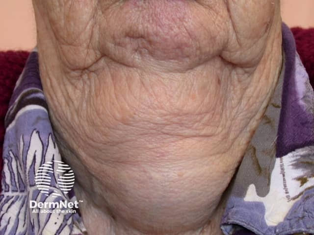
This clinical photograph shows the anterior neck of an adult patient with diffuse thyroid enlargement consistent with goiter. The image modality is clinical photography, captured in a frontal/anterior neck view with the patient seated or standing, natural lighting, and minimal magnification. The thyroid gland is visibly enlarged, producing a prominent cervical contour in the midline and bilateral lobes that extend laterally toward the sternocleidomastoid regions. Skin over the gland is wrinkled and thin, without evident ulceration or focal nodularity on the presented surface; there is modest venous prominence posteriorly. The enlargement appears diffuse rather than a single palpable nodule, suggesting a diffuse goiter or multinodular process that may be chronic. No tracheal deviation or airway compromise is clearly visible in this image, though clinical correlation is necessary to evaluate for compressive symptoms. Clinically, goiter etiologies include iodine deficiency, autoimmune thyroiditis (Hashimoto), Graves disease, or benign/multinodular thyroid neoplasms. Diagnostic significance: clinical confirmation of thyroid enlargement; subsequent assessment should include thyroid function tests (TSH, free T4), thyroid autoantibodies, and high-resolution neck ultrasound or CT/MRI as indicated. Differential considerations include diffuse toxic goiter, thyroiditis, and focal neoplasms. This image is educational for endocrine pathology, ENT, and radiology training, illustrating external thyroid enlargement and its gross appearance in situ.
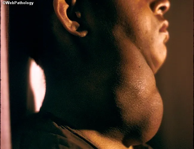
This image is a gross pathology photograph of the anterior neck demonstrating marked thyroid enlargement consistent with nodular hyperplasia (multinodular goiter). Acquired from a lateral profile view, the external contour shows a diffuse, enlarged thyroid gland occupying the lower neck with conspicuous nodularity and prominence of the isthmus. The patient is clinically euthyroid, a common feature of nodular goiter, with thyroid hormone levels typically within normal range despite substantial gland enlargement. Endemic forms in iodine-deficient regions result from chronic stimulation by elevated TSH, leading to hyperplastic nodules that create mass effect. The goiter here is sufficiently large to cause disfigurement, and in advanced cases may impinge on the trachea, producing potential airway compromise. Grossly, the thyroid tissue demonstrates uneven surface topology that may correspond to coarse nodularity; internal histology would typically reveal variably-sized follicles with fibrous septa and colloid depletion depending on the nodule. Clinically, this condition is distinguished from solitary adenomas and thyroid carcinomas by its multinodular architecture, diffuse enlargement, and euthyroid state. Diagnostic significance lies in recognizing goiter-related airway compression risk, cosmetic deformity, and iodine deficiency history; management ranges from observation in mild cases to thyroidectomy in obstructive or cosmetically unacceptable disease. This image serves educational value for endocrinology, pathology, radiology correlation, and clinical exam review.
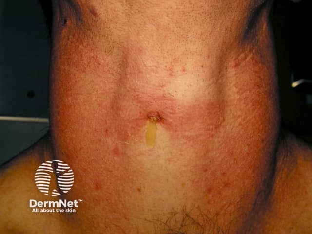
Clinical photograph of the anterior neck illustrating diffuse enlargement of the thyroid region consistent with a goiter. The image is a frontal view capturing the patient from the chin to the upper chest, suitable for documentation and educational purposes in endocrinology and dermatology. The thyroid gland appears expanded with smooth contour and midline projection; there is mild erythema and skin textural change over the lower neck, with a small vertical scar and minimal hair growth noted on the chest. Visual assessment alone cannot differentiate diffuse goiter from nodular disease; no discrete nodules are clearly identifiable in this image. The photograph emphasizes external neck anatomy, including the sternocleidomastoid borders and thyroid isthmus region just below the laryngeal prominence. This image is most applicable to clinical evaluation of neck swelling, patient education, and baseline documentation prior to ultrasound or other thyroid imaging. Potential clinical uses include tracking goiter progression, educating patients about thyroid enlargement, and serving as a visual aid in differential diagnosis discussions with Graves disease, Hashimoto thyroiditis, endemic iodine deficiency, and multinodular goiter. When combined with laboratory tests (TSH, free T4), and neck ultrasonography, this photo supports diagnosis and management planning, including consideration of fine-needle aspiration if focal nodularity is detected or suspicion of malignancy arises.
Thyroid Swelling - Treatment
1. Classification of Thyroid Swelling
| Type | Examples |
|---|---|
| Diffuse non-toxic | Simple/colloid goiter, iodine deficiency |
| Diffuse toxic | Graves' disease |
| Nontoxic multinodular goiter (MNG) | Multinodular, euthyroid |
| Toxic MNG | Autonomously functioning nodules |
| Solitary thyroid nodule | Benign adenoma, cyst, malignancy |
| Thyroiditis | Hashimoto's, subacute de Quervain's |
| Thyroid cancer | Papillary, follicular, medullary, anaplastic |
2. Diffuse Non-Toxic (Simple) Goiter
- Iodine deficiency: Iodine replacement causes variable regression, depending on the duration and degree of fibrosis or autonomous function that may have developed.
- Levothyroxine (LT4): TSH suppression is rarely effective for reducing size and risks inducing subclinical/overt thyrotoxicosis.
- Surgery: Reserved for tracheal compression, obstruction of the thoracic inlet, or cosmetic reasons. Subtotal or near-total thyroidectomy followed by LT4 replacement. - Harrison's Principles, p. 3083
3. Nontoxic Multinodular Goiter (MNG)
- Conservative management is appropriate for most asymptomatic euthyroid patients.
- T4 suppression is rarely effective and risks thyrotoxicosis, especially if autonomy is present.
- Avoid iodine-containing contrast agents - risk of Jod-Basedow effect (enhanced hormone production by autonomous nodules).
- Radioiodine (¹³¹I): Used where surgery is contraindicated; can decrease goiter volume and ablate areas of autonomy. Commonly used in Europe/Brazil for large nodular goiters.
- Surgery: Indicated for compressive symptoms (tracheal/esophageal compression), substernal extension, cosmetic concerns, or suspected malignancy. - Harrison's Principles, p. 3084
4. Toxic MNG (Plummer's Disease)
- Antithyroid drugs (ATDs): Normalize function and are particularly useful in the elderly or those with limited life span - though not curative.
- Radioiodine (¹³¹I): Definitive treatment of choice in many patients; selectively ablates autonomous nodules.
- Surgery: Near-total or total thyroidectomy - used when malignancy is suspected (cold nodules with indeterminate/suspicious FNA cytology direct therapy to surgery). - Harrison's Principles, p. 3086
5. Graves' Disease (Diffuse Toxic Goiter)
| Treatment | Details |
|---|---|
| Antithyroid drugs (ATDs) | Methimazole preferred (longer half-life, lower adverse effects); PTU used in 1st trimester pregnancy. Euthyroid state in 3-8 weeks. |
| Radioactive iodine (RAI / ¹³¹I) | Most commonly used in USA; ablates thyroid tissue; hypothyroidism expected afterward requiring LT4 replacement. |
| Surgery (thyroidectomy) | Near-total/total thyroidectomy; preferred for large goiters, ophthalmopathy, suspected malignancy, pregnancy planning soon. |
- Beta-blockers (propranolol, atenolol, metoprolol): Control symptoms (palpitations, tremor, tachycardia). Do not affect hormone levels.
- Iodide (Lugol's solution / SSKI): Pre-operative use - reduces gland vascularity. Short-term only (Wolff-Chaikoff effect lasts only days). - Lippincott Pharmacology, p. 785
6. Solitary Thyroid Nodule
| Bethesda Category | Management |
|---|---|
| I - Nondiagnostic | Repeat FNA |
| II - Benign | Observation / ultrasound follow-up |
| III - Atypia of undetermined significance | Repeat FNA / molecular testing |
| IV - Follicular neoplasm | Diagnostic lobectomy |
| V - Suspicious for malignancy | Near-total thyroidectomy |
| VI - Malignant | Total thyroidectomy ± RAI ablation |
- Cystic nodules: Ultrasound-guided aspiration ± percutaneous ethanol injection (PEI) for recurrent cysts.
- ACR TI-RADS ultrasound scoring guides which nodules need FNA based on size and sonographic pattern.
7. Thyroid Cancer (if malignant cause)
- Papillary/Follicular (differentiated): Total thyroidectomy + RAI ablation + TSH-suppressive LT4 therapy.
- Medullary: Total thyroidectomy + central neck dissection; vandetanib/cabozantinib for advanced disease.
- Anaplastic: Multimodal (surgery where feasible + chemoradiation); extremely poor prognosis.
- Per 2023 ETA guidelines and the 2024 Japan AES guidelines, molecular testing increasingly guides management of indeterminate nodules.
8. Hashimoto's Thyroiditis
- If hypothyroid: Levothyroxine (LT4) replacement - goal to normalize TSH.
- If euthyroid: Observation; LT4 may be given to reduce goiter size.
- Surgery: Rarely needed; reserved for compressive symptoms or suspicion of lymphoma.
9. Pre-operative Preparation for Thyroid Surgery
- ATDs (methimazole 4-6 weeks) + beta-blockers
- Lugol's iodine for 10 days pre-op (reduces gland vascularity and bleeding risk)
- Scott-Brown's Otorhinolaryngology, p. 1040
Quick Reference Summary
Thyroid Swelling
├── Euthyroid diffuse goiter → Iodine replacement / observe / surgery if compressive
├── Nontoxic MNG → Observe; RAI or surgery if symptomatic
├── Toxic MNG → ATDs (short-term) → RAI or surgery
├── Graves' disease → ATDs / RAI / thyroidectomy
├── Solitary nodule → FNA → Bethesda-guided management
├── Thyroid cancer → Surgery ± RAI ± targeted therapy
└── Hypothyroid (Hashimoto's) → Levothyroxine
Note: All treatment decisions should factor in patient age, comorbidities, goiter size, functional status, and nodule cytology. Referral to an endocrinologist and/or thyroid surgeon is recommended for definitive management.
Thyroid
What aspect of the thyroid would you like to learn about? · What level of detail do you need?
Which condition(s) do you want in-depth treatment detail for?
"thyroid" treatment guidelines
thyroid gland anatomy diagram
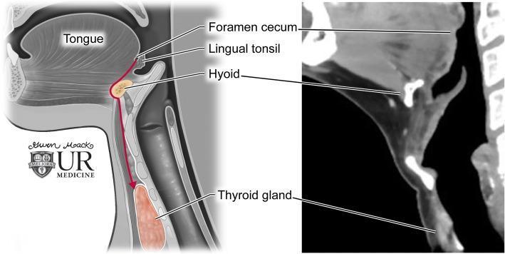
This dual-panel educational graphic illustrates thyroglossal duct anatomy and the embryological descent of the thyroid gland. On the left is a sagittal anatomical diagram showing the path of descent (indicated by a red arrow) from the foramen cecum at the base of the tongue, passing through the hyoid bone, to its final position at the thyroid gland anterior to the trachea. Key labeled structures include the tongue, lingual tonsil, foramen cecum, hyoid, and thyroid gland. The right panel displays a corresponding sagittal contrast-enhanced CT image of the neck. The CT scan highlights the radiologic appearance of these landmarks: the hyoid bone appears as a high-attenuation U-shaped structure, and the thyroid gland exhibits homogenous enhancement inferior to the thyroid cartilage. This comparison demonstrates the clinical relevance of embryological pathways in understanding the location of thyroglossal duct cysts, which can occur anywhere along this midline tract.
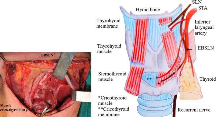
A composite medical visual consisting of a clinical intraoperative photograph (left) and a corresponding anatomical diagram (right) illustrating the anatomy of the neck and larynx. The content focuses on the anatomical variations of the External Branch of the Superior Laryngeal Nerve (EBSLN) in relation to the Superior Thyroid Artery (STA) and the thyroid gland, specifically representing Cernea Type IIb and Kierner Type III classifications. In the diagram, the EBSLN is shown descending alongside the STA and crossing it at a level below the upper pole of the thyroid gland, terminating in the cricothyroid muscle. Key labeled structures include the hyoid bone, thyrohyoid membrane/muscle, sternothyroid muscle, and cricothyroid membrane. The intraoperative photo displays the surgical field during thyroid surgery, with tissue retracted to expose the EBSLN in a clinical context. This material is designed for surgical education, highlighting the high-risk positioning of the nerve during the ligation of superior thyroid vessels.
Thyroid Disease - Comprehensive Treatment & Management (Exam-Level)
Overview of Thyroid Conditions
| Condition | Functional Status | Key Cause |
|---|---|---|
| Simple/colloid goiter | Euthyroid | Iodine deficiency |
| Hashimoto's thyroiditis | Hypothyroid (usually) | Autoimmune (TPO-Ab) |
| Graves' disease | Hyperthyroid | TSH-receptor antibody (TRAb) |
| Toxic MNG | Hyperthyroid | Autonomous nodules |
| Subacute (de Quervain's) thyroiditis | Triphasic | Viral / post-viral |
| Silent / postpartum thyroiditis | Triphasic | Autoimmune |
| Thyroid nodule / MNG | Euthyroid (usually) | Hyperplasia / neoplasia |
| Thyroid cancer | Usually euthyroid | Malignant transformation |
| Thyroid storm | Severe hyperthyroid | Precipitated thyrotoxicosis |
1. Hypothyroidism
Causes
- Hashimoto's thyroiditis (most common in iodine-sufficient areas)
- Post-radioiodine or post-thyroidectomy
- Iodine deficiency (most common globally)
- Secondary/tertiary (pituitary or hypothalamic failure)
- Drug-induced (lithium, amiodarone, tyrosine kinase inhibitors)
Treatment: Levothyroxine (LT4) Replacement
- Mechanism: Acts via nuclear receptors; peripheral conversion of T4 → T3 (via Dio1, Dio2) provides T3. - Goodman & Gilman's, p. 2451
- Standard adult dose: 1.7 mcg/kg/day (based on lean body weight)
- Elderly / cardiac disease: Start low at 12.5-50 mcg/day, increase by 25 mcg every 6 weeks
- Monitoring: Check TSH 6-8 weeks after any dose change (reflects the ~7-day T4 half-life)
- Goal: Normalize serum TSH in primary hypothyroidism; normalize free T4 in secondary/tertiary hypothyroidism
- Pregnancy: LT4 dose requirement increases (increased TBG from estrogen + placental Dio3 expression + small transplacental T4 passage); inadequately treated maternal hypothyroidism causes miscarriage, fetal distress, preterm delivery, and impaired neurodevelopment. Per ATA 2026 pregnancy guidelines, proactive dose adjustment is recommended.
- Hashimoto's + goiter: LT4 150-200 mcg/day suppresses TSH, causing slow goiter regression. - Katzung, p. 1304
- Goitrogenic diet: Eliminate goitrogen or add enough T4 to suppress TSH stimulation.
- Goiter from iodine deficiency: Prophylactic/therapeutic iodide supplementation (optimal: 150-200 mcg/day).
- Vast majority of patients do well on LT4 monotherapy alone.
- Combination T4/T3 therapy is not recommended as standard - evidence does not show consistent benefit over T4 alone; however, a small subgroup (particularly those with Dio2 Thr92Ala polymorphism) may feel better on combination therapy. - Goodman & Gilman's, p. 2453
2. Hyperthyroidism / Thyrotoxicosis
2A. Graves' Disease
Option 1: Antithyroid Drugs (ATDs) - Thionamides
| Drug | Dose | Half-life | Notes |
|---|---|---|---|
| Methimazole | Initial: 10-20 mg q12h; Maintenance: 2.5-10 mg/day | 6 h | Drug of choice - once-daily dosing possible; lower toxicity |
| Carbimazole | Similar to methimazole (prodrug) | - | Not available in USA |
| Propylthiouracil (PTU) | 100-200 mg q6-8h; Maintenance: 50-100 mg/day | 90 min | Reserved: 1st trimester pregnancy, thyroid storm, minor methimazole reaction |
- Titration regimen (preferred): Gradually reduce dose as euthyroidism is restored; provides index of treatment response.
- Block-replace regimen: Fixed high ATD dose + LT4 added to prevent hypothyroidism.
- Minor: Rash, urticaria, arthralgia, GI disturbance (switch to alternative drug)
- Major (rare): Agranulocytosis (0.2-0.5%) - stop drug immediately; PTU also carries black box warning for hepatotoxicity (potentially fatal)
- Methimazole: Teratogenicity (scalp defects, choanal atresia) - avoid in 1st trimester
- PTU: Preferred in 1st trimester (crosses placenta less, fewer fetal malformations)
Option 2: Radioiodine (¹³¹I / RAI)
- Most commonly used first-line in North America.
- Administered as oral dose of radioactive iodine - concentrated in thyroid, causing radiation-induced thyroid cell destruction over weeks to months.
- Outcome: Intentional hypothyroidism in most patients (followed by lifelong LT4 replacement).
- Contraindications: Pregnancy, breastfeeding (absolute); moderate-severe active Graves' ophthalmopathy (relative - may worsen).
- Pre-treatment: ATDs to achieve euthyroid state before RAI in elderly or those with cardiac disease (to prevent thyroid storm from RAI-induced hormone release).
- Post-RAI: Monitor for hypothyroidism and start LT4.
Option 3: Surgery (Thyroidectomy)
- Near-total or total thyroidectomy - preferred to avoid recurrence and the higher complication rates of reoperation. - Schwartz's Surgery
- Indications: Large goiter causing compressive symptoms, active Graves' ophthalmopathy, desire for prompt definitive treatment, pregnancy planning soon, suspected malignancy, failed ATDs/RAI.
- Pre-operative preparation: Render euthyroid with ATDs (4-6 weeks) + beta-blockers. Give Lugol's iodine (Wolff-Chaikoff effect) 10 days pre-op to reduce gland vascularity.
- Post-operative: LT4 replacement.
- Complications: Hypoparathyroidism (hypocalcemia), recurrent laryngeal nerve injury (hoarseness), bleeding, hypothyroidism.
Adjunct / Symptomatic Treatment
- Beta-blockers (propranolol 40-120 mg/day, atenolol, metoprolol): Rapid control of tachycardia, palpitations, tremor, heat intolerance. Do NOT reduce thyroid hormone levels.
- Iodide (Lugol's / SSKI): Pre-operative use only; blocks hormone release (Wolff-Chaikoff effect) and reduces gland vascularity. Not for long-term use (effect lasts only days). - Lippincott Pharmacology, p. 652
2B. Toxic Multinodular Goiter (Plummer's Disease)
- ATDs: Normalize function, especially for elderly/limited life expectancy (not curative).
- RAI (¹³¹I): Definitive - reduces goiter volume and ablates autonomous regions.
- Surgery: Indicated if malignancy suspected on FNA, or if compressive symptoms.
2C. Thyroid Storm (Thyrotoxic Crisis)
| Drug | Purpose |
|---|---|
| PTU (preferred over methimazole) | Blocks new synthesis + T4→T3 conversion |
| Iodide (given 1 hour AFTER PTU) | Blocks hormone release |
| Beta-blockers (propranolol IV) | Controls sympathetic hyperactivity |
| Corticosteroids (hydrocortisone / dexamethasone) | Blocks T4→T3 conversion, treats relative adrenal insufficiency |
| Cholestyramine | Interrupts enterohepatic circulation of thyroid hormones |
| Antipyretics (paracetamol) | Fever control (avoid aspirin - displaces T4 from protein) |
| Supportive | IV fluids, treat precipitant (infection, surgery) |
3. Thyroiditis
3A. Subacute (de Quervain's) Thyroiditis
| Phase | Treatment |
|---|---|
| Thyrotoxicosis | Beta-blockers (propranolol 20-40 mg TID/QID). ATDs have NO role (not synthesis-driven). |
| Pain / inflammation | Aspirin 600 mg q4-6h or NSAIDs (+ gastroprotection); if severe → Prednisone 15-40 mg/day, taper over 6-8 weeks |
| Hypothyroid phase | LT4 if prolonged; keep dose low (50-100 mcg/day) to allow TSH-mediated recovery |
3B. Silent / Postpartum Thyroiditis
- Glucocorticoids NOT indicated (unlike subacute thyroiditis)
- Severe thyrotoxic symptoms: brief course of propranolol 20-40 mg TID/QID
- Hypothyroid phase: LT4 (withdraw after 6-9 months - recovery expected)
- Annual follow-up (permanent hypothyroidism develops in some)
3C. Hashimoto's (Chronic Autoimmune) Thyroiditis
- Hypothyroid: LT4 replacement (target: normal TSH)
- Euthyroid + goiter: LT4 to suppress TSH and reduce goiter size
- Euthyroid, no goiter: Observation
- Surgery: Reserved for compressive symptoms or suspicion of thyroid lymphoma
3D. Drug-Induced Thyroiditis
- Treatment same as silent thyroiditis
- Monitor thyroid function routinely during these therapies (ASCO recommendation)
4. Thyroid Nodule / Multinodular Goiter
| Bethesda Category | Risk of Malignancy | Management |
|---|---|---|
| I - Nondiagnostic | N/A | Repeat FNA |
| II - Benign | <3% | Ultrasound surveillance |
| III - AUS/FLUS | 6-18% | Repeat FNA / molecular testing |
| IV - Follicular neoplasm | 10-40% | Diagnostic lobectomy |
| V - Suspicious for malignancy | 45-75% | Near-total thyroidectomy |
| VI - Malignant | 97-99% | Total thyroidectomy ± RAI |
- HIFU (High-Intensity Focused Ultrasound): 2023 meta-analysis (PMID 37478539) supports HIFU for benign thyroid nodules.
- Radiofrequency ablation (RFA): Per 2023 ETA guidelines, thermal ablation techniques (RFA, laser, HIFU) are recommended alternatives to surgery for benign nodules causing symptoms.
5. Thyroid Cancer
Types & Prognosis
| Type | Frequency | Origin | Prognosis |
|---|---|---|---|
| Papillary | 80% | Follicular cell | Excellent (10-yr survival >90%) |
| Follicular | 10-15% | Follicular cell | Good |
| Medullary | 3-5% | Parafollicular C cells (calcitonin) | Intermediate |
| Anaplastic | <2% | Follicular cell (dedifferentiated) | Very poor (<6 months median survival) |
Treatment of Differentiated Thyroid Cancer (Papillary/Follicular)
- Total thyroidectomy is standard for most differentiated thyroid cancer.
- Hemithyroidectomy may be sufficient for low-risk papillary cancers (<1 cm, no extrathyroidal extension, no nodal disease).
- Central neck dissection if nodal disease present.
- Ref: 2025 ATA Differentiated Thyroid Cancer Guidelines
- Destroys residual thyroid tissue (remnant ablation) and any metastatic disease.
- Used selectively - not all patients need RAI; risk stratification determines use.
- Prior to RAI: TSH must be elevated to drive ¹³¹I uptake. Two options:
- Withdraw LT4 for 4-6 weeks (patient becomes hypothyroid)
- Recombinant human TSH (rhTSH / Thyrogen) IM x 2 days - avoids hypothyroidism - Katzung, p. 1314
- LT4 post-surgery kept at dose sufficient to suppress TSH (reduces TSH-driven tumor recurrence).
- Degree of suppression based on risk: high-risk patients → fully suppressed TSH; low-risk → TSH low-normal.
- Serum thyroglobulin (Tg) is the tumor marker - rise indicates recurrence.
- Positive ¹³¹I scan on TSH stimulation confirms recurrence.
- Sorafenib, lenvatinib (tyrosine kinase inhibitors) for RAI-refractory disease.
Medullary Thyroid Cancer (MTC)
- Total thyroidectomy + central neck dissection.
- Tumor marker: Calcitonin and CEA for surveillance.
- Advanced/metastatic: Vandetanib or Cabozantinib (RET kinase inhibitors).
- Screen for RET mutation (familial MTC / MEN2).
Anaplastic Thyroid Cancer
- Surgery if resectable; most are unresectable at diagnosis.
- Multimodal: chemoradiation (doxorubicin + cisplatin).
- Dabrafenib + trametinib for BRAF V600E-mutated anaplastic thyroid cancer (targeted therapy).
- Median survival < 6 months; extremely poor prognosis.
6. Drug Summary Table
| Drug | Class | Use | Key Points |
|---|---|---|---|
| Levothyroxine (T4) | Thyroid hormone | Hypothyroidism, cancer TSH suppression | 1.7 mcg/kg/day; check TSH at 6-8 weeks |
| Liothyronine (T3) | Thyroid hormone | Short-term (pre-RAI scan); thyroid storm | Short half-life |
| Methimazole | Thioamide ATD | Hyperthyroidism (Graves', toxic MNG) | Drug of choice; once-daily; teratogenic in 1st trimester |
| Propylthiouracil (PTU) | Thioamide ATD | 1st trimester, thyroid storm | Hepatotoxic (black box); also blocks T4→T3 |
| ¹³¹I (Radioiodine) | Ablative | Graves', toxic MNG, thyroid cancer | CI in pregnancy; most used in N. America |
| Lugol's iodine | Inorganic iodide | Pre-op Graves', thyroid storm | Short-term only; Wolff-Chaikoff effect |
| Propranolol | Beta-blocker | Symptomatic hyperthyroidism, thyroid storm | Does NOT lower thyroid hormone levels |
| Prednisone | Corticosteroid | Subacute thyroiditis (severe) | Taper over 6-8 weeks |
| Recombinant hTSH (Thyrogen) | TSH analog | Pre-RAI scan/ablation | Avoids LT4 withdrawal hypothyroidism |
Exam High-Yield Pearls
- Methimazole is preferred over PTU except in 1st trimester pregnancy and thyroid storm.
- PTU carries FDA black box warning for liver failure - use only when specifically indicated.
- Agranulocytosis with ATDs: tell patients to stop the drug and seek urgent medical attention if they develop fever or sore throat.
- Radioiodine uptake differentiates causes of thyrotoxicosis: HIGH uptake = Graves'/toxic MNG (synthesis-driven); LOW uptake = thyroiditis, exogenous thyroid hormone.
- TSH is the first and most sensitive screening test for thyroid dysfunction.
- TSH remains suppressed for months after starting ATDs - use free T4 to monitor treatment response in the first few months.
- In subacute thyroiditis, ATDs have NO role in the thyrotoxic phase (not a synthesis problem).
- Amiodarone contains 37% iodine by weight and can cause both hypo- and hyperthyroidism.
- In thyroid cancer follow-up: rising thyroglobulin on LT4 therapy or positive RAI scan = recurrence.
- Pregnancy: LT4 dose requirements increase; untreated maternal hypothyroidism impairs fetal neurodevelopment.
Colloid cyst
Which colloid cyst are you asking about?
"thyroid colloid cyst" OR "thyroid cyst" management
thyroid colloid cyst ultrasound histology
"thyroid cyst" ethanol injection aspiration treatment
thyroid nodule cystic ultrasound sonography
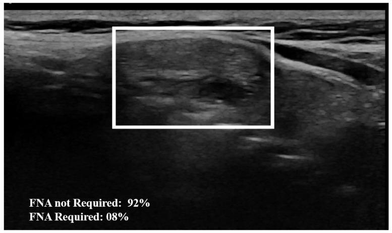
This diagnostic image is a B-mode grayscale ultrasound of the neck, specifically targeting a thyroid nodule. The central visual element is a solitary thyroid nodule highlighted within a white bounding box. The nodule exhibits a heterogeneous echo texture and mixed echogenicity, characterized by irregular internal grayscale variations. Several small hypoechoic (dark) focal areas are visible within the nodule, suggesting cystic components or internal tissue complexity. The surrounding thyroid parenchyma and adjacent anatomical structures, likely including strap muscles and connective tissue planes, appear more homogeneous and demonstrate a smoother texture compared to the coarse interior of the nodule. The image includes an overlaid AI-based classification score at the bottom, indicating a 92% probability that the nodule does not require fine-needle aspiration (FNA). This visual is intended for medical education on thyroid sonography, nodule characterization, and the integration of artificial intelligence in radiological risk assessment.
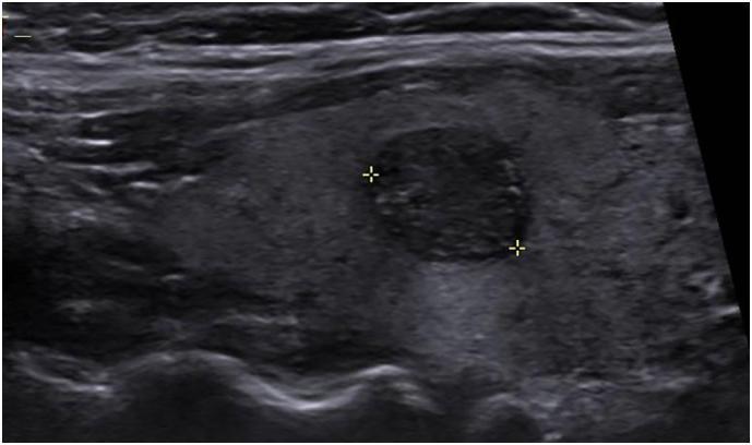
This grayscale ultrasound image of the neck displays a longitudinal view of the right thyroid lobe. A focal, well-defined solid nodule is demarcated by two yellow electronic calipers (crosshairs) representing its maximal diameter. The nodule exhibits a heterogeneous, predominantly hypoechoic echotexture compared to the adjacent normal thyroid parenchyma, which appears more echogenic and uniform. The lesion displays relatively smooth margins and lacks significant cystic components or macrocalcifications in this plane of view. Clinically, such findings in the context of a solid thyroid mass require further risk stratification, such as the TI-RADS (Thyroid Imaging Reporting and Data System) classification, to determine the necessity of fine-needle aspiration (FNA) biopsy for excluding papillary thyroid carcinoma or other malignancies. This diagnostic image serves as an educational example of thyroid sonography used to evaluate incidental nodules or those identified via PET-CT imaging.
Thyroid Colloid Cyst - Comprehensive Notes
Definition
Pathology
Normal Thyroid Follicle (Context)
- The thyroid follicle is the basic structural and functional unit of the thyroid gland.
- Each follicle = a roughly spherical, cyst-like compartment lined by follicular epithelium (thyrocytes), with a central colloid lumen rich in thyroglobulin.
- Surrounding stroma contains parafollicular (C) cells, capillaries, connective tissue, and lymphatics.
Colloid Cyst Formation
- Colloid cysts arise from distension and accumulation of colloid within one or more thyroid follicles - often as part of a multinodular goiter process.
- Many result from intranodular ischemia causing tissue necrosis and liquefaction (degenerating adenoma or hyperplastic nodule).
- True epithelial-lined cysts are rare; most "thyroid cysts" are colloid-rich degenerated nodules or partial cysts.
- Approximately 15-25% of all thyroid nodules are cystic or have a cystic component. - Cummings Otolaryngology, p. 3848
Classification of Thyroid Cysts (Pathological)
| Type | Features |
|---|---|
| Colloid cyst | Distended follicle(s) with thick colloid; most common benign type |
| Simple/haemorrhagic cyst | Degenerated nodule with old blood (brown/amber fluid on FNA) |
| Follicular adenoma | Encapsulated benign tumor; can undergo cystic change |
| Parathyroid cyst | Clear fluid; contains parathyroid hormone |
| Thyroglossal duct cyst | Developmental; columnar epithelium; midline neck |
Clinical Features
- Age/Sex: More common in women; increases with age.
- Presentation: Usually a painless, soft, smooth-surfaced swelling in the neck, moves with swallowing.
- Functional status: Typically euthyroid - does not produce excess thyroid hormone.
- Symptoms: Usually asymptomatic; large cysts may cause:
- Dysphagia (esophageal compression)
- Dyspnoea / stridor (tracheal compression)
- Cosmetic concern
- Sudden pain if hemorrhage occurs into the cyst
- May be discovered incidentally on imaging (ultrasound, CT, MRI).
Investigations
1. Thyroid Function Tests
- TSH (first-line) - typically normal in a euthyroid colloid cyst.
- Free T4, T3 if TSH abnormal.
2. Ultrasound (US) - Most Important Imaging
- Anechoic or hypoechoic well-defined lesion
- "Comet tail" artifact - pathognomonic of colloid: bright echogenic focus with posterior reverberation artifact from colloid crystals
- Smooth walls, thin capsule
- Purely cystic or predominantly cystic with minimal solid component
- TR1 (benign) - spongiform nodule with >50% microcystic areas = very low malignancy risk, no FNA needed unless >2 cm
- TR2 - pure cyst = no FNA
3. Fine-Needle Aspiration Cytology (FNAC / FNA)
- The single most important diagnostic test for thyroid masses. - Schwartz's Principles of Surgery, p. 1671
- Uses a 23-gauge needle; multiple passes with aspiration; slides stained with Papanicolaou or Wright's stain.
- US guidance is recommended for cystic, recurrent, or difficult-to-palpate nodules.
| Fluid Color | Interpretation |
|---|---|
| Clear/pale yellow, watery or thick colloid | Colloid cyst (benign) |
| Brown / dark "motor-oil" fluid | Old hemorrhage into adenoma (Bethesda II - benign) |
| Red / bloody | More suspicious for carcinoma |
| Clear, colorless | Parathyroid cyst (check PTH levels in fluid) |
| Category | Finding | Malignancy Risk | Management |
|---|---|---|---|
| I | Nondiagnostic / acellular cyst fluid | 1-4% | Repeat FNA with US guidance |
| II | Benign - colloid nodule, follicular nodule | <3% | Observe / aspirate |
| III | AUS / FLUS | 5-15% | Repeat FNA or molecular testing |
| IV | Follicular neoplasm | 10-40% | Lobectomy |
| V | Suspicious for malignancy | 45-75% | Near-total/total thyroidectomy |
| VI | Malignant | 97-99% | Total thyroidectomy |
4. Thyroid Scintigraphy (Isotope Scan)
- Colloid cysts typically appear as "cold" nodules (non-functioning, reduced isotope uptake).
- Limited diagnostic value; largely replaced by FNA.
- Still useful if TSH is suppressed (to identify "hot"/autonomous nodules).
5. CT / MRI
- For substernal extension, airway compression assessment, or if malignancy suspected.
Important: Is Every Cystic Thyroid Nodule Benign?
- Papillary carcinomas may manifest as cystic masses and are present in 14-32% of all cystic thyroid nodules. - Cummings Otolaryngology
- A cystic appearance alone does NOT confirm benignity.
- Suspicion for carcinoma increases if:
- Cyst reaccumulates after 3 drainage attempts
- Rapid reaccumulation after drainage
- Solid component on US (especially hypoechoic, irregular margins, microcalcifications)
- Red/bloody aspirate
- History of radiation or family history of thyroid cancer
Management
Management Algorithm (from Schwartz's Surgery):
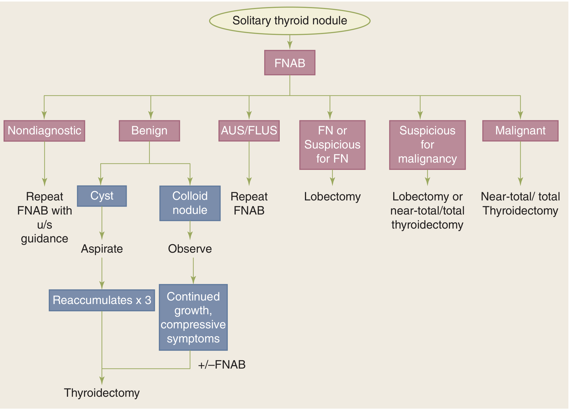
1. Asymptomatic, Confirmed Benign Colloid Cyst
- Observation with periodic ultrasound surveillance (6-12 monthly, then less frequently if stable).
- No treatment required if truly benign and asymptomatic.
- Levothyroxine (T4) suppression is NOT recommended for benign nodules in iodine-sufficient areas (no evidence of benefit; risk of thyrotoxicosis).
2. Symptomatic / Enlarging Cyst - Aspiration (FNAC Drainage)
- Drain the cyst completely at time of FNA - this may be curative for simple cysts.
- One or two additional drainage procedures may be required (cysts commonly reaccumulate).
- If the cyst reaccumulates after 3 drainage attempts → increased suspicion for malignancy → surgery.
3. Percutaneous Ethanol Injection (PEI) - Sclerotherapy
- For recurrent cystic nodules after repeated aspiration failure.
- Ethanol is injected into the cyst cavity under ultrasound guidance, causing sclerosis of the cyst wall.
- A 2024 systematic review and meta-analysis (PMID 39030844) confirmed PEI is effective for cystic thyroid nodules with a low overall complication rate (~2.5%), most commonly transient pain or mild burning sensation.
- Note: Some textbooks (e.g. Cummings) advise caution with sclerosing agents due to potential thyroid carcinoma being missed, and prefer surgery over injection in suspicious cases.
4. Surgery
- Persistent cyst after 3 aspiration attempts
- Rapid reaccumulation
- Suspicious FNA cytology (Bethesda III-VI)
- Solid component with suspicious US features
- Compressive symptoms (dysphagia, stridor) not relieved by aspiration
- Cosmetically unacceptable
- Clinical suspicion of carcinoma (bloody aspirate, hard fixed lesion, lymphadenopathy, hoarseness)
- Unilateral thyroid lobectomy (hemithyroidectomy): For a unilateral cyst - provides definitive diagnosis
- Near-total / total thyroidectomy: If bilateral disease, or confirmed malignancy on frozen section
5. Newer Ablation Techniques (for symptomatic benign cysts)
- Radiofrequency ablation (RFA): Effective for reducing benign cystic/solid thyroid nodules; endorsed by 2023 ETA guidelines.
- HIFU (High-intensity focused ultrasound): Supported by a 2023 meta-analysis (PMID 37478539) for benign thyroid nodules.
- These are alternatives to surgery for patients who decline or are unfit for surgery.
Differential Diagnosis of a Cystic Thyroid Mass
| Condition | Key Distinguishing Feature |
|---|---|
| Colloid cyst | "Comet-tail" artifact on US; colloid fluid on FNA; Bethesda II |
| Thyroglossal duct cyst | Midline; moves with tongue protrusion; columnar epithelium on FNA |
| Parathyroid cyst | Clear fluid with elevated PTH; lateral neck |
| Cystic papillary carcinoma | Solid component; microcalcifications; psammoma bodies on cytology |
| Cystic follicular adenoma | Thin capsule; may look identical to colloid cyst |
| Hemorrhagic cyst | Brown "motor-oil" fluid; hemosiderin-laden macrophages on cytology |
Prognosis
- True colloid cysts are entirely benign with excellent prognosis.
- Most resolve or remain stable with aspiration.
- Recurrence after single aspiration is common (~50-75%).
- Malignant transformation does not occur in a true colloid cyst.
- The key clinical task is reliably excluding malignancy - which requires FNA cytology (and sometimes surgery).
Exam High-Yield Pearls
- Colloid cyst is a Bethesda II lesion on FNA - management is aspirate and observe.
- "Comet-tail" artifact on ultrasound = strongly suggests colloid - low malignancy risk.
- A cyst that reaccumulates ≥3 times after aspiration → suspect malignancy → surgery.
- Red/bloody aspirate from a thyroid cyst = more suspicious for carcinoma.
- 14-32% of cystic thyroid nodules may harbor papillary carcinoma - never assume all cysts are benign.
- Levothyroxine suppression is NOT recommended for benign thyroid nodules.
- Parathyroid cyst: clear fluid + high PTH in fluid - do not inject it thinking it is a thyroid cyst.
- PEI (percutaneous ethanol injection) is an effective non-surgical option for recurrent benign cysts.
Is thyroid colloid cyst dangerous
thyroid colloid cyst dangerous malignant risk prognosis
Is a Thyroid Colloid Cyst Dangerous?
The Reassuring Facts
| Feature | Risk Level |
|---|---|
| Purely cystic thyroid nodule | Cancer risk <2% |
| Spongiform / colloid nodule (>50% microcystic) | Extremely low malignancy risk - ACR TI-RADS 1 |
| Confirmed Bethesda II colloid on FNA | <3% malignancy risk |
| FNA showing thick colloid fluid | Strongly benign |
- About 95% of all thyroid nodules (including colloid cysts) are non-cancerous.
- A colloid cyst confirmed on FNA is classified as Bethesda Category II (Benign) - this is essentially a "do not worry" result.
- Malignant transformation of a true colloid cyst does not occur.
When It Could Be a Problem
1. Misidentification - It May Not Be a Pure Colloid Cyst
- 14-32% of cystic thyroid nodules can harbor papillary carcinoma inside them - particularly those with a solid component. - Cummings Otolaryngology
- A cystic appearance on ultrasound alone cannot rule out malignancy.
- This is why FNA (fine-needle aspiration) is essential before concluding a cyst is benign.
2. Warning Signs That Raise Concern
| Warning Sign | Why It Matters |
|---|---|
| Solid component on ultrasound | Must be biopsied - may contain cancer |
| Rapid reaccumulation after drainage | Suspicious for malignancy |
| Reaccumulates after 3 aspirations | Surgery needed to exclude cancer |
| Red or bloody aspirate | More suspicious for carcinoma |
| Hard or fixed lump on examination | May suggest cancer |
| Hoarseness or difficulty swallowing | Possible nerve/local invasion |
| Lymph node enlargement in neck | Potential nodal spread |
| History of radiation to the neck | Higher cancer risk overall |
| Family history of thyroid cancer or MEN2 | Genetic risk |
| Age <20 or male sex | Thyroid nodules in these groups have higher malignancy rates |
3. Compressive Effects (Large Cysts)
- A large colloid cyst or multinodular goiter may, over time, press on nearby structures causing:
- Difficulty swallowing (dysphagia)
- Breathing difficulty or stridor (tracheal compression)
- Voice changes (laryngeal nerve pressure)
- These mechanical effects can be problematic even in a fully benign cyst.
4. Hemorrhage Into the Cyst
- Sudden pain in a previously painless thyroid lump often signals hemorrhage into a colloid nodule.
- This is not dangerous long-term but causes acute discomfort and rapid enlargement.
- Resolves spontaneously or with aspiration.
Bottom Line - Practical Guidance
Thyroid colloid cyst confirmed by FNA (Bethesda II)
→ BENIGN - not dangerous
→ Periodic ultrasound surveillance (6-12 months, then yearly)
→ No surgery needed unless it causes compressive symptoms
   or reaccumulates after 3 aspirations

Thyroid cyst NOT yet evaluated by FNA
→ Needs proper workup (ultrasound + FNA)
→ Do NOT assume it is safe until confirmed
Key message: A confirmed thyroid colloid cyst is benign and carries an excellent prognosis. The real clinical task is making sure it IS genuinely benign - which requires ultrasound assessment and FNA cytology. Once confirmed, it can be safely monitored without surgery in most cases.