
Lytic lesion with hyperclacemia in hematological condition differentials
Lytic Lesions + Hypercalcemia in Hematological Conditions: Differentials
1. Multiple Myeloma (PRIMARY and most common)
- Myeloma cells secrete MIP-1α (CCL3) and upregulate RANKL while simultaneously suppressing OPG - this shifts the RANKL/OPG ratio strongly toward osteoclast activation
- Myeloma cells also produce Wnt pathway inhibitors that block osteoblast function, creating a purely lytic pattern with no new bone formation
- Net result: massive uncoupled osteolysis, hypercalcemia, and pathologic fractures
- (Robbins, Cotran & Kumar Pathologic Basis of Disease, p. 570; Goldman-Cecil Medicine, RANKL/OPG section)
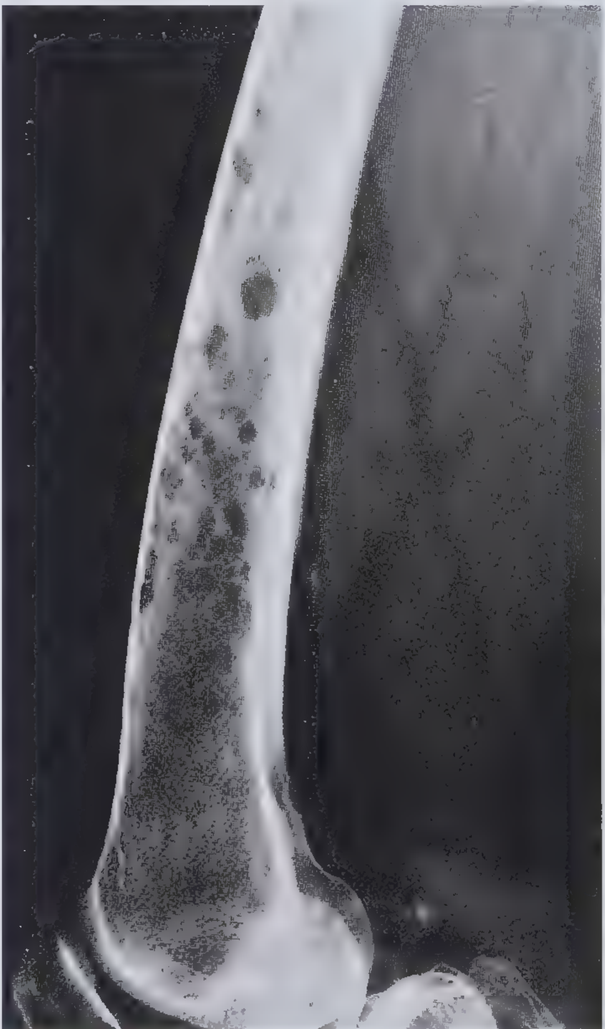
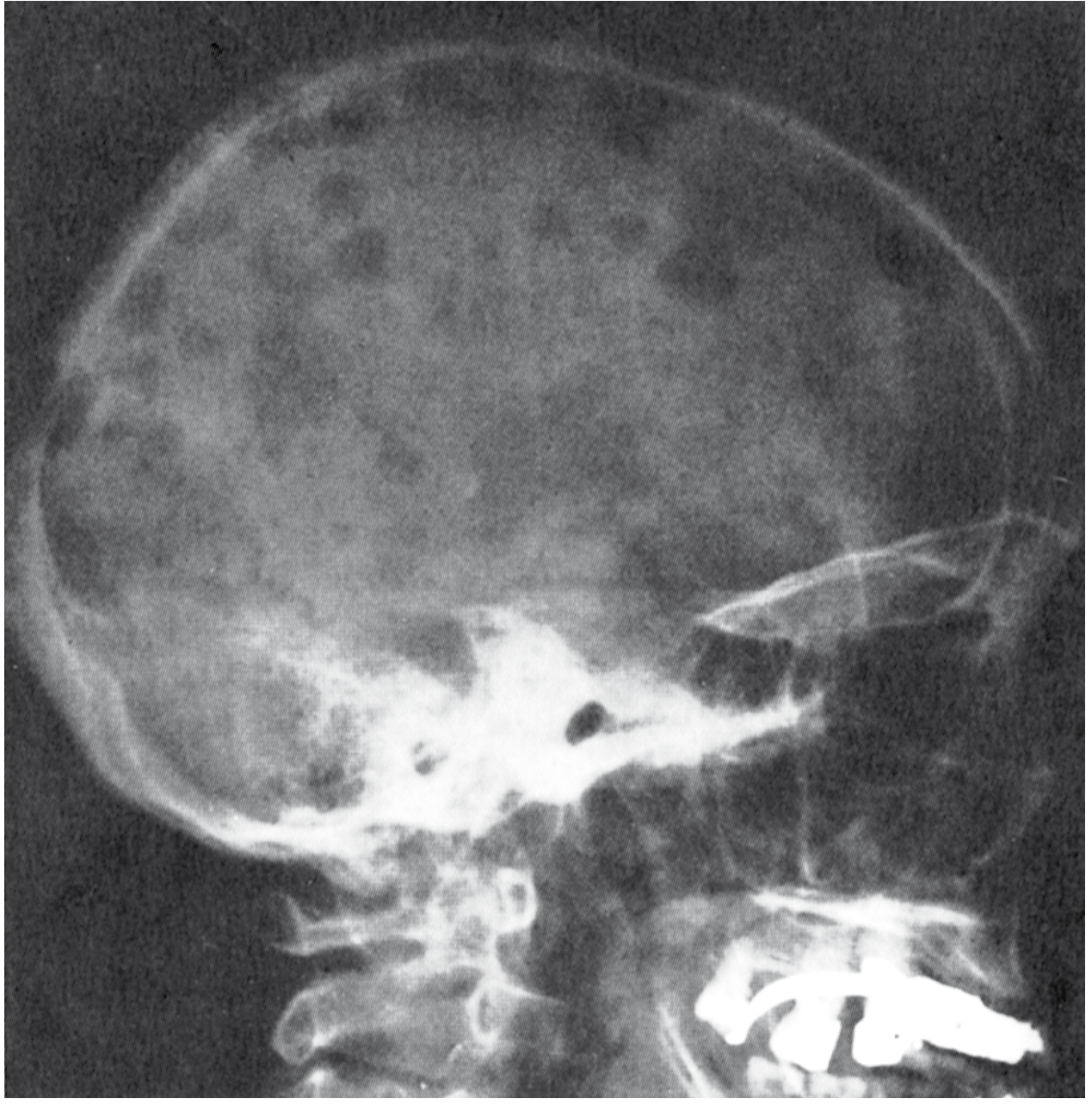
- "Punched-out" lytic defects, classically 1-4 cm, with no surrounding sclerosis (the absence of reactive new bone is characteristic)
- "Moth-eaten" appearance in long bones; calvaria lesions are highly characteristic
| Letter | Manifestation |
|---|---|
| C | Hypercalcemia (>11 mg/dL or >1 mg/dL above upper normal) |
| R | Renal insufficiency (creatinine >2 mg/dL) |
| A | Anemia (Hb <10 g/dL) |
| B | Bone lesions (lytic lesions on skeletal survey/CT/PET) |
2. Adult T-Cell Leukemia/Lymphoma (ATLL) - HTLV-1 Associated
- Driven by HTLV-1 (most prevalent in Japan and the Caribbean basin)
- Patients present with circulating disease, bone marrow involvement, hypercalcemia, lytic bone lesions, lymphadenopathy, hepatosplenomegaly, skin lesions, and opportunistic infections
- Pathognomonic: "flower cell" (CD4+, CD25+, CD2+, CD3+, CD5+, CD7-)
- 4 variants: acute (worst, median survival 6 months), lymphomatous, chronic, smoldering
- (Harrison's Principles of Internal Medicine 22E, 2025)
3. Non-Hodgkin Lymphoma (NHL)
| Mechanism | Details |
|---|---|
| Local osteolysis | Direct lytic bone involvement - all types of NHL can cause this |
| Ectopic 1,25(OH)₂D production | Malignant lymphocytes (especially in Hodgkin's and some NHL subtypes) convert 25(OH)D to active calcitriol via 1α-hydroxylase - this is PTHrP-independent |
4. Leukemias (less common, but recognized)
- AML (acute myeloid leukemia) - rare but reported
- CLL - uncommon, but B-cell infiltration of bone can produce lytic change
- Mechanism involves locally produced osteoclast-activating cytokines including PTHrP, IL-1, IL-6, and IL-8
- (Brenner & Rector's The Kidney - Malignancy section)
Summary Table: Hematological Differentials
| Condition | Lytic Lesions | Hypercalcemia | Key Mechanism | Distinguishing Feature |
|---|---|---|---|---|
| Multiple Myeloma | +++ | +++ | Local osteolysis (RANKL↑, OPG↓) | CRAB criteria, M-protein, BJ protein, punched-out lesions |
| ATLL (HTLV-1) | ++ | +++ | PTHrP + osteoclast activation | Flower cells, HTLV-1 serology, CD4+/CD25+/CD7- |
| NHL | ++ | ++ | Local osteolysis OR ectopic 1,25(OH)₂D | Elevated 1,25(OH)₂D (calcitriol), PTH suppressed |
| Hodgkin Lymphoma | + | + | Ectopic 1,25(OH)₂D | Reed-Sternberg cells, calcitriol-mediated |
| Leukemia (AML/CLL) | + | + | Osteoclast-activating cytokines | Peripheral blasts, bone marrow infiltration |
| Plasma cell leukemia | ++ | ++ | Same as myeloma | >20% circulating plasma cells, aggressive |
Key Differentiating Lab Workup
- SPEP/UPEP + immunofixation - M-protein = myeloma/plasmacytoma
- Serum free light chains - kappa/lambda ratio
- PTH - suppressed in all malignancy-related hypercalcemia
- PTHrP - elevated in ATLL, some NHL; low in myeloma
- 1,25(OH)₂D (calcitriol) - elevated in lymphoma-related hypercalcemia
- HTLV-1 serology - ATLL
- Bone marrow biopsy - plasma cell % (myeloma), T-cell immunophenotype (ATLL)
- Skeletal survey / Whole body low-dose CT - distribution and morphology of lytic lesions
- CRAB = the four cardinal myeloma manifestations (Calcium, Renal, Anemia, Bone)
- The absence of bone scan uptake (cold lesions) in myeloma vs. hot lesions in metastatic carcinoma is a classic radiological differentiator
Tell me more differentials and perfect like hematologist minimum 5
Lytic Lesions + Hypercalcemia in Hematological Conditions: Complete Differential (Hematologist Level)
TIER 1 - Plasma Cell Dyscrasias (Most Likely)
1. Multiple Myeloma
- Bone scan is characteristically cold (purely lytic, no reactive new bone = no osteoblast activity = no uptake) - use skeletal survey or PET/CT/whole-body low-dose CT instead
- RANKL↑ / OPG↓ ratio is the core pathophysiologic driver
- IgG (52%) > IgA (21%) > light chain only (16%) > IgD (2%) > IgE (rare) - IgD and IgE myelomas have disproportionately high rates of plasma cell leukemia transformation
- Bones affected descending: vertebrae > ribs > skull > pelvis > femur > clavicle > scapula
- Robbins, Cotran & Kumar; Harrison's 22E (2025)
2. Solitary Plasmacytoma of Bone
| Feature | Detail |
|---|---|
| Definition | Single bony lytic mass of clonal plasma cells, no evidence of systemic myeloma |
| Bone marrow | <10% plasma cells at non-affected sites |
| M-protein | May be present, but without suppression of normal immunoglobulins |
| CRAB | Absent by definition (if CRAB present → reclassify as myeloma) |
| Hypercalcemia | Can occur, but far less common than in myeloma |
| Treatment | Radiation therapy (curative intent) - local control achieved in most |
| Key caveat | Two-thirds eventually progress to overt myeloma - lifelong surveillance is mandatory |
3. Plasma Cell Leukemia (PCL)
- Diagnostic threshold: >5% plasma cells in peripheral blood differential and/or absolute plasma cell count ≥500/µL
- Primary PCL (60%): de novo presentation in leukemic phase - younger patients, more hepatosplenomegaly, lymphadenopathy, higher platelet count, fewer bone lesions, smaller M-protein, but better survival than secondary PCL
- Secondary PCL (40%): leukemic transformation of known myeloma - older patients, more bone disease, rapidly fatal
- Immunophenotype: unlike typical myeloma, CD56 is often absent in PCL
- IgD (12%) and IgE (25%) myelomas are over-represented among PCL transformations
- Hypercalcemia present due to same RANKL/osteoclast mechanism
- Treatment: aggressive induction (VDT-PACE or daratumumab-VRd) → ASCT if response achieved
- Goldman-Cecil Medicine; Harrison's 22E; Robbins Cotran
4. Nonsecretory Myeloma
- 3% of myeloma cases - no detectable M-protein in serum or urine by SPEP/UPEP/immunofixation
- Bone lesions and hypercalcemia are present and identical to secretory myeloma
- Diagnosis relies on bone marrow biopsy with immunoperoxidase/flow cytometry confirming clonal plasma cells
- Serum free light chain (sFLC) assay is abnormal in >60% - this is the key monitoring tool
- Pitfall: can be missed if sFLC not ordered; the clinician must have high suspicion when CRAB features present but standard protein studies are negative
- Goldman-Cecil Medicine
5. POEMS Syndrome (Osteosclerotic Myeloma)
| Component | Detail |
|---|---|
| P | Polyneuropathy (chronic inflammatory demyelinating - predominantly motor) |
| O | Organomegaly (liver, spleen, lymph nodes) |
| E | Endocrinopathy (hypogonadism, hypothyroidism, adrenal insufficiency, diabetes) |
| M | M-protein (almost always λ light chain) |
| S | Skin changes (hyperpigmentation, hypertrichosis, hemangiomas, clubbing) |
- Hypercalcemia and renal insufficiency are rare in POEMS (key differentiator from myeloma)
- Bone marrow usually has <5% plasma cells
- The diagnosis is confirmed by biopsy of a sclerotic lesion showing monoclonal plasma cells
- If lesions are limited: radiation therapy (>50% neuropathy improvement)
- If widespread: ASCT or systemic therapy
- Goldman-Cecil Medicine
TIER 2 - Lymphoid Malignancies
6. Adult T-Cell Leukemia/Lymphoma (ATLL) - HTLV-1 Driven
- The hematologist must ask about origin: Japan, Caribbean basin, West Africa, Middle East - HTLV-1 endemic regions
- Hypercalcemia in ATLL is mediated by PTHrP secreted by malignant T-cells, often dramatically elevated, can reach severe levels (>14 mg/dL)
- Lytic lesions + hypercalcemia + flower cells on smear = pathognomonic triad
- CD4+, CD25+, CD2+, CD3+, CD5+, CD7- (the CD7 loss is a key negative)
- FOXP3+ (resembles regulatory T-cells - contributes to profound immunosuppression)
- Skin involvement: erythroderma, nodules, papules (not just rash - actual leukemic skin infiltration)
- Opportunistic infections mirror AIDS because CD4 cells are dysfunctional despite being numerous
- Acute form: survival 6 months; treatment with zidovudine + interferon-α ± arsenic, mogamulizumab (anti-CCR4)
- Harrison's Principles of Internal Medicine 22E (2025)
7. Non-Hodgkin Lymphoma (NHL) with Bone Involvement
- Direct bone marrow infiltration → cytokine-mediated osteoclast activation (PTHrP, IL-1, IL-6, IL-8)
- Lytic lesions on imaging; PTH suppressed, PTHrP may be elevated
- Less commonly produces discrete punched-out defects (more permeative/infiltrative pattern)
- Malignant lymphocytes express 1α-hydroxylase (same enzyme as granulomatous macrophages)
- Converts 25(OH)D → active 1,25(OH)₂D without PTH regulation
- Lab pattern: PTH suppressed ↓, PTHrP normal, serum 1,25(OH)₂D elevated ↑
- This is the most important distinguishing lab from myeloma (where calcitriol is NOT elevated)
- Can occur even without direct bone involvement
- All types of lymphoma can cause this syndrome
- (Brenner & Rector's The Kidney; Frameworks for Internal Medicine)
8. Hodgkin Lymphoma (HL)
- Lytic bone lesions and hypercalcemia occur but are less common than in NHL/myeloma
- Mechanism primarily ectopic 1,25(OH)₂D production (same as NHL above)
- Bone involvement in HL is usually part of stage IV disease
- Lytic lesions in HL can cause cord compression, pathologic fractures
- Key clinical point: HL is one of the few metastatic bone diseases that is potentially curable even with bone involvement - must not be mistaken for myeloma and written off as palliative
- (Harrison's 22E; Goldman-Cecil)
TIER 3 - Histiocytic Neoplasms
9. Langerhans Cell Histiocytosis (LCH)
| Disease Spectrum | Features |
|---|---|
| Eosinophilic granuloma (unifocal) | Single lytic bone lesion, usually asymptomatic |
| Hand-Schüller-Christian (multifocal unisystem) | Classic triad: exophthalmos + diabetes insipidus + skull lytic lesions; 15-40% of cases |
| Letterer-Siwe (multisystem) | Fulminant multiorgan disease; skin, bone, marrow, liver, spleen; often fatal without chemo |
| Multisystem with risk-organ involvement | Worst prognosis; liver/spleen/bone marrow ("risk organs") determine prognosis |
- "Punched-out" lytic lesions of the skull - can resemble myeloma radiologically
- CT shows sharp, well-defined lytic defect with beveled edges (geographic lysis)
- Preferential involvement of the calvaria, mandible, and temporal bone (causes aural polyp, conductive hearing loss, postauricular swelling)
- Hypercalcemia occurs but is less prominent than in myeloma
- Key differentiator: age (children/young adults), Birbeck granules on EM, CD1a+ / CD207 (Langerin)+ / S100+ on IHC
- BRAF V600E mutation - has therapeutic implications (vemurafenib responsive)
- (Goldman-Cecil Medicine; Robbins & Kumar Basic Pathology; Grainger & Allison's Diagnostic Radiology)
TIER 4 - Rare/Advanced Differentials (Complete the Hematologist's List)
10. Erdheim-Chester Disease (ECD)
- Non-Langerhans cell histiocytosis (CD68+, CD163+, CD1a-, S100-)
- BRAF V600E in ~54% of cases
- Bilateral and symmetrical long bone involvement (femora, tibiae, humeri) - the PET-CT pattern is distinctive (bilateral periosteal uptake)
- Lytic lesions less common than sclerotic lesions, but mixed patterns occur
- Hypercalcemia can occur
- Retroperitoneal fibrosis ("hairy kidney"), periaortic cuffing, diabetes insipidus, xanthelasma are hallmarks
- (Goldman-Cecil Medicine - described as a differential to LCH in the same section)
11. Hemophagocytic Lymphohistiocytosis (HLH) secondary to hematological malignancy
- Bone marrow infiltration by underlying lymphoma/leukemia triggers the syndrome
- Hypercalcemia via direct bony involvement + cytokine storm (IL-6, TNF-α)
- Context: ferritin >10,000, fever, splenomegaly, cytopenias, hypofibrinogenemia, elevated sCD25
Master Comparison Table (Hematologist Reference)
| Condition | Lytic Lesion | Hypercalcemia | Key Mechanism | Unique Lab/Feature | Bone Pattern |
|---|---|---|---|---|---|
| Multiple Myeloma | +++ | ++ | RANKL↑/OPG↓, MIP-1α | M-protein, BJ protein, CD138+ | Punched-out, NO sclerosis |
| Solitary Plasmacytoma | + (single) | +/- | Same as myeloma | Single lesion; 2/3 → myeloma | Single punched-out |
| Plasma Cell Leukemia | ++ | ++ | Same as myeloma | >5% plasma cells in blood, CD56- | Fewer lesions than MM |
| Nonsecretory Myeloma | +++ | ++ | Same as myeloma | Normal SPEP, sFLC assay key | Same as MM |
| POEMS | + (sclerotic) | Rare | - | λ-chain, neuropathy, VEGF↑ | Sclerotic not lytic |
| ATLL | ++ | +++ | PTHrP from T-cells | HTLV-1+, flower cells, CD4+/CD7- | Diffuse infiltrative |
| NHL | ++ | ++ | Osteolysis OR ectopic 1,25(OH)₂D | 1,25(OH)₂D elevated (not in MM) | Permeative |
| Hodgkin Lymphoma | + | + | Ectopic 1,25(OH)₂D | Reed-Sternberg, calcitriol↑ | Stage IV, variable |
| LCH | ++ | + | Cytokine-mediated osteolysis | CD1a+, Langerin+, BRAF V600E | Beveled-edge, geographic |
| ECD | +/- | +/- | Histiocyte infiltration | CD68+, CD1a-, bilateral long bone | Sclerotic > lytic |
Diagnostic Algorithm - Hematologist Approach
Lytic lesion(s) + Hypercalcemia
│
├── Check PTH → Suppressed in ALL malignancy-related causes
│
├── SPEP/UPEP + Immunofixation + sFLC ratio
│ ├── M-protein present → Plasma cell dyscrasia spectrum
│ │ ├── Multiple lesions + CRAB → MULTIPLE MYELOMA
│ │ ├── Single lesion, no systemic disease → SOLITARY PLASMACYTOMA
│ │ ├── >5% plasma cells in blood → PLASMA CELL LEUKEMIA
│ │ ├── No M-protein on SPEP but sFLC abnormal → NONSECRETORY MYELOMA
│ │ └── Sclerotic lesions + neuropathy + λ-chain → POEMS
│ │
│ └── No M-protein → Consider lymphoid or histiocytic cause
│ ├── Check PTHrP elevated? → ATLL, some NHL
│ ├── Check 1,25(OH)₂D elevated? → NHL, Hodgkin lymphoma
│ ├── HTLV-1 serology positive + flower cells → ATLL
│ ├── Lymph node biopsy / PET-CT → NHL / Hodgkin
│ └── Young patient + skull/temporal lytic lesions → LCH (CD1a, BRAF)
│
└── Bone marrow biopsy in all cases
Key Calcium Mechanism Summary for Exams & Boards
| Mechanism | Conditions | Lab Signature |
|---|---|---|
| Local osteolysis (RANKL/cytokines) | MM, PCL, ATLL, NHL, LCH | PTH↓, PTHrP normal, 1,25(OH)₂D normal |
| PTHrP-mediated osteolysis | ATLL, some NHL | PTH↓, PTHrP↑, 1,25(OH)₂D normal |
| Ectopic 1,25(OH)₂D | Hodgkin + NHL | PTH↓, PTHrP normal, 1,25(OH)₂D↑ |
If patient becomes allergic to l aspariginase then how to proceed
"asparaginase" AND "hypersensitivity" AND "ALL" AND "management"
"asparaginase" AND "hypersensitivity" AND "Erwinia"
PMID: 35107320
PMID: 36795447
L-Asparaginase Allergy: How to Proceed
Step 1 - Classify the Reaction First
| Reaction Type | Mechanism | Clinical Features | Action |
|---|---|---|---|
| Overt clinical hypersensitivity | IgE/IgG-mediated immune response to foreign bacterial protein | Urticaria, angioedema, bronchospasm, anaphylaxis during/after infusion | Stop current formulation → switch |
| Silent inactivation (subclinical) | Antibody-mediated enzyme inactivation WITHOUT clinical symptoms | No allergic signs, but asparaginase activity level undetectable | Clinically misleading - treatment failing silently |
| Grade 1-2 infusion reaction | Mild cytokine release or local reaction | Flushing, mild urticaria only | May attempt premedication first (see below), then reassess |
Key Pearl: Silent inactivation is more common than overt hypersensitivity and is associated with a negative clinical outcome in high-risk ALL because the enzyme appears to be given but is actually not working. Measuring serum asparaginase activity (SAA) is the only way to detect this.
Step 2 - Immediate Management of the Acute Reaction
- Stop the infusion immediately
- Administer:
- Epinephrine (for anaphylaxis: 0.01 mg/kg IM, max 0.5 mg)
- IV antihistamines (diphenhydramine)
- IV corticosteroids (hydrocortisone or methylprednisolone)
- IV fluids; oxygen; bronchodilators as needed
- Observe for minimum 1-2 hours post-reaction
- Document the reaction grade per CTCAE criteria (Grade 1-4)
- Check serum asparaginase activity level - if reaction occurred after a previous dose, retroactively check if prior doses had adequate activity
Step 3 - The Formulation Switch Strategy
The Three Formulations
| Formulation | Source | t½ | Dosing | Role |
|---|---|---|---|---|
| Native E. coli L-asparaginase (Elspar) | Escherichia coli | ~1 day | 6,000-10,000 IU every 3 days | Historical first-line (now rarely used as monotherapy) |
| Pegaspargase (PEG-asparaginase) (Oncaspar) | E. coli + PEG conjugate | 6-7 days | IM/IV every 14 days | Current first-line standard for ALL |
| Calaspargase pegol (Asparlas) | E. coli + different PEG linkage | >14 days | IV every 21 days | Newer, longer-acting; approved for ALL |
| Asparaginase Erwinia chrysanthemi (Erwinaze / JZP458/crisantaspase) | Erwinia chrysanthemi | ~16 hours (very short) | IM 3x/week or IV | For patients allergic to E. coli-derived formulations |
The Decision Tree
Patient develops hypersensitivity or silent inactivation to:
│
├── NATIVE E. COLI asparaginase
│ └── Switch to PEGASPARGASE (PEG-asparaginase)
│ (PEG coating reduces immunogenicity: <20% develop antibodies)
│
├── PEGASPARGASE (first-line) → allergic reaction
│ └── Switch to ERWINIA asparaginase (Erwinaze / JZP458)
│ (Completely different bacterial source = no cross-reactivity)
│ - Dose: 25,000 IU/m² IM 3× per week (Mon/Wed/Fri)
│ OR 25,000 IU/m² IV over 1 hour 3× per week
│ - Activity window is SHORT (t½ ~16h), hence the 3×/week dosing
│
├── ERWINIA asparaginase → shortage/unavailable
│ └── JZP458 (recombinant Erwinia-derived, produced in Pseudomonas)
│ - Approved 2021; closes the "asparaginase allergy gap"
│ - Produced recombinantly, not dependent on bacterial fermentation →
│ addresses supply shortage problem
│ [Blood, 2023 - PMID 36795447]
│
└── Allergic to ALL formulations (very rare)
└── Asparaginase discontinuation as LAST RESORT
- Intensify other components of regimen (consult protocol-specific guidance)
- Do NOT simply omit without expert hematology/oncology discussion
Step 4 - Why Cross-Reactivity Does NOT Occur Between Species
- Pegaspargase and native E. coli asparaginase: Both derived from E. coli - share the same protein epitopes. Antibodies raised against one will cross-react with the other. If pegaspargase causes hypersensitivity, switching to native E. coli asparaginase is not an option - both will be inactivated.
- Erwinia chrysanthemi asparaginase: Derived from a completely different organism with different protein structure and epitopes. Anti-E. coli asparaginase antibodies do not bind Erwinia enzyme. This is why the switch works.
Step 5 - Key Additional Considerations
Premedication Before Subsequent Doses
- Antihistamines (cetirizine or diphenhydramine)
- Acetaminophen
- Low-dose corticosteroids
Therapeutic Drug Monitoring (TDM) - Asparaginase Activity Levels
- Target serum asparaginase activity (SAA): ≥0.1 IU/mL at nadir (trough)
- Check SAA ~72 hours after E. coli preparations, ~7 days after pegaspargase
- If SAA <0.1 IU/mL (even without clinical reaction) = silent inactivation = switch formulation immediately
- (Future Oncology, Burke & Zalewska-Szewczyk, 2022 - PMID 35107320)
Managing Other Asparaginase Toxicities (Not Allergy)
| Toxicity | Mechanism | Management |
|---|---|---|
| Pancreatitis | Direct pancreatic damage | Hold asparaginase; if severe/necrotizing, may need permanent discontinuation; amylase/lipase monitoring |
| Coagulopathy/Thrombosis | ↓ antithrombin III, fibrinogen, protein C/S | Monitor coag profile; consider AT-III replacement; LMWH for thrombosis |
| Hemorrhage | ↓ clotting factors (fibrinogen, factors V, VII, VIII, X) | FFP if active bleeding; cryoprecipitate for fibrinogen <1 g/L |
| Hyperglycemia | ↓ insulin synthesis (protein synthesis inhibition) | Insulin sliding scale; monitor glucose closely |
| Hepatotoxicity | Protein synthesis inhibition → ↑ transaminases | Monitor LFTs; usually reversible |
| Hypoalbuminemia | ↓ albumin synthesis | Monitor albumin; affects pharmacokinetics of other drugs |
| Hyperammonemia | Ammonia release from asparagine hydrolysis | Rifaximin; lactulose; dietary protein restriction |
| Intracranial thrombosis/hemorrhage | AT-III ↓ + coagulopathy | Urgent MRI brain; anticoagulation for thrombosis; neurosurgery consult |
Summary Algorithm
L-Asparaginase allergy/silent inactivation confirmed
│
▼
Measure SAA → if <0.1 IU/mL = inactivation confirmed
│
▼
Identify source: E. coli-derived (native or PEG)?
│ │
Yes: SWITCH to Already on Erwinia?
ERWINIA asparaginase │
│ Yes: Use JZP458 (recombinant Erwinia)
▼ or asparaginase discontinuation
Continue ALL protocol as absolute last resort
with Erwinia dosing
(25,000 IU/m² 3×/week IM or IV)
│
▼
Monitor SAA 72h post-dose → target ≥0.1 IU/mL
Continue TDM throughout course
Should i give peg asparagonase next time then
The Core Principle: E. coli Cross-Reactivity
The Answer Depends on the Scenario
Scenario A: Patient reacted to native E. coli L-asparaginase
- The PEG conjugation significantly reduces immunogenicity
- Less than 20% of patients develop antibodies to pegaspargase vs. much higher rates with native E. coli form
- Goodman & Gilman explicitly states: "In hypersensitive patients [to native E. coli], pegaspargase is a safe and effective alternative"
- This switch is well-supported and widely practiced
Scenario B: Patient reacted to pegaspargase itself
- The antibodies raised against pegaspargase will still recognize and neutralize any E. coli-derived asparaginase, including calaspargase pegol
- Giving pegaspargase again risks anaphylaxis or guaranteed silent inactivation
- The only correct move here is Erwinia asparaginase (Erwinaze / JZP458/crisantaspase) - completely different bacterial source, no cross-reactivity
Decision Table
| Patient reacted to | Can you give Pegaspargase? | Correct next step |
|---|---|---|
| Native E. coli asparaginase | YES | Pegaspargase |
| Pegaspargase | NO - contraindicated | Erwinia asparaginase |
| Calaspargase pegol | NO | Erwinia asparaginase |
| Erwinia asparaginase | N/A | JZP458 (recombinant Erwinia) |
Why This Distinction Matters Clinically
- The patient was started on native E. coli asparaginase (older protocol, resource-limited setting)
- The allergy occurred during the older native formulation phase before PEG was introduced
One More Critical Point: Silent Inactivation
Treaatment of secondary itp dure to sle
Treatment of Secondary ITP due to SLE
Step 1 - Confirm It IS SLE-ITP (Rule Out Mimics)
| Mimic | Mechanism | Key Findings | Treatment difference |
|---|---|---|---|
| TMA / TTP | ADAMTS13 deficiency / microvascular thrombosis | Schistocytes on smear, elevated LDH, low ADAMTS13 | Plasma exchange - NOT steroids alone |
| Catastrophic APS (CAPS) | Antiphospholipid antibodies → multi-organ micro-thrombosis | Triple aPL positivity, multi-organ involvement | Anticoagulation + steroids + IVIG ± plasma exchange |
| APS-associated thrombocytopenia | Platelet-directed antiphospholipid antibodies (mild, rarely <50k) | LA/anti-β2GPI positive, history of thrombosis | Hydroxychloroquine, not aggressive IS |
| Drug-induced (e.g., hydroxychloroquine, NSAIDs) | Direct drug effect | Timing with drug introduction | Withdraw offending drug |
| Bone marrow suppression | Azathioprine, MMF toxicity | Pancytopenia, check drug levels | Dose reduction |
Step 2 - When to Treat
| Platelet Count | Clinical Status | Action |
|---|---|---|
| >50,000/mm³ | No bleeding | Monitor; optimize background SLE therapy (HCQ) |
| 20,000-50,000/mm³ | No active bleeding | Treatment generally warranted |
| <20,000-30,000/mm³ | ± bleeding | Treatment required |
| Any count | Active significant bleeding | Emergency treatment |
The Management Algorithm
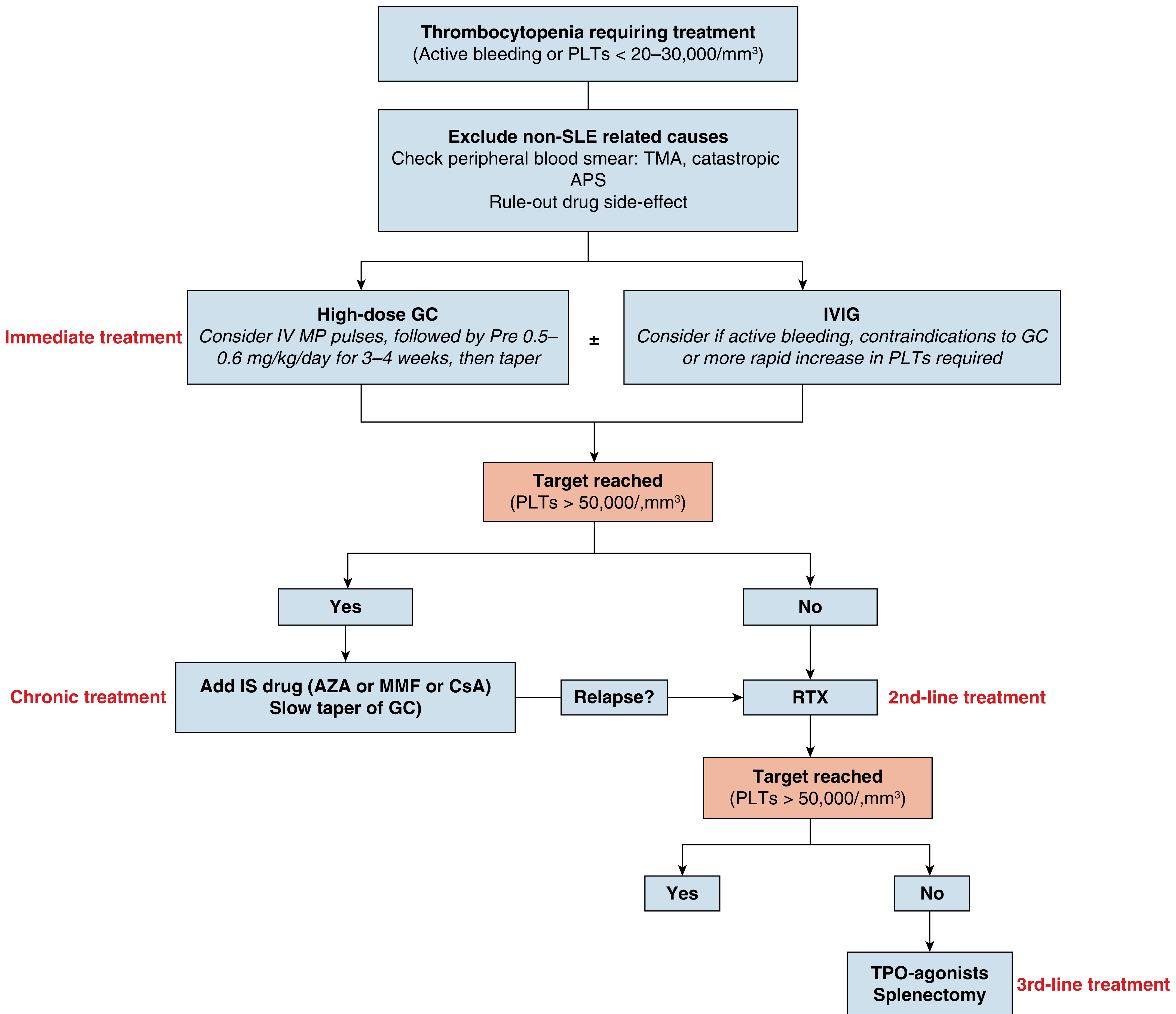
Structured Treatment by Line
FIRST-LINE: Glucocorticoids ± IVIG
| Regimen | Dose | Use |
|---|---|---|
| IV methylprednisolone pulse | 1 g/day × 3 days | Severe/life-threatening thrombocytopenia |
| Oral prednisolone | 0.5-0.6 mg/kg/day for 3-4 weeks, then taper | Standard first-line |
| High-dose dexamethasone | 40 mg/day × 4 days (pulse) | Preferred by some experts, borrowing from primary ITP data; good for rapid response |
- Active bleeding risk or ongoing significant hemorrhage
- Pre-surgical/procedural coverage (platelet count needs rapid rise)
- Contraindication to glucocorticoids (severe infection, uncontrolled diabetes)
- More rapid platelet increase required
- Dose: 1 g/kg/day × 1-2 days (or 0.4 g/kg/day × 5 days)
CHRONIC/MAINTENANCE: Steroid-Sparing Immunosuppressants
| Drug | Dose | Notes |
|---|---|---|
| Azathioprine (AZA) | 1-3 mg/kg/day | First choice steroid-sparing agent; also treats other SLE manifestations; check TPMT before starting |
| Mycophenolate mofetil (MMF) | 1.5-3 g/day | Alternative to AZA; evidence from MMF-for-primary-ITP trial (NEJM 2021); also useful if concurrent nephritis |
| Cyclosporine A (CsA) | 2-5 mg/kg/day | Calcineurin inhibitor; useful in refractory cases; monitor BP and renal function |
| Hydroxychloroquine (HCQ) | 5 mg/kg/day (max 400 mg/day) | Should be used in ALL SLE patients as background therapy unless contraindicated; helps stabilize disease activity including thrombocytopenia |
Critical note: HCQ is not a primary ITP treatment, but it reduces overall SLE disease activity, prevents flares, and reduces the risk of progression. It should be the background agent in ALL SLE-ITP patients.
SECOND-LINE: For Non-Response or Relapse After First-Line
- Chimeric anti-CD20 monoclonal antibody, depletes B-cells
- Dose: 375 mg/m² weekly × 4 doses (lymphoma protocol) OR 1000 mg × 2 doses 2 weeks apart (RA protocol)
- Response rate: 65-73% in SLE-related thrombocytopenia at 12 months
- Effective both for thrombocytopenia specifically and for overall SLE hematological domain
- Should be considered even before cyclophosphamide in most cases due to better safety profile
- Harrison's 22E (2025): "Anti-CD20 agents such as rituximab are used for lupus-related severe thrombocytopenia"
- IV pulse: 500-1000 mg/m² monthly
- Reserve due to toxicity (gonadotoxicity, hemorrhagic cystitis, malignancy risk)
- Third-line in some algorithms
- Addresses underlying SLE pathogenesis by reducing B-cell survival
- Growing evidence in SLE-related cytopenia; formally third-line
THIRD-LINE / REFRACTORY: TPO Receptor Agonists and Splenectomy
| Drug | Route | Dose |
|---|---|---|
| Eltrombopag | Oral | 25-75 mg/day |
| Romiplostim | SC | 1-10 mcg/kg/week |
| Avatrombopag | Oral | 20-40 mg/day |
- Response rate in ITP: 70-90% (primary ITP data); SLE-specific data from case reports and small series
- Currently considered experimental therapy in SLE-ITP per Rheumatology textbook (2022)
- Caution: theoretical concern about thrombosis risk in APS-positive patients (TPO-RAs can increase platelet count in a prothrombotic milieu)
- Dose: 200-1200 mg/day
- Historically used for refractory cases; also effective in Evans syndrome and AIHA
- Relatively well tolerated; does not increase infection risk
- Now less used due to availability of TPO-RAs and biologic options
- Not routinely recommended in SLE-ITP
- Concerns: less durable therapeutic effect than in primary ITP, high infection risk (SLE patients already immunocompromised), risk of thrombosis post-splenectomy especially if aPL-positive
- Reserve as absolute last resort if all medical management fails
- Pre-splenectomy vaccinations (pneumococcal, meningococcal, Hib) mandatory
Structured Treatment Table (Rheumatology 2022 - Table 142.3)
| Line | Therapies |
|---|---|
| First-line | Glucocorticoids (IV MP pulse or oral prednisolone 0.5-0.6 mg/kg/day) |
| Second-line | Azathioprine, IVIG, MMF, Cyclosporine A |
| Third-line | Rituximab, Cyclophosphamide, Belimumab |
| Experimental | TPO receptor agonists (eltrombopag, romiplostim, avatrombopag) |
| Last resort | Splenectomy |
Special Clinical Scenarios
APS-Associated Thrombocytopenia within SLE
- Usually mild (rarely drops below 50,000)
- Does not typically require aggressive immunosuppression
- Hydroxychloroquine is particularly important - reduces thrombotic risk
- Anticoagulation is indicated for thrombotic events, but is not the treatment for thrombocytopenia itself
- Avoid aggressive platelet transfusion as it can fuel thrombosis
Evans Syndrome (SLE + ITP + AIHA)
- Combination of autoimmune hemolytic anemia AND thrombocytopenia
- More aggressive - treat both cytopenias simultaneously
- Rituximab particularly useful; danazol reported effective
- High-dose steroids + IVIG as bridge
Pregnancy-Associated SLE-ITP
- Avoid MMF (teratogenic - Category D/X)
- Safe options: prednisolone, AZA (low dose), HCQ, IVIG
- Rituximab: evidence of fetal B-cell depletion, avoid in pregnancy
- IVIG recommended pre-delivery if PLT <50,000 or planned cesarean/epidural
Key Monitoring Parameters
| Parameter | Frequency | Target |
|---|---|---|
| CBC with differential | Weekly until stable, then monthly | PLT >50,000 (safe target) |
| Complement (C3/C4), anti-dsDNA | Every 3 months | Disease activity monitoring |
| Renal function (creatinine, urine protein:creatinine) | Monthly on IS drugs | Rule out nephritis flare |
| LFTs | Monthly on AZA/MMF/danazol | Hepatotoxicity screening |
| TPMT genotype | Before AZA initiation | Prevent myelotoxicity |
| HCQ eye exam | Annual after 5 years | Maculopathy screening |
Summary Flow
SLE + PLT <20-30,000 or active bleeding
↓
Rule out TMA, CAPS, drug effect (smear!)
↓
FIRST LINE: IV MP pulse → oral prednisolone 0.5-0.6 mg/kg ± IVIG
(Add HCQ as background in ALL patients)
↓
PLT >50,000? → YES: Add AZA/MMF, taper steroids slowly
→ NO: Move to second line
↓
SECOND LINE: Rituximab (best evidence) ± CYC
↓
Still failing? → Belimumab / Danazol / TPO-RAs
↓
All medical therapy failed → Splenectomy (last resort)
Generate a patient education handout on SLE-ITP management doses of dexona 40 mg has been given
ls /home/daytona/workspace/ && ls /home/daytona/skills/docx/
mkdir -p /home/daytona/workspace/sle-itp-handout
~/sle-itp-handout/create_handout.js
const {
Document, Packer, Paragraph, TextRun, Table, TableRow, TableCell,
AlignmentType, HeadingLevel, BorderStyle, WidthType, ShadingType,
VerticalAlign, PageNumber, Header, Footer, UnderlineType,
TableOfContents
} = require('docx');
const fs = require('fs');
// Color palette
const DARK_PURPLE = "4A235A";
const LIGHT_PURPLE = "E8D5F0";
const TEAL = "1A7A8A";
const LIGHT_TEAL = "D0EEF2";
const ORANGE = "D35400";
const LIGHT_ORANGE = "FAE0CC";
const LIGHT_GRAY = "F5F5F5";
const RED = "C0392B";
const GREEN = "1E8449";
const DARK_TEXT = "1A1A1A";
const WHITE = "FFFFFF";
function makeHeading(text, color = DARK_PURPLE) {
return new Paragraph({
children: [
new TextRun({
text,
bold: true,
size: 28,
color: WHITE,
font: "Calibri",
}),
],
shading: { type: ShadingType.SOLID, color: color, fill: color },
spacing: { before: 200, after: 100 },
indent: { left: 150, right: 150 },
});
}
function makeSubHeading(text, color = TEAL) {
return new Paragraph({
children: [
new TextRun({
text: `■ ${text}`,
bold: true,
size: 24,
color: color,
font: "Calibri",
}),
],
spacing: { before: 180, after: 60 },
});
}
function makeBullet(text, bold_part = "", rest = "") {
const children = [
new TextRun({ text: " • ", bold: true, size: 22, color: TEAL, font: "Calibri" }),
];
if (bold_part) {
children.push(new TextRun({ text: bold_part, bold: true, size: 22, color: DARK_TEXT, font: "Calibri" }));
}
if (rest) {
children.push(new TextRun({ text: rest, size: 22, color: DARK_TEXT, font: "Calibri" }));
} else if (!bold_part) {
children.push(new TextRun({ text, size: 22, color: DARK_TEXT, font: "Calibri" }));
}
return new Paragraph({ children, spacing: { before: 40, after: 40 } });
}
function makeWarningBox(text) {
return new Paragraph({
children: [
new TextRun({ text: "⚠ " + text, bold: true, size: 22, color: RED, font: "Calibri" }),
],
shading: { type: ShadingType.SOLID, color: LIGHT_ORANGE, fill: LIGHT_ORANGE },
spacing: { before: 100, after: 100 },
indent: { left: 150, right: 150 },
border: {
left: { style: BorderStyle.THICK, size: 12, color: ORANGE },
},
});
}
function makeGreenBox(text) {
return new Paragraph({
children: [
new TextRun({ text: "✓ " + text, bold: true, size: 22, color: GREEN, font: "Calibri" }),
],
shading: { type: ShadingType.SOLID, color: "D5F5E3", fill: "D5F5E3" },
spacing: { before: 80, after: 80 },
indent: { left: 150, right: 150 },
border: {
left: { style: BorderStyle.THICK, size: 12, color: GREEN },
},
});
}
function makeBodyText(text) {
return new Paragraph({
children: [new TextRun({ text, size: 22, color: DARK_TEXT, font: "Calibri" })],
spacing: { before: 40, after: 40 },
});
}
function makeSpacer() {
return new Paragraph({ children: [new TextRun({ text: "" })], spacing: { before: 60, after: 60 } });
}
function makeInfoTable(rows) {
return new Table({
width: { size: 100, type: WidthType.PERCENTAGE },
rows: rows.map(([label, value, highlight]) =>
new TableRow({
children: [
new TableCell({
children: [new Paragraph({
children: [new TextRun({ text: label, bold: true, size: 22, color: WHITE, font: "Calibri" })],
})],
shading: { type: ShadingType.SOLID, color: TEAL, fill: TEAL },
width: { size: 35, type: WidthType.PERCENTAGE },
margins: { top: 80, bottom: 80, left: 120, right: 120 },
}),
new TableCell({
children: [new Paragraph({
children: [new TextRun({
text: value,
size: 22,
bold: !!highlight,
color: highlight ? RED : DARK_TEXT,
font: "Calibri"
})],
})],
shading: { type: ShadingType.SOLID, color: LIGHT_TEAL, fill: LIGHT_TEAL },
width: { size: 65, type: WidthType.PERCENTAGE },
margins: { top: 80, bottom: 80, left: 120, right: 120 },
}),
],
})
),
margins: { top: 0, bottom: 0, left: 0, right: 0 },
});
}
function makeMedicineTable(medicines) {
const headerRow = new TableRow({
children: ["Medicine", "Dose / Schedule", "Purpose"].map(h =>
new TableCell({
children: [new Paragraph({
children: [new TextRun({ text: h, bold: true, size: 22, color: WHITE, font: "Calibri" })],
alignment: AlignmentType.CENTER,
})],
shading: { type: ShadingType.SOLID, color: DARK_PURPLE, fill: DARK_PURPLE },
margins: { top: 80, bottom: 80, left: 120, right: 120 },
})
),
});
const dataRows = medicines.map(([med, dose, purpose], i) =>
new TableRow({
children: [med, dose, purpose].map(val =>
new TableCell({
children: [new Paragraph({
children: [new TextRun({ text: val, size: 22, color: DARK_TEXT, font: "Calibri" })],
})],
shading: { type: ShadingType.SOLID, color: i % 2 === 0 ? LIGHT_PURPLE : WHITE, fill: i % 2 === 0 ? LIGHT_PURPLE : WHITE },
margins: { top: 60, bottom: 60, left: 120, right: 120 },
})
),
})
);
return new Table({
width: { size: 100, type: WidthType.PERCENTAGE },
rows: [headerRow, ...dataRows],
});
}
function makeDivider() {
return new Paragraph({
children: [new TextRun({ text: "" })],
border: { bottom: { style: BorderStyle.SINGLE, size: 6, color: LIGHT_PURPLE } },
spacing: { before: 120, after: 120 },
});
}
const titlePara = new Paragraph({
children: [
new TextRun({
text: "Patient Education Guide",
bold: true,
size: 40,
color: WHITE,
font: "Calibri",
}),
],
shading: { type: ShadingType.SOLID, color: DARK_PURPLE, fill: DARK_PURPLE },
alignment: AlignmentType.CENTER,
spacing: { before: 0, after: 0 },
indent: { left: 0, right: 0 },
});
const subtitlePara = new Paragraph({
children: [
new TextRun({
text: "SLE-Related Low Platelet Count (Immune Thrombocytopenia)",
bold: true,
size: 26,
color: WHITE,
font: "Calibri",
}),
],
shading: { type: ShadingType.SOLID, color: TEAL, fill: TEAL },
alignment: AlignmentType.CENTER,
spacing: { before: 0, after: 200 },
});
const doc = new Document({
sections: [{
properties: {
page: {
margin: { top: 720, bottom: 720, left: 900, right: 900 },
},
},
headers: {
default: new Header({
children: [
new Paragraph({
children: [
new TextRun({ text: "SLE-ITP Patient Education | Prepared by Your Care Team | Date: _______________", size: 18, color: "888888", font: "Calibri" }),
],
alignment: AlignmentType.RIGHT,
border: { bottom: { style: BorderStyle.SINGLE, size: 4, color: LIGHT_PURPLE } },
}),
],
}),
},
footers: {
default: new Footer({
children: [
new Paragraph({
children: [
new TextRun({ text: "This handout is for educational purposes only. Always follow your doctor's instructions. Questions? Contact your rheumatology/hematology team.", size: 16, color: "888888", italics: true, font: "Calibri" }),
],
alignment: AlignmentType.CENTER,
border: { top: { style: BorderStyle.SINGLE, size: 4, color: LIGHT_PURPLE } },
}),
],
}),
},
children: [
titlePara,
subtitlePara,
// ─── SECTION 1: What is SLE-ITP ───
makeHeading("1. What Is SLE-ITP?", DARK_PURPLE),
makeBodyText("You have been diagnosed with Immune Thrombocytopenia (ITP) related to your Systemic Lupus Erythematosus (SLE). This means your immune system — which usually protects you from infection — is mistakenly attacking your own platelets (the tiny blood cells that help stop bleeding)."),
makeSpacer(),
makeInfoTable([
["SLE", "A chronic autoimmune disease where your immune system attacks your own body"],
["ITP", "Immune Thrombocytopenia — low platelet count caused by your immune system"],
["Platelets", "Small blood cells that form clots and stop bleeding. Normal: 150,000–400,000/mm³"],
["Your Target", "Platelet count > 50,000/mm³ (safe level to prevent serious bleeding)"],
]),
makeSpacer(),
makeWarningBox("Low platelets can cause bruising, bleeding gums, prolonged bleeding from cuts, or rarely, dangerous internal bleeding. Report any unusual bleeding immediately."),
makeDivider(),
// ─── SECTION 2: Your Treatment ───
makeHeading("2. Your Treatment: What You Have Received", DARK_PURPLE),
makeBodyText("Your doctor has started treatment to raise your platelet count quickly and safely. Here is what has been given:"),
makeSpacer(),
makeSubHeading("Dexamethasone (Dexona) — High-Dose Pulse Therapy", TEAL),
makeInfoTable([
["Drug Name", "Dexamethasone (Dexona)"],
["Dose Given", "40 mg per day", true],
["Duration", "4 days (Day 1 to Day 4)"],
["How Given", "Intravenous (IV drip) or oral tablet — as prescribed by your doctor"],
["Why This Dose?", "This high-dose 'pulse' quickly calms your overactive immune system and signals your bone marrow to make more platelets"],
]),
makeSpacer(),
makeGreenBox("Dexamethasone 40 mg × 4 days is a well-proven, internationally recommended first-line treatment for SLE-ITP."),
makeSpacer(),
makeSubHeading("How Does It Work?", TEAL),
makeBullet("", "Suppresses immune attack: ", "Stops your immune system from destroying platelets"),
makeBullet("", "Reduces inflammation: ", "Lowers overall SLE disease activity"),
makeBullet("", "Stimulates platelet production: ", "Encourages your bone marrow to produce more platelets"),
makeSpacer(),
makeSubHeading("What To Expect During Treatment", TEAL),
makeBullet("Platelet count usually begins to rise within 3–7 days of treatment"),
makeBullet("You may feel better energy as inflammation reduces"),
makeBullet("Blood tests will be checked regularly to monitor your response"),
makeDivider(),
// ─── SECTION 3: Side Effects ───
makeHeading("3. Side Effects of Dexamethasone — What To Watch For", ORANGE),
makeBodyText("High-dose steroids are very effective but can cause side effects. Most are temporary and manageable. Know what to watch for:"),
makeSpacer(),
makeMedicineTable([
["High Blood Sugar", "Steroids raise glucose levels — especially if you are diabetic", "Check blood sugar daily; take prescribed diabetic medication"],
["Increased Appetite / Weight Gain", "Common with steroids — usually temporary", "Eat a balanced low-salt, low-sugar diet"],
["Difficulty Sleeping (Insomnia)", "High-dose dexamethasone can cause restlessness at night", "Take the dose in the morning if possible; tell your doctor"],
["Mood Changes", "You may feel anxious, irritable, or unusually energetic", "This is normal; it resolves after treatment ends"],
["Fluid Retention / Swelling", "Mild ankle swelling or puffy face", "Reduce salt intake; elevate legs when resting"],
["Stomach Upset / Acidity", "Steroids irritate the stomach lining", "Take with food; use prescribed antacid (e.g., omeprazole/pantoprazole)"],
["Increased Infection Risk", "Immune suppression makes infections more likely", "Avoid crowded places; report fever, chills, or cough immediately"],
["High Blood Pressure", "May rise temporarily", "Monitor BP at home if possible; report if >140/90"],
]),
makeSpacer(),
makeWarningBox("CALL YOUR DOCTOR IMMEDIATELY if you develop: Fever >38°C, Severe stomach pain, Chest pain, Sudden confusion, Signs of active bleeding (blood in urine/stool, coughing blood, heavy bruising)"),
makeDivider(),
// ─── SECTION 4: Medicines After Dexamethasone ───
makeHeading("4. Medicines You May Continue After the Pulse", DARK_PURPLE),
makeBodyText("After the 4-day dexamethasone pulse, your doctor will likely prescribe additional medicines to keep your platelet count stable and prevent relapse. These may include:"),
makeSpacer(),
makeMedicineTable([
["Hydroxychloroquine (HCQ / Plaquenil)", "200–400 mg once daily (long-term)", "Controls SLE disease activity; reduces flares; ALL SLE patients should take this unless contraindicated"],
["Prednisolone (oral steroid taper)", "As prescribed — usually starting at 0.5–0.6 mg/kg/day, then slowly tapered", "Maintains remission after the dexamethasone pulse; dose is gradually reduced"],
["Azathioprine (AZA)", "Dose as prescribed (usually 1–2 mg/kg/day)", "Steroid-sparing drug; helps reduce the amount of steroid needed long-term"],
["Mycophenolate Mofetil (MMF)", "Dose as prescribed", "Alternative to AZA; especially useful if you also have kidney involvement"],
["Omeprazole/Pantoprazole", "20–40 mg once daily", "Stomach protection — must be taken while on steroids"],
["Calcium + Vitamin D", "Calcium 500 mg + Vit D 400–800 IU daily", "Protects your bones from steroid-related weakening (osteoporosis)"],
]),
makeSpacer(),
makeWarningBox("DO NOT stop any medicine — especially steroids — without asking your doctor. Stopping steroids suddenly can cause a dangerous 'adrenal crisis'."),
makeDivider(),
// ─── SECTION 5: Monitoring Schedule ───
makeHeading("5. Your Follow-Up & Monitoring Schedule", TEAL),
makeBodyText("Regular blood tests and doctor visits are essential to make sure the treatment is working and adjust doses as needed."),
makeSpacer(),
makeInfoTable([
["During Treatment (Days 1–4)", "Platelet count daily or every alternate day; blood sugar; BP monitoring"],
["Week 1–2 after treatment", "CBC (platelet count), blood sugar, kidney function (creatinine), liver tests"],
["Monthly (first 3 months)", "CBC, complement levels (C3/C4), anti-dsDNA, kidney function, urine protein"],
["Every 3 months", "Full SLE disease activity assessment, complete blood count, metabolic panel"],
["Annually (on HCQ)", "Eye examination (ophthalmology review) to screen for HCQ-related eye changes"],
]),
makeSpacer(),
makeGreenBox("Keep a personal record of your platelet counts at each visit. This helps your doctor track your progress and adjust treatment."),
makeDivider(),
// ─── SECTION 6: Lifestyle & Self-Care ───
makeHeading("6. Lifestyle & Self-Care During Treatment", TEAL),
makeSubHeading("Protect Yourself From Bleeding (When Platelets Are Low)", TEAL),
makeBullet("Use a soft toothbrush and gentle flossing"),
makeBullet("Avoid contact sports or activities with risk of injury"),
makeBullet("Avoid aspirin, ibuprofen, and other NSAIDs — these thin the blood and increase bleeding risk"),
makeBullet("Apply gentle pressure for longer (5–10 minutes) if you get a cut"),
makeBullet("Wear protective footwear; avoid barefoot walking outdoors"),
makeSpacer(),
makeSubHeading("Diet & Nutrition", TEAL),
makeBullet("", "Low salt: ", "Reduces fluid retention and blood pressure from steroids"),
makeBullet("", "Low sugar/refined carbs: ", "Controls steroid-induced blood sugar rise"),
makeBullet("", "High calcium foods: ", "Milk, yoghurt, cheese, sesame seeds, green vegetables — protect bones"),
makeBullet("", "Adequate protein: ", "Supports healing and immune function"),
makeBullet("", "Avoid raw/undercooked food: ", "Infection risk is higher on immunosuppression"),
makeSpacer(),
makeSubHeading("Infection Prevention", TEAL),
makeBullet("Wash hands frequently with soap and water"),
makeBullet("Avoid contact with people who have colds, flu, or fever"),
makeBullet("Wear a mask in crowded or healthcare settings"),
makeBullet("Ensure vaccinations are up to date — ask your doctor before any live vaccines"),
makeBullet("Report any fever (>38°C / 100.4°F) to your doctor immediately — do not wait"),
makeSpacer(),
makeSubHeading("Sun Protection (Important in SLE)", TEAL),
makeBullet("SLE can flare with sun (UV) exposure — use SPF 50+ sunscreen daily"),
makeBullet("Wear protective clothing, hats, and UV-protective sunglasses outdoors"),
makeBullet("Avoid peak sun hours (10 AM – 4 PM) when possible"),
makeDivider(),
// ─── SECTION 7: Red Flags ───
makeHeading("7. When To Seek Emergency Care Immediately", RED),
makeSpacer(),
makeWarningBox("GO TO EMERGENCY or CALL YOUR DOCTOR NOW if you experience any of the following:"),
makeSpacer(),
makeBullet("", "Sudden severe headache ", "— may indicate bleeding in the brain"),
makeBullet("", "Vomiting blood or blood in stool ", "— GI bleeding"),
makeBullet("", "Blood in urine (pink/red/cola-coloured) "),
makeBullet("", "Heavy or non-stop bleeding ", "from any wound or during menstruation"),
makeBullet("", "Fever above 38°C (100.4°F) ", "— risk of serious infection"),
makeBullet("", "Severe chest pain or difficulty breathing "),
makeBullet("", "Sudden weakness, confusion, or vision changes "),
makeBullet("", "Large sudden bruising ", "especially on the trunk, without injury"),
makeSpacer(),
makeInfoTable([
["Emergency Contact", "____________________________"],
["Your Doctor's Name", "____________________________"],
["Hospital / Clinic", "____________________________"],
["Appointment Date", "____________________________"],
]),
makeDivider(),
// ─── SECTION 8: Questions ───
makeHeading("8. Questions to Ask Your Doctor at Your Next Visit", DARK_PURPLE),
makeBullet("What is my platelet count today and is it improving?"),
makeBullet("Do I need another cycle of dexamethasone?"),
makeBullet("When will the steroid dose be reduced?"),
makeBullet("Should I start azathioprine or mycophenolate?"),
makeBullet("Is my SLE otherwise well-controlled right now?"),
makeBullet("Are my kidneys being affected? (urine protein test)"),
makeBullet("Can I get vaccinated against pneumococcus and flu?"),
makeBullet("When should I have my eye check for hydroxychloroquine?"),
makeSpacer(),
new Paragraph({
children: [
new TextRun({
text: "You are not alone in managing this condition. Your rheumatology and hematology team will work with you every step of the way. The goal is long-term remission — a stable platelet count and well-controlled SLE so you can live a full, healthy life.",
size: 22,
italics: true,
color: DARK_PURPLE,
font: "Calibri",
}),
],
shading: { type: ShadingType.SOLID, color: LIGHT_PURPLE, fill: LIGHT_PURPLE },
alignment: AlignmentType.CENTER,
spacing: { before: 200, after: 200 },
indent: { left: 200, right: 200 },
}),
],
}],
});
Packer.toBuffer(doc).then(buffer => {
fs.writeFileSync("/home/daytona/workspace/sle-itp-handout/SLE_ITP_Patient_Education.docx", buffer);
console.log("Document created successfully.");
}).catch(err => {
console.error("Error:", err);
process.exit(1);
});
cd /home/daytona/workspace/sle-itp-handout && node create_handout.js 2>&1
python3 /home/daytona/skills/docx/scripts/office/validate.py /home/daytona/workspace/sle-itp-handout/SLE_ITP_Patient_Education.docx 2>&1
ls -lh /home/daytona/workspace/sle-itp-handout/SLE_ITP_Patient_Education.docx && python3 -c " import zipfile with zipfile.ZipFile('/home/daytona/workspace/sle-itp-handout/SLE_ITP_Patient_Education.docx') as z: names = z.namelist() print('Files in docx:', names[:10]) print('Valid ZIP/DOCX structure confirmed.') "
SLE ITP Patient Education
Word Document · DOCX
| Section | Content |
|---|---|
| 1. What Is SLE-ITP? | Plain-language explanation of SLE, ITP, platelets, and the safe target count (>50,000) |
| 2. Your Treatment | Dexamethasone (Dexona) 40 mg × 4 days - dose, schedule, how it works, what to expect |
| 3. Side Effects | Full table of steroid side effects with what to do for each (blood sugar, insomnia, infection, BP) |
| 4. Medicines After Dexona | Prednisolone taper, hydroxychloroquine, AZA/MMF, omeprazole, calcium + Vit D - with doses and purposes |
| 5. Monitoring Schedule | Follow-up blood tests and timing (daily during treatment → monthly → quarterly) |
| 6. Lifestyle & Self-Care | Bleeding precautions, low-salt/low-sugar diet, infection prevention, sun protection |
| 7. Red Flags | Emergency warning signs (headache, blood in stool/urine, fever, chest pain) with fillable contact fields |
| 8. Questions for Your Doctor | Ready-made checklist of 8 questions to ask at the next visit |
Write detailed very detailed on t cell lymphoma ..include every topic and differentiate indolent and aggressive types of it..not to miss anything ..also including all landmark and latest trial..write like the perfect hematologist of world..not to misss anything ..its a exam type so in very great detail of each and every subtype..make sure this becimes sufficient to study
"peripheral T-cell lymphoma" AND "ECHELON-2" AND brentuximab
"T-cell lymphoma" AND ("brentuximab" OR "ECHELON-2" OR "romidepsin" OR "belinostat")
"T-cell lymphoma" AND ("prognosis" OR "frontline" OR "CHOP" OR "consolidation")
"ECHELON-2" AND "brentuximab vedotin" AND "CHP"
PMID: 34921960
"mycosis fungoides" AND ("mogamulizumab" OR "brentuximab" OR "romidepsin" OR "pembrolizumab")
mkdir -p /home/daytona/workspace/tcell-lymphoma
Since i couldnt download the file write evwrything written here
T-CELL LYMPHOMAS: THE COMPLETE HEMATOLOGIST'S GUIDE
CHAPTER 1: INTRODUCTION & EPIDEMIOLOGY
CHAPTER 2: WHO CLASSIFICATION (2022 - 5th Edition)
MATURE T-CELL AND NK-CELL NEOPLASMS
- Mycosis Fungoides (MF)
- Sézary Syndrome (SS)
- Primary Cutaneous CD4+ Small/Medium T-cell Lymphoproliferative Disorder
- Primary Cutaneous Acral CD8+ T-cell Lymphoma
- Subcutaneous Panniculitis-like T-cell Lymphoma (αβ type - indolent)
- T-cell Large Granular Lymphocytic Leukemia (T-LGL)
- NK-cell Large Granular Lymphocytic Leukemia (Chronic NK-cell lymphoproliferative disorder)
- ATLL - Chronic and Smoldering subtypes
- PTCL, Not Otherwise Specified (PTCL-NOS)
- Angioimmunoblastic T-cell Lymphoma (AITL) / Nodal TFH lymphoma
- Follicular T-cell Lymphoma
- Anaplastic Large Cell Lymphoma, ALK+ (ALCL ALK+)
- Anaplastic Large Cell Lymphoma, ALK- (ALCL ALK-)
- Primary Cutaneous ALCL (pc-ALCL) — relatively indolent
- Extranodal NK/T-cell Lymphoma, Nasal Type (ENKTL)
- ATLL — Acute and Lymphomatous subtypes
- Hepatosplenic T-cell Lymphoma (HSTCL)
- Enteropathy-Associated T-cell Lymphoma (EATL, Type I)
- Monomorphic Epitheliotropic Intestinal T-cell Lymphoma (MEITL, Type II)
- Subcutaneous Panniculitis-like T-cell Lymphoma (γδ type — aggressive)
- Primary Cutaneous γδ T-cell Lymphoma
- T-cell Prolymphocytic Leukemia (T-PLL)
- Adult T-cell Leukemia/Lymphoma, Acute/Lymphomatous
- AITL, follicular T-cell lymphoma, and nodal PTCL with TFH phenotype are grouped under "Nodal TFH-cell lymphomas"
- Breast implant-associated ALCL (BIA-ALCL) is now a distinct entity
- MEITL (Type II EATL) is definitively separated from EATL (Type I)
CHAPTER 3: NORMAL T-CELL BIOLOGY & ONCOGENESIS
T-Cell Development
- Pre-T cells arise in bone marrow → migrate to thymus
- In thymus: TCR rearrangement → positive and negative selection → mature αβ or γδ T-cells
- CD4+ T-cells: T-helper functions (Th1, Th2, Th17, Tfh, Treg)
- CD8+ T-cells: cytotoxic functions
- NK cells: innate immune cytotoxicity, do NOT rearrange TCR
Oncogenic Mechanisms by Subtype
| Subtype | Key Oncogenic Driver |
|---|---|
| ALCL ALK+ | t(2;5)(p23;q35) → NPM1-ALK fusion → constitutive ALK kinase activation |
| AITL/TFH | TET2 (76%), DNMT3A (33%), IDH2 R172 (20%), RHOA G17V (50-70%), CD28 amplification |
| HSTCL | Isochromosome 7q [i(7q)], trisomy 8; JAK/STAT pathway |
| ENKTL | EBV latent infection (all cases); 6q deletion; JAK3, STAT3/5B mutations |
| MF/SS | Loss of CDKN2A; gains of MYC; DNMT3A mutations |
| PTCL-NOS | Heterogeneous; TP53 mutations; MYC amplification; GATA3 vs TBX21 subgroups |
| ATLL | HTLV-1 integration → Tax protein → NF-κB activation, IL-2Rα (CD25) upregulation |
| T-PLL | ATM deletion/mutation; inv(14)(q11q32) or t(14;14) → TCL1 overexpression; JAK3 mutations |
CHAPTER 4: DIAGNOSTIC APPROACH TO T-CELL LYMPHOMA
Initial Workup (All T-cell lymphomas)
- H&E morphology
- Immunohistochemistry panel: CD2, CD3, CD4, CD5, CD7, CD8, CD10, CD20, CD25, CD30, CD45, CD56, ALK, TIA-1, granzyme B, perforin, BCL6, CXCL13, PD-1, EBV-LMP1, EBER-ISH
- Flow cytometry: aberrant loss of pan-T markers (CD5, CD7 loss most sensitive)
- TCR gene rearrangement (PCR for TCRβ and TCRγ): confirms clonality
- Cytogenetics (FISH): ALK rearrangement, i(7q), 6q deletion
- CT chest/abdomen/pelvis with contrast
- PET-CT (FDG-avid in most aggressive subtypes)
- Bone marrow biopsy (bilateral trephine + aspirate)
- Lumbar puncture: CNS involvement in blastic variants, ENKTL, HSTCL
- HTLV-1 serology (all patients in endemic regions / clinical suspicion of ATLL)
- EBV serology + EBER-ISH on tissue
Prognostic Scoring - IPI for T-cell lymphoma
- Age >60
- LDH > normal
- ECOG PS ≥ 2
- Stage III-IV
-
1 extranodal site
- Age >60, LDH elevation, ECOG PS ≥2, bone marrow involvement
- Better discriminatory power than IPI in PTCL-NOS
CHAPTER 5: AGGRESSIVE T-CELL LYMPHOMAS (IN DETAIL)
5.1 PTCL, Not Otherwise Specified (PTCL-NOS)
- Diffuse effacement of lymph node architecture by pleomorphic T-cells (small, medium, and large)
- High mitotic index; prominent inflammatory background (eosinophils, plasma cells, histiocytes)
- Immunophenotype: CD4+ (most common); loss of CD5 and/or CD7 (aberrant T-cell phenotype)
- CD30 expression variable (20-30% of cases — relevant for BV eligibility)
- EBV negative
- GATA3+ subgroup (~40%): GATA3, CCR4, IL18RA expression; worse prognosis; associated with copy number alterations in 7q
- TBX21+ subgroup (~35%): T-bet, CXCR3, IFN-γ expression; somewhat better outcome; associated with cytotoxic gene expression
- Neither subgroup changes standard therapy yet, but trials are ongoing
- Peripheral lymphadenopathy (most common)
- B symptoms (fever, night sweats, >10% weight loss) in ~50%
- Extranodal involvement: skin, liver, spleen, bone marrow, GI tract
- Advanced stage (III-IV) at diagnosis in >70%
- Elevated LDH in ~60%
- CHOP (cyclophosphamide, doxorubicin, vincristine, prednisone) q21 days × 6 cycles: historically standard; ORR ~70%, but CR rate only 40-50%; 5-year OS ~32%
- CHOEP (CHOP + etoposide): superior to CHOP in patients <60 years based on German NHL-B1 trial; NOT recommended in elderly due to excess toxicity
- BV-CHP (brentuximab vedotin + CHP): FDA-approved for CD30+ PTCL (≥1% expression) based on ECHELON-2 (see landmark trials); vincristine replaced by BV
- Autologous stem cell transplantation (ASCT) in CR1 is recommended for eligible patients — improves PFS in retrospective analyses
- Prospective data limited but ASCT in CR1 is guideline standard in most centers (NCCN, ESMO)
- Romidepsin (HDAC inhibitor): ORR ~25-35%; FDA-approved for relapsed PTCL
- Belinostat (HDAC inhibitor): ORR ~26%; FDA-approved
- Pralatrexate (antifolate): ORR ~29%; FDA-approved (first drug specifically approved for PTCL)
- Brentuximab vedotin (anti-CD30 ADC): ORR ~33-41% in CD30+ PTCL
- Alisertib (Aurora A kinase inhibitor): ORR ~30% in PTCL; Phase III LUMIERE trial ongoing
- Duvelisib (PI3K-δ/γ inhibitor): ORR ~32% in R/R PTCL
- Salvage chemotherapy (GDP, ICE, DHAP) followed by allogeneic SCT for eligible patients
- 5-year OS: 25-32% with CHOP-based therapy
- Median OS: ~18-24 months
5.2 Angioimmunoblastic T-cell Lymphoma (AITL) / Nodal TFH Lymphoma
- Arises in germinal center microenvironment
- EBV-positive B-immunoblasts are commonly present (not the neoplastic clone, but a bystander population)
- These EBV+ cells can give rise to secondary EBV+ B-cell lymphomas (occurs in ~5% of cases)
- Polyclonal hypergammaglobulinemia is almost universal
| Gene | Frequency | Significance |
|---|---|---|
| TET2 | 76% | Epigenetic regulator; also seen in myeloid neoplasms; acquired early (clonal hematopoiesis) |
| DNMT3A | 33% | DNA methyltransferase mutation; pre-malignant event |
| RHOA G17V | 50-70% | Dominant negative mutation of RhoA; gain of function for TFH signaling |
| IDH2 R172 | 20% | IDH2 mutant; creates oncometabolite 2-HG; targeted by enasidenib |
| CD28 | 10-15% | CD28 amplification activates PI3K/Akt/mTOR |
- Generalized lymphadenopathy (virtually all patients)
- Hepatosplenomegaly (~50%)
- Skin rash (morbilliform, pruritic) — very characteristic
- B symptoms: fever, weight loss, night sweats
- Polyarthritis (~30%)
- Pleural effusions, ascites
- Polyclonal hypergammaglobulinemia (near-universal)
- Positive Coombs test / autoimmune hemolytic anemia (~30%)
- Eosinophilia
- Elevated LDH
- Advanced stage (III-IV) in >80%
- Opportunistic infections (due to immune dysregulation)
- Partial or complete effacement of lymph node architecture
- Prominent high endothelial venules (HEV)
- Expanded follicular dendritic cell (FDC) meshwork around vessels
- Polymorphous infiltrate: neoplastic TFH cells (pale cytoplasm), plus reactive eosinophils, plasma cells, histiocytes, B-immunoblasts
- EBV+ large B-immunoblasts present
- Neoplastic cells: CD3+, CD4+, CD10+, BCL6+, CXCL13+, PD-1+, ICOS+ — these TFH markers are required for diagnosis
- Similar to PTCL-NOS in general
- A subset (~15-20%) may respond to immunosuppression alone (glucocorticoids, cyclosporine, methotrexate) — especially for "inflammatory" presentations
- Standard: CHOP or BV-CHP for CD30+ cases
- IDH2-mutant AITL: enasidenib (IDH2 inhibitor) has shown activity in pilot studies
- Romidepsin has been combined with CHOP in ongoing trials
- ASCT in CR1 recommended for eligible patients
- PRISM-CRC trial: Romidepsin + CHOP (RomCHOP) showed no benefit over CHOP alone in a Phase III trial — negative result but important to know
- Median OS: 15-36 months (Harrison's 22E)
- 5-year OS: ~32%
- Complete remissions are uncommon and short-lived
- Worse than PTCL-NOS in some series despite "inflammatory" appearance
5.3 Anaplastic Large Cell Lymphoma (ALCL)
5.3a ALCL, ALK-Positive (sALCL ALK+)
- Other ALK translocations: t(1;2) → TPM3-ALK; t(2;3) → TFG-ALK; inv(2) → ATIC-ALK; t(2;17) → CLTC-ALK
- All result in constitutive ALK kinase activation → JAK/STAT3, PI3K/Akt, RAS/ERK pathway activation
- 40-60% of all ALCL cases
- Younger patients: median age 34 years; most common in children/adolescents (up to 10-15% of pediatric NHL)
- Male predominance (male:female ~3:1)
- Characteristic "hallmark cells": large pleomorphic cells with horseshoe- or kidney-shaped (reniform) nuclei with prominent nucleoli and perinuclear eosinophilic cytoplasm
- Sinusoidal (intrasinusoidal) pattern of growth in lymph nodes — cells pack the sinuses
- Immunophenotype: CD30+ (strong, uniform), ALK+, EMA+
- Variable: CD3+/-, CD4+, CD43+, CD25+, CD45+ (weak)
- CD15 negative (distinguishes from Hodgkin lymphoma)
- Hallmark cell + CD30+ + ALK+ = diagnosis
- Cytotoxic markers (TIA-1, granzyme B): variable
- Rapidly growing peripheral and/or mediastinal lymphadenopathy
- Extranodal involvement (skin, bone, soft tissue, liver) in ~60%
- B symptoms common
- Less CNS involvement than B-cell counterparts
- Occasionally presents with "soft tissue masses"
- CHOP × 6 cycles (standard)
- BV-CHP (brentuximab vedotin + CHP): ECHELON-2 showed superior PFS and OS vs CHOP in CD30+ PTCL including sALCL; now preferred for eligible patients
- CHOEP: may benefit younger patients (ALK+); add etoposide to CHOP
- Crizotinib (ALK inhibitor): ORR ~90% in heavily pretreated ALK+ ALCL; remarkable responses but not curative
- Alectinib, brigatinib, lorlatinib (next-gen ALK inhibitors): active in crizotinib-resistant cases
- Brentuximab vedotin (anti-CD30): ORR ~86% in relapsed sALCL — landmark SG035-0004 trial
- Salvage chemo → autologous SCT for chemo-sensitive relapse
- Allogeneic SCT for multiply relapsed disease
- 5-year OS: 70-80% with CHOP
- 8-year OS: 82% (Harrison's 22E data)
- IPI-Low risk: 5-year OS >90%
- Favorable prognostic factors: ALK+, young age, complete remission
5.3b ALCL, ALK-Negative (sALCL ALK-)
- DUSP22-IRF4 rearrangement (~30%): associated with relatively favorable prognosis (closer to ALK+ ALCL)
- TP63 rearrangement (~8%): associated with very poor prognosis
- JAK1/STAT3 mutations (~18-20%): potentially targetable
- No ALK rearrangement
- Older patients: median age 58 years
- Male predominance
- Accounts for ~60% of all ALCL in adults
- CD30+, ALK-, EMA variable
- Must exclude ALK+ by FISH (not just IHC, as some ALK rearrangements show weak staining)
- Must exclude Hodgkin lymphoma (CD15-, CD20-)
- BV-CHP: now standard first-line for CD30+ disease (ECHELON-2)
- CHOP remains an option
- ASCT in CR1 strongly recommended (worse prognosis requires consolidation)
- Relapsed: Brentuximab vedotin (ORR ~56-86%); crizotinib has no role (ALK-negative)
- Duvelisib, romidepsin, belinostat for multiply relapsed disease
- Allogeneic SCT for eligible multiply relapsed patients
- Significantly worse than ALK+
- 5-year OS: ~49% with CHOP (Harrison's 22E)
- DUSP22+ subgroup: ~90% 5-year OS (excellent)
- TP63+ subgroup: ~17% 5-year OS (devastating)
5.3c Primary Cutaneous ALCL (pc-ALCL)
- Related to lymphomatoid papulosis (LyP) — both are in the CD30+ lymphoproliferative disease spectrum
- Solitary or grouped skin nodules/tumors, often with ulceration
- Usually on trunk or extremities
- May wax and wane (spontaneous regression possible)
- Regional lymph node involvement does NOT change the diagnosis (still pc-ALCL, not sALCL) unless generalized
- Rarely disseminates systemically
- Solitary/localized: Radiotherapy (curative intent, ~95% response rate) OR surgical excision
- Multiple lesions: Brentuximab vedotin — ALCANZA trial showed superior response to physician's choice
- Low-dose methotrexate (weekly oral) for multiple/recurring lesions
- Spontaneous regression observed — watchful waiting is appropriate for regressing lesions
- Systemic chemotherapy rarely needed
- 5-year OS: ~90-95%
- Disease-specific survival approaches that of the general population
5.3d Breast Implant-Associated ALCL (BIA-ALCL)
- Estimated incidence: 1 in 3,000-30,000 women with textured implants (varies with implant type)
- Textured implants >> smooth implants
- Median time from implantation to diagnosis: ~10 years (range 2-28 years)
- Late-onset periprosthetic seroma (most common — in 90%)
- Capsular mass (less common but worse prognosis)
- Usually confined to the capsule without lymph node involvement
- Rarely disseminates
- Complete surgical removal of implant + total capsulectomy = curative in most cases (seroma-type)
- If capsular mass or node+ disease: BV-CHP chemotherapy
- Radiotherapy for residual disease after surgery
- Disease-specific OS ~95% with complete capsulectomy
- Mass-forming/nodal disease: worse (similar to sALCL ALK-)
5.4 Extranodal NK/T-cell Lymphoma, Nasal Type (ENKTL)
- EBV latency type II (LMP1+, EBNA2-)
- EBV causes constitutive NF-κB activation, anti-apoptotic signaling (BCL2↑), immune evasion (CD274/PD-L1↑)
- 6q deletion (del 6q21-q25) — loss of PRDM1, ATG5 tumor suppressors
- JAK3 mutations (~35%)
- STAT3/STAT5B mutations
- TP53 mutations (~30%)
- DDX3X mutations
- RAS/MAPK pathway mutations
- Angiocentric and angioinvasive growth — tumor cells infiltrate and destroy blood vessel walls
- Coagulative necrosis (often extensive)
- Pleomorphic lymphoid cells admixed with inflammatory cells
- Immunophenotype: CD2+, CD56+, cytoplasmic CD3ε+ (surface CD3-), TIA-1+, granzyme B+, perforin+
- CD4-, CD8-, CD5- (NK-cell phenotype)
- EBER-ISH+ (mandatory for diagnosis)
- EBV DNA in plasma (very useful for monitoring)
- Nasal obstruction, epistaxis, nasal discharge — early symptoms
- Facial swelling, proptosis
- "Lethal midline granuloma" appearance — destruction of nasal cartilage/bone, perforation of palate, massive facial destruction in advanced disease
- Fever, B symptoms
- Other sites: skin, GI tract, testis, orbit, salivary gland (extranasal ENKTL — worse prognosis)
- Stage I: Single site, localized (e.g., nasal cavity)
- Stage II: Local extension with or without regional lymph nodes
- Stage III/IV: Disseminated disease (rare at presentation but rapidly fatal)
- EBV DNA levels (plasma) correlate with disease burden, response, and relapse
- Age >60
- Stage III/IV
- Distant lymph node involvement
- Non-nasal type
- Low/high risk based on score
- Elevated EBV DNA adds prognostic value to PINK score
- L-asparaginase-containing regimens + concurrent or sequential radiotherapy = standard of care
- SMILE regimen: Steroid (dexamethasone), Methotrexate, Ifosfamide, L-asparaginase, Etoposide — landmark regimen for ENKTL
- ORR: 80% in newly diagnosed; 79% in relapsed
- P-GEMOX: Pegaspargase + gemcitabine + oxaliplatin — another effective regimen, less toxic than SMILE
- DDGP: Cisplatin + dexamethasone + gemcitabine + pegaspargase — Chinese protocol with excellent results (ORR >90% in early stage)
- Radiotherapy dose: 50-54 Gy to involved field
- Concurrent chemoradiation (e.g., DeVIC + RT) is an option for Stage IE
- NK cells (unlike normal tissue) lack asparagine synthetase → dependent on exogenous asparagine
- L-asparaginase depletes asparagine → selective death of NK/T cells
- MDR (P-glycoprotein) overexpression makes ENKTL resistant to anthracyclines — this is why CHOP does NOT work
- Allogeneic SCT (only curative option for relapsed)
- Pembrolizumab / nivolumab (PD-1 inhibitors): ORR ~35-68% in R/R ENKTL; EBV+ tumors upregulate PD-L1 (LMP1 induces PD-L1) → rationale for checkpoint inhibition
- BV: limited activity (often CD30-)
- CAR-T targeting CD30 or CD7 under investigation
- Stage I-II with L-ASP + RT: 5-year OS ~70-80% (dramatically improved over CHOP era)
- Stage III-IV: 5-year OS ~10-20%; median OS <12 months
- Previously (CHOP era): 5-year OS <30% even for early stage → L-ASP changed everything
- EBV DNA clearance after treatment = CR and favorable outcome; persistent EBV = relapse
5.5 Hepatosplenic T-cell Lymphoma (HSTCL)
- Young men (median age 35 years); male:female ~9:1
- Strong association with chronic immunosuppression:
- IBD patients on azathioprine + infliximab (anti-TNF) combination — highest risk group
- Post-solid organ transplant (renal, hepatic)
- HIV infection
- Other immunosuppressive conditions
- Isochromosome 7q [i(7q)] — cardinal genetic alteration; gain of 7q22-q31
- Trisomy 8
- Ring chromosome 7
- JAK3 mutations; STAT5B mutations; TET3, SETD2 mutations
- PI3K/mTOR pathway activation
- Characteristic sinusoidal infiltration without destroying architecture
- Spleen: Red pulp infiltrated; white pulp relatively spared
- Liver: Sinusoidal infiltration (hepatic sinuses packed with neoplastic cells)
- Bone marrow: Sinusoidal infiltration; BM almost always involved
- Lymphadenopathy is ABSENT — this is a key clinical feature
- Cells: Small to medium-sized lymphocytes with moderate pale cytoplasm, slightly irregular nuclei
- Immunophenotype: CD3+, TCRδ1+, CD4-, CD8- (double negative), CD56+/-, CD5-, CD7+/-
- Granzyme B- and perforin- in most cases (despite being "cytotoxic" lineage — represents an immature NK-like cytotoxic cell)
- TCRβ negative (confirms γδ phenotype)
- EBV negative
- Massive splenomegaly — the dominant finding
- Hepatomegaly
- Cytopenias (thrombocytopenia, anemia, neutropenia) — from splenic sequestration and marrow involvement
- NO lymphadenopathy (absent in virtually all cases)
- Constitutional symptoms (fever, weight loss, fatigue)
- Leukocytosis or lymphocytosis occasionally (circulating tumor cells)
- Often misdiagnosed as infection/inflammatory disease initially
- Bone marrow biopsy with sinusoidal pattern + γδ phenotype = diagnostic
- Splenectomy/splenic biopsy if BM non-diagnostic
- PET-CT: spleen and liver FDG-avid; no significant nodal involvement
- No standard of care — very rare disease with no RCTs
- CHOP-based regimens: ORR ~60% but responses brief; median OS only 10-16 months
- ICE, IVAC, FLAG-Ida as second-line options
- Pentostatin (purine analog): some responses in case series
- Allogeneic SCT is the only potentially curative approach — should be pursued in all eligible patients achieving remission
- For patients with IBD + immunosuppression-related HSTCL: discontinue azathioprine and anti-TNF immediately
- Splenectomy: may improve cytopenias (not a curative measure but useful for palliation)
- Median OS: 10-16 months even with treatment
- Nearly uniformly fatal without allogeneic SCT
- Even with allogeneic SCT: long-term DFS ~25-35%
5.6 Enteropathy-Associated T-cell Lymphoma (EATL) — Type I
- ~65-95% of EATL patients have celiac disease (current or historical)
- Only 1 in 10,000 celiac patients develops EATL (rare complication)
- Refractory celiac disease type II (RCD II) is a precursor state — clonal proliferation of aberrant intraepithelial lymphocytes → may transform to EATL over years
- Risk can be reduced (but not eliminated) by strict gluten-free diet
- Gain of chromosome 9q (MYC amplification) common
- Gain of 1q
- Loss of 8p, 13q
- JAK1/STAT3 mutations
- Large, pleomorphic T-cells with prominent nucleoli
- Ulceration and necrosis of small bowel
- Adjacent mucosa shows villous atrophy (celiac-type changes)
- Background inflammation (histiocytes, eosinophils)
- Immunophenotype: CD3+, CD7+, CD8+/-, CD4-, CD103+, CD30+, TIA-1+, granzyme B+
- Most cases are CD56-
- Abdominal pain, weight loss, diarrhea (often in a celiac patient whose symptoms have changed)
- Small bowel obstruction or perforation (presenting emergency in ~50%)
- Malabsorption
- B symptoms
- Often diagnosed at emergency laparotomy
- May present with new diagnosis of celiac disease in elderly
- Surgical resection (mandatory for perforation/obstruction)
- CHOP or CHOEP: ORR ~60% but brief responses
- IVE/MTX protocol (ifosfamide, vincristine, etoposide + high-dose methotrexate): used in UK (LATTE study); ORR ~50%
- ASCT in CR1 — recommended for eligible patients; only curative option aside from allogeneic
- Nutritional support critical (often severely malnourished)
- 5-year OS: ~10-20% with conventional therapy
- Most patients are elderly, malnourished, and PS-poor → cannot tolerate intensive therapy
- Median OS: 7-10 months
5.7 Monomorphic Epitheliotropic Intestinal T-cell Lymphoma (MEITL) — Type II
- Monotonous small to medium lymphoid cells (NOT large/pleomorphic like EATL)
- "Monomorphic" appearance
- CD3+, CD8+, CD56+, CD103+, TIA-1+ — the CD56+ is a key distinguisher from EATL
- MATK (HCK) overexpression — characteristic molecular finding
- JAK1 and STAT5B mutations common
- EBV negative
5.8 Adult T-cell Leukemia/Lymphoma (ATLL) — Acute & Lymphomatous Subtypes
- Endemic: Japan (southwest), Caribbean, West Africa, South America
- Transmission: breast milk (most important), sexual contact, blood transfusion
- Latency: 20-60 years from infection to malignancy; <5% of infected develop ATLL
| Subtype | % | Key Features | Prognosis |
|---|---|---|---|
| Acute | 60% | Leukemia, skin lesions, lytic bones, hypercalcemia, organomegaly | Median OS 6 months |
| Lymphomatous | 20% | Lymphadenopathy predominant, no leukemia | Median OS 10 months |
| Chronic | 15% | Exfoliative skin disease, mild lymphocytosis, no hypercalcemia | Median OS 2 years |
| Smoldering | 5% | Minimal disease, skin patches/papules only | Median OS >10 years |
- CD4+, CD25+, CD2+, CD3+, CD5+ but CD7- and FOXP3+
- CCR4 overexpression (targetable by mogamulizumab)
- VCAP-AMP-VECP (LSG15 regimen, Japan): standard in Japan; complex 8-drug regimen
- CHOP (Western centers — limited efficacy)
- Zidovudine (AZT) + interferon-α: effective particularly for chronic/smoldering; also used in acute
- Mogamulizumab (anti-CCR4 mAb): approved in Japan; ORR ~50% in relapsed ATLL; ADCP mechanism
- Allogeneic SCT: only curative approach; recommended for all eligible patients achieving remission
- Lenalidomide: activity in R/R ATLL
- Valemetostat (EZH1/2 dual inhibitor): recent approval in Japan for R/R ATLL (2021); first epigenetic drug for ATLL
5.9 T-cell Prolymphocytic Leukemia (T-PLL)
- ATM deletion/mutation (del 11q22.3) — cardinal alteration; seen in >75%
- TCL1 overexpression: inv(14)(q11q32) or t(14;14)(q11;q32) in ~80%; activates AKT signaling
- JAK3 mutations: ~30%; constitutive JAK/STAT activation
- STAT5B mutations: activating mutations
- Trisomy 8q, gains of 8q24 (MYC)
- Small to medium lymphocytes with round/irregular nuclei and distinct nucleolus
- Prominent cytoplasmic protrusions (blebs/knobs) visible by EM
- CD3+, CD7+, CD5+, CD52 (strongly, important for therapy), CD4+CD8- in 60%, CD4+CD8+ in 25%, CD4-CD8+ in 15%
- TCL1 IHC positive
- Marked lymphocytosis (often >100 × 10⁹/L)
- Splenomegaly, hepatomegaly
- Lymphadenopathy
- Skin infiltration (~25%)
- Serosal effusions
- Rare CNS involvement
- Alemtuzumab (anti-CD52 mAb): ORR ~70-80%; CR ~60% — the most active agent; this was the drug that transformed T-PLL outcomes
- IV alemtuzumab superior to SC for T-PLL
- Alemtuzumab induction → allogeneic SCT = only curative strategy; mandatory for eligible patients achieving CR
- Ibrutinib: active in some cases (TCL1-driven cases activate BTK); used in combination strategies
- Venetoclax: BCL2 overexpressed in T-PLL; clinical trials ongoing
- Pentostatin + mitoxantrone + cyclophosphamide (PMitCEBO): some activity
- Median OS without treatment: ~7 months
- With alemtuzumab: median OS ~21 months
- With alemtuzumab + allogeneic SCT: median OS ~3-4 years; some long-term survivors
CHAPTER 6: INDOLENT T-CELL LYMPHOMAS (IN DETAIL)
6.1 Mycosis Fungoides (MF)
- Annual incidence: ~0.5 per 100,000
- Median age at diagnosis: 50-60 years (but can occur at any age)
- Male:female ~2:1; higher incidence in Black individuals
- Flat, non-indurated, erythematous patches (resembling eczema, psoriasis, or tinea)
- Fine wrinkling of skin within the patch ("cigarette paper" texture)
- Predilection for non-sun-exposed areas: buttocks, lower trunk, breast, inner thigh (the "bathing trunk distribution")
- May persist for years to decades before progressing
- Histology: atypical lymphocytes in epidermis (epidermotropism); Pautrier's microabscesses may be absent at this stage
- Indurated, raised, variably erythematous plaques
- Often pruritic
- Well-demarcated borders
- Histology: dense band-like infiltrate in upper dermis; Pautrier's microabscesses (collections of atypical T-cells within the epidermis — pathognomonic)
- Epidermotropism well established
- Ulcerating skin tumors, often >1 cm diameter
- Rapid progression
- Extracutaneous spread begins (lymph nodes, viscera)
- Histology: loss of epidermotropism; large transformed cells may predominate (large cell transformation — 20% risk; associated with poor prognosis, median OS <2 years)
- T1: Patches/plaques <10% body surface area (BSA); T1a = patches only, T1b = plaques ± patches
- T2: Patches/plaques ≥10% BSA; T2a/b as above
- T3: Skin tumors (≥1 tumor ≥1cm diameter)
- T4: Erythroderma (≥80% BSA involvement)
- N0: No clinical abnormality
- N1: Clinically abnormal but histologically normal (Dutch grade 1/NCI LN0-2)
- N2: Clinically abnormal, dermatopathic changes with clusters (Dutch grade 2/NCI LN3)
- N3: Histologically involved (Dutch grade 3-4/NCI LN4)
- NX: Clinically abnormal, not biopsied
- M0: No visceral organ involvement
- M1: Visceral involvement
- B0: <5% Sézary cells; B0a <250/µL clone, B0b ≥250/µL clone
- B1: >5% Sézary cells but not B2
- B2: ≥1000/µL Sézary cells with positive clone (Sézary syndrome criterion)
| Stage | T | N | M | B |
|---|---|---|---|---|
| IA | T1 | N0-1 | M0 | B0-1 |
| IB | T2 | N0-1 | M0 | B0-1 |
| IIA | T1-2 | N1-2 | M0 | B0-1 |
| IIB | T3 | N0-2 | M0 | B0-1 |
| IIIA | T4 | N0-2 | M0 | B0 |
| IIIB | T4 | N0-2 | M0 | B1 |
| IVA1 | T1-4 | N0-2 | M0 | B2 |
| IVA2 | T1-4 | N3 | M0 | B0-2 |
| IVB | T1-4 | N0-3 | M1 | B0-2 |
- Stage IA: 5-year OS >95% (near-normal life expectancy)
- Stage IB-IIA: 5-year OS ~60-80%
- Stage IIB: 5-year OS ~40-50%
- Stage IV: 5-year OS <20%
- Pautrier's microabscesses: Collections of atypical T-cells within the epidermis (pathognomonic; seen in ~30-50% of biopsies)
- Epidermotropism: lymphocytes aligning along the dermal-epidermal junction and infiltrating into epidermis
- Cerebriform nuclei (highly irregular, convoluted) — Lutzner cells
- Dense band-like (lichenoid) infiltrate in papillary dermis
- CD3+, CD4+, CD45RO+
- Aberrant loss of CD7 (most sensitive marker) and/or CD5
- CD8- (in most cases; rare CD8+ variant exists)
- CCR4+ (targeted by mogamulizumab)
- Clinical appearance
- Histopathology (minimum 2 biopsies from different sites)
- Immunophenotype (aberrant CD4+ T-cells)
- TCR gene rearrangement (clonal T-cell population)
- Blood involvement assessment (B staging)
- Topical corticosteroids (Class I-II): First-line for limited disease; ORR ~90% for T1, ~60% for T2
- Topical mechlorethamine (HN2/nitrogen mustard) (Valchlor gel): ORR ~60-70% in early MF; approved
- Topical bexarotene (retinoid): ORR ~40-60% in T1-T2
- Topical imiquimod (TLR7 agonist): For limited patches
- Narrowband UVB (NB-UVB): For patch-stage MF without skin thickening; ORR ~80% for Stage IA; CR possible
- PUVA (psoralen + UVA): Superior to UVB for plaque-stage disease; ORR ~90% for Stage IA-IIA; photochemotherapy
- Localized radiotherapy: Single plaques/tumors; MF is extremely radiosensitive; doses of 20-36 Gy effective
- Total Skin Electron Beam Therapy (TSEBT): Electrons to entire skin surface (4-36 Gy); ORR >90%; CR ~50-75%; used for T3-T4 or extensive T2 disease; modern low-dose TSEBT (12 Gy) effective with fewer toxicities (ACE-LY-004 trial framework)
- Bexarotene (oral RXR-retinoid): ORR ~55%; FDA-approved; causes hypertriglyceridemia + central hypothyroidism (prophylactic fenofibrate + levothyroxine required)
- HDAC inhibitors:
- Vorinostat (SAHA): ORR ~30%; FDA-approved; first HDAC inhibitor for MF/SS
- Romidepsin: ORR ~34-38%; FDA-approved
- Mogamulizumab (anti-CCR4 mAb): ORR ~28% (skin), ~37% (blood); FDA-approved 2018 for relapsed MF/SS; MAVORIC Phase III trial: mogamulizumab superior to vorinostat in PFS (7.7 vs 3.1 months; p<0.0001) [PMID 30100375] — landmark trial
- Brentuximab vedotin (anti-CD30 ADC): ORR ~73% in CD30+ MF/SS; ALCANZA Phase III trial: BV superior to physician's choice (MTX or bexarotene) in CD30+ CTCL; ORR 56.3% vs 12.5%; median PFS 16.7 vs 3.5 months [PMID 34507350] — landmark trial
- Extracorporeal photopheresis (ECP): For erythrodermic MF/SS (T4, B0-1); blood removed, treated with UVA + psoralen, returned; immunomodulatory; ORR ~60% for blood; response in skin ~40%
- Alemtuzumab (anti-CD52): Activity in erythrodermic MF/SS; significant infection risk
- Pralatrexate: ORR ~54% in relapsed CTCL (PROPEL study)
- Duvelisib (PI3Kδ/γ inhibitor): Active in R/R CTCL
- Pembrolizumab (PD-1 inhibitor): Activity in MF; caution — PD-1 is expressed on neoplastic cells; rare paradoxical skin flare
- Multi-agent chemotherapy (CHOP, ICE)
- Consider allogeneic SCT for eligible patients (only curative)
- Autologous SCT: short responses, not generally recommended
6.2 Sézary Syndrome (SS)
- Erythroderma (T4 skin involvement)
- Lymphadenopathy
- Sézary cells in peripheral blood (≥1000/µL Sézary cells with clonal TCR)
- MF: upregulation of PD-1, TOX2
- SS: higher expression of CCR4, CCR10; different cytokine milieu (Th2-skewed)
- SS has more chromosomal complexity than MF
- Erythroderma: Total body erythema, warmth, scaling, pruritus (often severe, intractable)
- Leonine facies: Thickened, furrowed facial skin
- Ectropion: Eversion of eyelids
- Alopecia, onychodystrophy (nail changes)
- Profound pruritus (most debilitating symptom)
- Palpable lymphadenopathy
- Marked immune impairment: recurrent bacterial/viral/fungal infections
- Hypereosinophilia common
- Elevated IgE
- Sézary cells: lymphocytes with cerebriform (convoluted) nuclei — looks like a brain on EM
- Clonal TCR rearrangement in blood (mandatory criterion)
- CD4:CD8 ratio >10 (due to expansion of CD4+ Sézary clone)
- Loss of CD7 on circulating T-cells (most sensitive blood marker)
- Low NK cell counts → susceptibility to viral infections (especially herpes)
- ECP + bexarotene or interferon = most common backbone for blood control
- Mogamulizumab: Highly effective at clearing blood disease (B2 → B0); ORR in blood ~47%; MAVORIC trial landmark
- ECP + mogamulizumab: synergistic
- Skin-directed: TSEBT, PUVA
- Note: mogamulizumab depletes Tregs — must be cautious before allogeneic SCT (increased GvHD risk)
- Median OS: 2-4 years
- Worse than patch/plaque MF due to systemic involvement
- CD4:CD8 ratio, blood disease burden, LDH are prognostic factors
6.3 T-cell Large Granular Lymphocytic Leukemia (T-LGL)
- Rheumatoid arthritis (~30% of T-LGL patients; strong association)
- Other autoimmune diseases (Sjögren, SLE, vasculitis)
- Post-HSCT (clonal expansion of donor T-cells)
- Felty's syndrome = RA + splenomegaly + neutropenia — often driven by T-LGL
- STAT3 mutations (~40%): Y640F most common; gain-of-function → constitutive JAK/STAT signaling
- STAT5B mutations (~5-10%): associated with more aggressive disease
- BCL2 overexpression (STAT3-driven)
- Survival signals: FasL dysregulation; Mcl-1 overexpression
- Large lymphocytes with abundant pale cytoplasm and azurophilic granules (visible on Wright-Giemsa)
- Immunophenotype: CD3+, CD8+, CD57+, CD16+/-, NK markers variable
- CD4-; CD56-/+
- TCR gene rearrangement (clonal)
- PB count: Usually 2,000-20,000 LGL/µL (may be <500 — "aleukemic LGL")
- Usually asymptomatic or discovered incidentally
- Neutropenia (most common clinically significant finding) → recurrent bacterial infections
- Anemia (sometimes pure red cell aplasia — PRCA)
- Splenomegaly (moderate)
- Associated autoimmune features (RA, others)
- Lymphadenopathy uncommon
- Clonal LGL expansion in blood (>2 months duration; LGL count usually >500/µL)
- CD3+CD8+CD57+ immunophenotype
- Clonal TCR gene rearrangement
- Compatible bone marrow histology (interstitial infiltration)
- Severe neutropenia (ANC <500) causing recurrent infections
- Transfusion-dependent anemia/PRCA
- Symptomatic splenomegaly
- Associated with comorbid autoimmune disease requiring LGL treatment
- Methotrexate (MTX) 10 mg/m² weekly: ORR ~50-60%; response in 3-6 months; first-line in most centers
- Cyclosporine A (CsA) 5-10 mg/kg/day: ORR ~50%; particularly useful for PRCA; faster response than MTX
- Cyclophosphamide (oral low-dose 50-100 mg/day): ORR ~50%; particularly for autoimmune manifestations
- Choice depends on: neutropenia → MTX; PRCA → CsA; RA coexisting → MTX
- Alemtuzumab: Active but high infection risk
- Pentostatin: Active in T-LGL (purine analog)
- JAK inhibitors (ruxolitinib, tofacitinib): Rationale based on STAT3 mutations; clinical trials ongoing; responses reported
- Anti-CD52 strategies
- 10-year OS: ~70%
- Most patients live near-normal lifespan
- Transformation to aggressive lymphoma is rare (<5%)
- STAT5B-mutated T-LGL: more aggressive (worse OS)
6.4 Primary Cutaneous CD4+ Small/Medium T-cell Lymphoproliferative Disorder
- Usually presents as a solitary skin papule or nodule (face, neck, trunk)
- Indolent; often self-limiting
- Immunophenotype: CD4+, CD8-, PD-1+, CD10+, BCL6+/- (TFH-like)
- Clonal TCR rearrangement present
- No systemic disease
- Treatment: Excision or observation; rarely needs systemic treatment
- Excellent prognosis: 5-year OS >95%; spontaneous regression can occur
6.5 Primary Cutaneous Acral CD8+ T-cell Lymphoma
- Rare, indolent
- Presents as solitary nodule on ear (most characteristic) or acral sites (feet, hands, nose)
- CD3+, CD8+, TIA-1+, granzyme B-
- Indolent clinical course; no systemic spread
- Treatment: Radiotherapy or surgery
- 5-year OS >95%
6.6 Subcutaneous Panniculitis-like T-cell Lymphoma (SPTCL)
- CD3+, CD8+, αβ TCR+, TIA-1+, granzyme B+
- Usually presents as multiple subcutaneous nodules (trunk, extremities)
- NO hemophagocytic syndrome (HPS) typically — when HPS absent, prognosis excellent
- Treatment: Immunosuppression (cyclosporine, steroids); avoid chemotherapy if possible
- 5-year OS: >80% (excellent without HPS)
- CD3+, CD56+, γδ TCR+, TIA-1+
- More aggressive; often accompanied by HPS
- Systemic chemotherapy required
- Worse prognosis: 5-year OS <30%
- HPS in SPTCL: 5-year OS drops to <15%
CHAPTER 7: EXTRANODAL NK/T-CELL LYMPHOMA — SPECIAL TOPICS
"Aggressive NK-cell Leukemia" (ANKL)
- Fulminant NK-cell neoplasm; distinct from chronic NK-cell LPD
- EBV+ in >95%
- Rapid multi-organ failure; HPS nearly universal
- Peripheral blood: large granular lymphocytes flooding the blood
- Median OS: weeks to months
- Treatment: L-ASP-based regimens (SMILE); allogeneic SCT if achievable
CHAPTER 8: LANDMARK CLINICAL TRIALS
Trial 1: ECHELON-2 (Horwitz et al., Lancet 2019; 5-year update Ann Oncol 2022 [PMID 34921960])
- 5-year PFS: 51.4% (BV-CHP) vs 43.0% (CHOP); HR 0.70 (p = 0.0098)
- 5-year OS: 70.1% (BV-CHP) vs 61.0% (CHOP); HR 0.72 (p = 0.0244)
- Subgroup analysis: Benefit greatest in sALCL subtype; also seen in CD30+ PTCL-NOS and AITL
- Peripheral neuropathy: 67% (BV-CHP) vs 52% (CHOP); resolved/improved in 72-78%
- Retreatment with BV after BV-CHP: ORR 59% (still active)
- Benefit was primarily driven by sALCL subgroup
- CD30 threshold: ≥1% expression required (FDA label); real-world patients with <10% CD30 may have less benefit
- Vincristine is replaced — concerns about loss of vincristine efficacy
- ASCT post-BV-CHP data analyzed in companion paper [PMID 35470385]
Trial 2: MAVORIC (Kim et al., Lancet Oncol 2018 [PMID 30100375])
- Median PFS: 7.7 months (mogamulizumab) vs 3.1 months (vorinostat); HR 0.53 (p<0.0001)
- ORR: 28% vs 5%
- Blood response: 37% vs 7%
- Skin response: 42% vs 24%
- Grade ≥3 AEs: 41% vs 61% (mogamulizumab better tolerated)
- Characteristic mogamulizumab rash (drug eruption) in ~24%
Trial 3: ALCANZA (Prince et al., Lancet 2017; final data Blood Adv 2021 [PMID 34507350])
- ORR lasting ≥4 months: 56.3% (BV) vs 12.5% (physician's choice); p<0.0001
- Median PFS: 16.7 months (BV) vs 3.5 months (PC); HR 0.27
- CR rate: 16% (BV) vs 2% (PC)
- 5-year OS: 43% (BV) vs 33% (PC) — long-term benefit
- Subgroup analysis: Benefit in both MF and pc-ALCL
Trial 4: SG035-0004 (Pro et al., J Clin Oncol 2012; updated 2014)
- ORR: 86% (highest single-agent ORR in any lymphoma trial at that time)
- CR rate: 57%
- Durable responses in a subset
- Median OS: not reached at initial analysis
Trial 5: PROPEL (O'Connor et al., J Clin Oncol 2011)
- ORR: 29%; CR rate: 11%
- Median duration of response: 10.1 months
Trial 6: RomCHOP (Bachy et al., Lancet Oncol 2022 — NEGATIVE TRIAL)
- No improvement in PFS or OS with romidepsin + CHOP vs CHOP
- Increased toxicity with combination
Trial 7: LUMIERE (Phase III, AITL-specific — ongoing as of 2024)
Trial 8: BELIV (IDH2 inhibitor in AITL — early phase)
Trial 9: ACE-LY-004 (Brentuximab + TSEB in MF)
Trial 10: HTLV-1 / AZT + IFN (Bazarbachi et al.)
- For chronic and smoldering ATLL: AZT/IFN superior to chemotherapy (OS benefit)
- For acute/lymphomatous ATLL: AZT/IFN + chemotherapy appears superior to chemotherapy alone
- First randomized-level evidence supporting antiviral approach in ATLL
CHAPTER 9: TARGETED THERAPIES AND NOVEL AGENTS
Antibody-Drug Conjugates (ADCs)
| Drug | Target | Indication | Key Data |
|---|---|---|---|
| Brentuximab vedotin (BV) | CD30-MMAE | ALCL, CD30+ PTCL, MF/SS, pc-ALCL | ECHELON-2, SG035-0004, ALCANZA |
| Camidanlumab tesirine (Cami-T) | CD25-SG3249 (pyrrolobenzodiazepine) | R/R PTCL, CTCL | ORR ~54% in CTCL Phase II (ADCT-301) |
HDAC Inhibitors
| Drug | Subtype | Approved Indication | ORR |
|---|---|---|---|
| Vorinostat | Pan-HDAC | MF/SS | ~30% |
| Romidepsin | Class I HDAC | PTCL, MF/SS | ~25-35% |
| Belinostat | Pan-HDAC | PTCL | ~26% |
| Tucidinostat (chidamide) | Class I/IIb | PTCL (China approved) | ~40% |
Monoclonal Antibodies
| Drug | Target | Indication | Mechanism |
|---|---|---|---|
| Mogamulizumab | CCR4 | MF/SS, ATLL | ADCC + Treg depletion |
| Alemtuzumab | CD52 | T-PLL, MF/SS, PTCL | ADCC + CDC |
| Pembrolizumab | PD-1 | MF/SS, ENKTL | Checkpoint inhibition |
Targeted Pathway Inhibitors
| Drug | Target | Context |
|---|---|---|
| Crizotinib | ALK | Relapsed ALK+ ALCL (ORR ~90%) |
| Alectinib/brigatinib/lorlatinib | ALK (next-gen) | Crizotinib-resistant ALK+ ALCL |
| Enasidenib | IDH2 R172 | IDH2-mutant AITL/PTCL |
| Ruxolitinib | JAK1/2 | T-LGL (STAT3-driven); HSTCL |
| Duvelisib | PI3Kδ/γ | R/R T-cell lymphoma (ORR ~32%) |
| Ibrutinib | BTK | T-PLL (TCL1-driven signaling) |
| Venetoclax | BCL2 | T-PLL (BCL2 overexpression) |
| Valemetostat | EZH1/2 | R/R ATLL (approved Japan 2021) |
CAR-T Cell Therapy in T-cell Lymphoma
- Allogeneic "off-the-shelf" CAR-T using gene-edited cells (knock-out of CD7 in donor T-cells before engineering)
- CD7 CAR-T with CD7 knocked out (CRISPR-edited): Phase I data showing ORR ~64% in R/R T-ALL/PTCL
- CD5-directed CAR-T: Under investigation
- CD30 CAR-T: Active in ALCL and other CD30+ PTCL (Phase I ongoing)
CHAPTER 10: PROGNOSIS COMPARISON TABLE
| Subtype | Median OS | 5-year OS | Curative potential |
|---|---|---|---|
| PTCL-NOS | 18-24 months | 25-32% | Low (ASCT in CR1) |
| AITL | 15-36 months | 30-35% | Low |
| ALCL ALK+ | Not reached at 5y | 70-80% | Yes (CHOP/BV-CHP) |
| ALCL ALK- | ~24-36 months | 49% | Moderate (BV-CHP + ASCT) |
| pc-ALCL | Not reached | >90% | Yes (RT/surgery) |
| BIA-ALCL | Not reached | >95% | Yes (surgery) |
| ENKTL Stage I-II | Not reached | 70-80% | Yes (L-ASP + RT) |
| ENKTL Stage III-IV | <12 months | 10-20% | Rare |
| HSTCL | 10-16 months | <20% | Rare (alloSCT) |
| EATL | 7-12 months | 10-20% | Rare |
| ATLL Acute | 6 months | <15% | Only alloSCT |
| T-PLL | 7-21 months | <20% | Rare (alemtuz + alloSCT) |
| MF Stage IA | >20 years | >95% | Functionally curable |
| MF Stage IIB | ~5 years | 40-50% | Possible (early treatment) |
| MF/SS Stage IV | <3 years | <20% | Rare |
| Sézary Syndrome | 2-4 years | <30% | Rare (alloSCT) |
| T-LGL | Near-normal | ~70% at 10y | Not needed (indolent) |
| SPTCL αβ (no HPS) | >10 years | >80% | Yes (immunosuppression) |
CHAPTER 11: KEY IMMUNOPHENOTYPE QUICK REFERENCE
| Subtype | Positive Markers | Negative/Absent |
|---|---|---|
| PTCL-NOS | CD3, CD4 (most) | Aberrant CD5/CD7 loss |
| AITL | CD3, CD4, CD10, BCL6, CXCL13, PD-1, ICOS | CD8 |
| ALCL | CD30 (strong), EMA, CD25 | CD15 (rules out HL) |
| ALCL ALK+ | Above + ALK | — |
| ENKTL | CD2, CD56, cyCD3ε | Surface CD3, CD4, CD8, CD5 |
| HSTCL | CD3, TCRδ1, CD56 | CD4, CD8 (double-neg), CD5 |
| MF/SS | CD3, CD4, CCR4 | Aberrant CD7 loss |
| ATLL | CD3, CD4, CD25, CD2, CD5, FOXP3 | CD7, CD8 |
| T-LGL | CD3, CD8, CD57 | CD4, CD56 |
| T-PLL | CD3, CD5, CD7, TCL1 | Variable CD4/CD8 |
| SPTCL αβ | CD3, CD8, αβTCR, granzyme B | CD4, CD56 |
| SPTCL γδ | CD3, CD56, γδTCR | CD4, CD8 |
CHAPTER 12: BOARD EXAM HIGH-YIELD MNEMONICS & PEARLS
Peripheral t cell lymphoma nos
Multiple myeloma diagnosis and treatment in great detailed hematologist with all trial