
Bell's palsy
"Bell palsy" treatment management
Bell's palsy facial nerve palsy clinical features
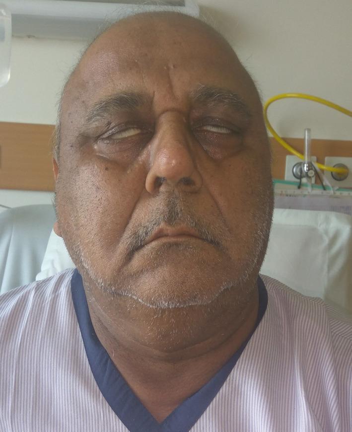
This clinical photograph shows the frontal view of an elderly male patient exhibiting signs of bilateral lower motor neuron facial nerve palsy (Bell's palsy). A prominent finding is Bell's phenomenon, where the patient's eyeballs rotate upward and outward when attempting to close the eyes, leaving the sclera visible due to incomplete eyelid closure (lagophthalmos). The facial features appear relatively symmetrical but mask-like, with a loss of normal expression lines. The mouth is slightly open with evidence of drooling at the commissures, consistent with orbicularis oris muscle weakness. This presentation is highly suggestive of cranial nerve VII involvement, often associated with systemic conditions such as Guillain-Barr syndrome, sarcoidosis, or leptomeningeal metastasis. The image is an educational resource for demonstrating neurological deficits and the physical examination findings of bilateral facial paralysis.
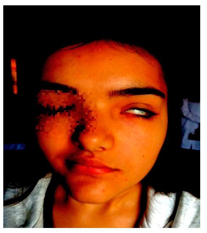
This clinical photograph of a pediatric patient demonstrates classic features of left-sided peripheral facial nerve palsy (Bell's palsy), commonly associated with Melkersson-Rosenthal Syndrome in this context. The image illustrates significant facial asymmetry: the left eye exhibits lagophthalmos (incomplete closure) with notable scleral show (Bell's phenomenon), where the eyeball rolls upward as the patient attempts to close the eye. On the lower face, there is a distinct obliteration of the left nasolabial fold and a compensatory right-sided deviation of the labial commissure, resulting from the loss of muscle tone on the paralyzed left side. These findings represent a House-Brackmann grade 4 dysfunction. The image serves as a visual diagnostic tool for identifying lower motor neuron facial weakness, characterized by the involvement of both the upper and lower facial muscles on the affected side.
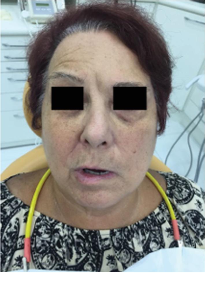
A frontal clinical photograph of an elderly female patient demonstrating symptoms of unilateral facial nerve paralysis, specifically Bell's palsy, in a pretreatment state. The image illustrates significant facial asymmetry localized to the left side. Key clinical findings include the flattening of the left forehead wrinkles, a slight drooping of the left lower eyelid (suggestive of orbicularis oculi weakness), and a pronounced drooping of the left labial commissure (mouth corner) compared to the right side. The nasolabial fold on the left appears effaced or less defined than on the right. Additional visible skin features consistent with the patient's age include solar lentigines (hyperpigmented age spots) on the cheeks and forehead, and dynamic rhytids (wrinkles) on the unaffected right side of the forehead. This visual serves as an educational example of House-Brackmann Grade V facial paralysis, where resting asymmetry is prominent and motor function is severely impaired.
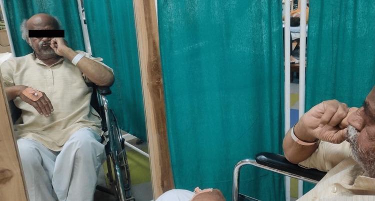
This clinical photograph illustrates an elderly male patient undergoing facial rehabilitation therapy for Bell's palsy or secondary facial nerve paralysis. The patient is seated in a wheelchair within a clinical setting, characterized by green privacy curtains and a large standing mirror used for visual biofeedback. The primary focus of the image is the patient performing a facial exercise: assisted closure of the mouth. He is using his right hand to manually manipulate the right side of his lips, compensating for muscular weakness and inability to close the mouth independently. Key clinical features visible in the patient's reflection include potential right-sided facial drooping and a dressing on the dorsum of the left hand, indicating recent intravenous access or monitoring. The educational focus of this visual is the demonstration of manual facilitation techniques in neuromuscular re-education for the facial muscles. This type of intervention is common in physical or occupational therapy to address functional deficits such as drooling and difficulty chewing during recovery from lower motor neuron lesions.
Bell's Palsy
Epidemiology & Risk Factors
- Affects men and women roughly equally at all ages
- Risk factors: Diabetes mellitus, pregnancy (especially the third trimester and first 2 weeks postpartum - up to 3x increased risk), and possibly hypertension
- Recurrence occurs in approximately 7-8% of cases; average interval between episodes ~10 years
Pathophysiology
- Herpes simplex virus type 1 (HSV-1) is the primary culprit. PCR has detected HSV-1 DNA in endoneurial fluid surrounding CN VII in the majority of Bell's palsy cases, and in the geniculate ganglion. Mouse models show that inoculating HSV into the ear/tongue replicates facial paralysis.
- Varicella-zoster virus (VZV) accounts for up to one-third of cases (when there is no visible rash, it is still Bell's palsy; with a rash it becomes Ramsay Hunt syndrome).
- SARS-CoV-2 and HIV seroconversion have also been implicated less commonly.
- The likely mechanism: viral reactivation in the geniculate ganglion → inflammation and edema of CN VII → nerve entrapment within the tight bony facial canal → ischemia and axonal injury.
Clinical Features
| Feature | Detail |
|---|---|
| Onset | Acute; maximum weakness by 48 hours in ~50%, and virtually all within 3-4 days |
| Weakness pattern | All ipsilateral facial muscles - upper AND lower face (frontalis, orbicularis oculi, lower face) |
| Retroauricular pain | Precedes paralysis by 1-2 days in many patients |
| Taste disturbance | Loss of taste on anterior 2/3 of tongue (chorda tympani involvement) - common but rarely persists beyond 2 weeks |
| Hyperacusis | Ipsilateral - indicates stapedius muscle paralysis |
| Facial numbness | Subjective fullness/numbness; rarely true hypoesthesia in trigeminal distribution |
| Eye | Lagophthalmos (incomplete eye closure), Bell's phenomenon (upward deviation of eyeball on attempted closure) |
Clinical images
MRI Findings
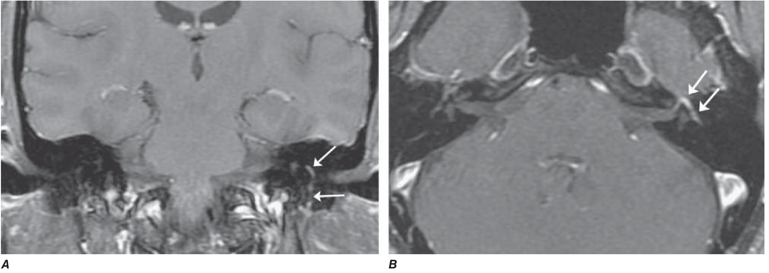
Differential Diagnosis
| Cause | Clue |
|---|---|
| Ramsay Hunt syndrome (VZV reactivation) | Vesicular rash in external auditory canal, palate, tongue; CN VIII often affected |
| Lyme disease | Tick exposure, erythema migrans, can cause bilateral palsy; CSF pleocytosis |
| HIV seroconversion | Risk factors; CSF pleocytosis |
| Sarcoidosis | Bilateral facial palsy, elevated ACE, bilateral hilar adenopathy |
| Guillain-Barré syndrome | Bilateral palsy, ascending weakness, CSF albuminocytologic dissociation |
| Parotid/acoustic tumors | Gradual onset, mass on exam |
| Leprosy | Endemic area exposure |
| Melkersson-Rosenthal syndrome | Recurrent palsy + facial edema + fissured tongue |
| Diabetes, connective tissue disease, amyloidosis | Systemic features |
Investigations
- ESR / CRP
- Fasting glucose (screen for diabetes)
- Lyme serology (endemic areas - accounts for ≥10% of facial palsy in endemic zones)
- HIV serology
- Serum ACE + chest imaging (sarcoidosis)
- MRI brain + temporal bones (with gadolinium)
- Lumbar puncture if Guillain-Barré suspected
- EMG/nerve conduction after 10 days: denervation potentials indicate axonal degeneration, predicting 3+ months to recovery
Treatment
1. Corticosteroids (First-line, ESSENTIAL)
- Prednisone 60-80 mg/day for 5 days, then taper over 5 days (Harrison's 22e)
- Alternatively: prednisolone 25 mg twice daily for 10 days (Goldman-Cecil)
- Must be started early (within 72 hours of onset ideally)
- Improves rate of complete recovery from 63% to 83% at 3 months
- Mechanism: reduces edema and swelling of the nerve in the facial canal
2. Antivirals
- Acyclovir (400 mg 5x/day for 10 days) or valacyclovir (1000 mg/day for 5-7 days)
- Most large RCTs (including Sullivan et al.) found no added benefit of antivirals over steroids alone in the general Bell's palsy population
- However, antivirals should be added when:
- Vesicular lesions are observed in the external ear canal or palate (Ramsay Hunt)
- Complete facial paralysis at presentation (some evidence for additive benefit - Hato et al.)
- A 2025 meta-analysis (PMID 39724817) re-examined glucocorticoids + antivirals vs. glucocorticoids alone across RCTs
3. Eye care (Mandatory)
- Artificial tears during the day
- Paper tape / eye patch at night to close the eyelid and prevent corneal exposure
- Lubricating ointment
4. Physiotherapy
- Facial exercises and neuromuscular re-education
- A 2025 systematic review (PMID 39647183) supports physical therapy for idiopathic facial paralysis
5. Surgery
- Surgical decompression of the facial nerve is not recommended and may be harmful (Adams & Victor)
- Cosmetic reconstruction (fascia lata slings, nerve grafting) reserved for permanent paralysis
Grading
Prognosis
| Outcome | Data |
|---|---|
| Complete recovery | ~70% within 1-2 months |
| Near-normal function | ~85% overall |
| Poor recovery | ~10% have little/no recovery |
| Recurrence | ~7-8% |
- Incomplete paralysis in the first week (strongest predictor)
- Return of taste in the first week
- Any voluntary motor movement within 5-7 days
- Complete paralysis from onset
- Pronounced gadolinium enhancement on MRI
- EMG evidence of denervation after 10 days (predicts delayed, potentially incomplete recovery over months to years)
- Synkinesis - involuntary co-contraction (e.g., eye closure when chewing - "jaw winking")
- Crocodile tears - lacrimation when eating (aberrant salivary fibers reinnervate lacrimal gland)