
Gate control theory of pain
gate control theory of pain spinal cord dorsal horn diagram
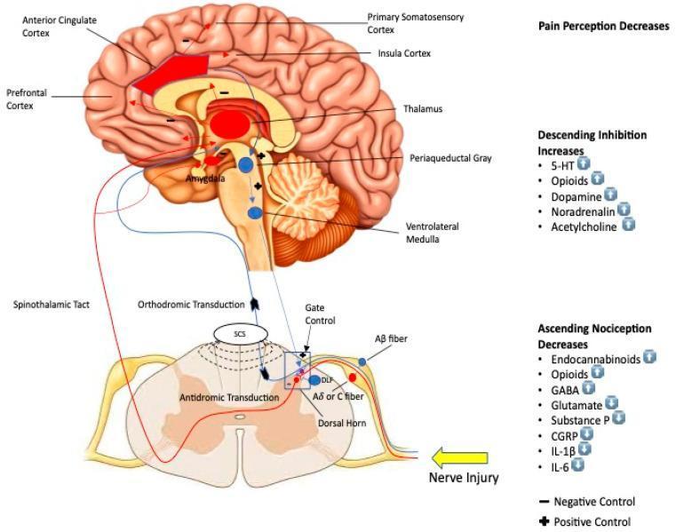
This pathophysiology diagram illustrates the neuromodulation mechanisms of Spinal Cord Stimulation (SCS) on pain pathways. The schematic is divided into two main sections: a sagittal view of the human brain and a cross-section of the spinal cord dorsal horn. Key brain regions involved in pain processing are highlighted, including the anterior cingulate cortex, prefrontal cortex, primary somatosensory cortex, insula cortex, thalamus, amygdala, periaqueductal gray, and ventrolateral medulla. The diagram depicts the spinothalamic tract carrying ascending signals and descending inhibitory pathways. At the spinal level, the 'gate control' mechanism is shown where SCS influences orthodromic and antidromic transduction, specifically targeting Aβ fibers to modulate Aδ or C fiber nociception. Accompanying text lists biochemical changes associated with stimulation: increases in descending inhibitors (5-HT, opioids, dopamine, noradrenalin, acetylcholine) and decreases in ascending nociceptive markers (glutamate, substance P, CGRP, IL-1β, IL-6), alongside increases in inhibitory molecules like endocannabinoids, opioids, and GABA. Symbols indicate positive and negative control nodes within the neuroanatomical circuit.
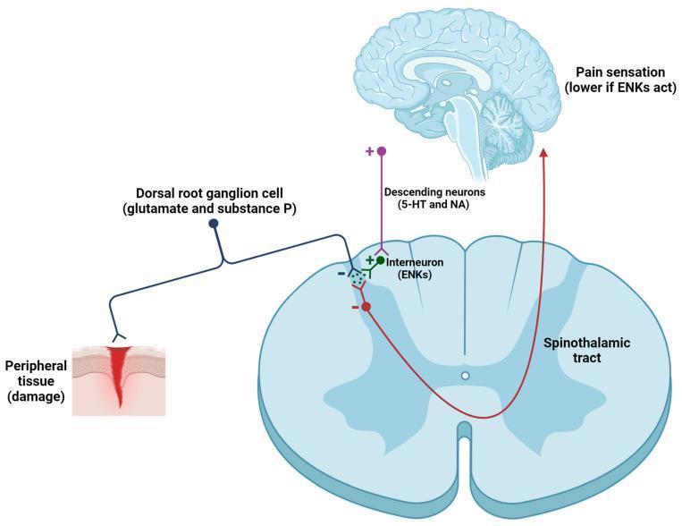
This pathophysiology diagram illustrates the modulation of the nociceptive pathway in the spinal cord, specifically the role of enkephalins (ENKs) in pain gating. The visual depicts three primary components: the ascending pain pathway, the descending inhibitory pathway, and the central nervous system structures (spinal cord cross-section and brain). The process begins with peripheral tissue damage activating a dorsal root ganglion (DRG) primary afferent neuron, which releases excitatory neurotransmitters glutamate and substance P into the dorsal horn. This signal is transmitted via the spinothalamic tract to the brain, resulting in pain sensation. Simultaneously, the diagram shows a descending inhibitory pathway originating from the brain (serotonergic 5-HT and noradrenergic NA neurons) that positively stimulates a spinal interneuron containing ENKs. These ENKs provide inhibitory feedback (noted by '-' signs) onto both the primary afferent terminal and the second-order ascending neuron. This enkephalinergic modulation effectively reduces the transmission of nociceptive signals to the cortex, resulting in a lower perception of pain. The diagram is an educational resource for understanding Gate Control Theory and opioid-mediated analgesia.
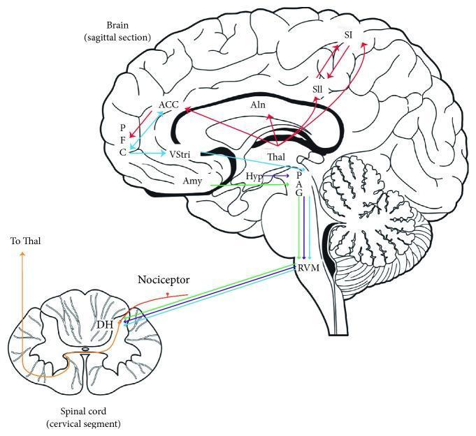
A medical anatomical diagram illustrating the human pain modulatory system through a sagittal section of the brain and a transverse section of the cervical spinal cord. The diagram highlights the ascending and descending pathways involved in pain processing. Nociceptive input is shown entering the dorsal horn (DH) and ascending via the spinothalamic tract to the thalamus (Thal). From the thalamus, pathways diverge into the sensory-discriminative system (primary and secondary somatosensory cortices: SI, SII) and the affective-motivational system (anterior cingulate cortex: ACC, prefrontal cortex: PFC, and anterior insula: AIn). Subcortical involvement is indicated by connections to the hypothalamus (Hyp), amygdala (Amy), and ventral striatum (VStri). The descending inhibitory control system is depicted originating from the periaqueductal gray (PAG) and rostroventromedial medulla (RVM), which project back to the dorsal horn to modulate spinal nociceptive transmission. This visualization serves as an educational resource for neuroanatomy and pain physiology.
"gate control theory" pain
Gate Control Theory of Pain
Historical Background
Core Concept
| Fiber Type | Diameter | Modality | Effect on Gate |
|---|---|---|---|
| A-beta (Aβ) | Large, myelinated | Touch, pressure, vibration | Closes the gate (inhibits pain) |
| A-delta (Aδ) / C fibers | Small, thin/unmyelinated | Pain, temperature, nociception | Opens the gate (promotes pain) |
Circuit Mechanism
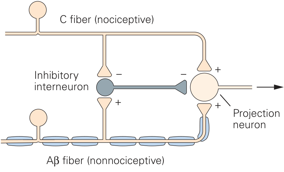
- C fibers (nociceptive) - synapse on projection neurons (+) and inhibit inhibitory interneurons (-)
- Aβ fibers (nonnociceptive) - synapse on projection neurons (+) and excite inhibitory interneurons (+)
- Inhibitory interneuron (the "gate") - when active, it suppresses the projection neuron output
- Projection neuron (T cell / transmission cell) - sends signals up the anterolateral/spinothalamic tract to the brain
- C fiber activity dominates → inhibitory interneurons are suppressed → projection neurons fire → pain signal ascends
- Aβ fiber activity dominates → inhibitory interneurons are activated → projection neurons are suppressed → pain signal is blocked
Descending (Supraspinal) Control
- Periaqueductal gray (PAG) - a midbrain structure that, when stimulated, produces powerful analgesia
- Rostroventromedial medulla (RVM) - relays descending inhibition to the dorsal horn
- Neurotransmitters involved: serotonin (5-HT), noradrenaline, endogenous opioids, GABA, dopamine, acetylcholine
Why You Rub a Bruise - The Classic Example
Clinical Applications
| Therapy | Mechanism |
|---|---|
| TENS (Transcutaneous Electrical Nerve Stimulation) | Electrically activates large Aβ fibers through skin electrodes → closes gate |
| Spinal Cord Stimulation (SCS) | Stimulates dorsal columns → activates large myelinated fibers → inhibits nociception via gate mechanism |
| Acupuncture / massage / vibration | Activate mechanoreceptors → Aβ fiber stimulation → gate closure |
| Opioid analgesics | Act on inhibitory interneurons and descending pathways |
| Cognitive-behavioral therapy | Modulates descending supraspinal control of the gate |
Limitations and Critiques
- The exact anatomical substrate for the "gate" (originally modeled as a simple 3-cell circuit) is more complex
- Pain relief from TENS and dorsal column stimulation does not always correlate strictly with large-fiber activation alone
- The theory does not fully account for all neuropathic pain states or phantom limb pain
- Phantom limb pain led Melzack himself to later develop the Neuromatrix Theory, positing a body-self neuromatrix in the brain that generates pain independently of peripheral input
Summary Table
| Component | Role |
|---|---|
| Aβ fibers | Large, myelinated; activate inhibitory interneuron → close gate |
| Aδ / C fibers | Small; inhibit interneuron → open gate |
| Inhibitory interneuron (SG cells) | Substantia gelatinosa; the physical gate |
| Projection neuron (T cell) | Sends signal up spinothalamic tract to brain |
| Descending pathways (PAG, RVM) | Supraspinal modulation; can open or close gate |
| TENS / SCS | Clinical application - preferentially activate Aβ fibers |
For MBBS 1st year student clear cut explanation with maximum flowcharts
🧠 Gate Control Theory of Pain
For MBBS 1st Year | Physiology
STEP 1 — What is Pain? (Basic Foundation)
TISSUE DAMAGE / NOXIOUS STIMULUS
            │
            ▼
    NOCICEPTORS activated
    (free nerve endings in skin/tissue)
            │
            ▼
    Nerve signals travel to SPINAL CORD
            │
            ▼
    Signal reaches BRAIN → Pain perceived
Before Gate Control Theory, pain was thought to be a simple one-way street. Melzack & Wall (1965) said: "Wait — there's a GATE in the spinal cord that controls whether pain reaches the brain!"
STEP 2 — The Two Types of Nerve Fibers (KEY CONCEPT)
┌─────────────────────────────────────────────────────────────────┐
│                    SENSORY NERVE FIBERS                         │
├─────────────────┬───────────────────────────────────────────────┤
│   Aβ FIBERS     │        Aδ / C FIBERS                         │
├─────────────────┼───────────────────────────────────────────────┤
│ Large diameter  │  Small diameter                               │
│ Myelinated      │  Aδ = thin myelinated / C = unmyelinated      │
│ Fast conduction │  Slower conduction                            │
│ Carry: TOUCH,   │  Carry: PAIN, temperature,                    │
│ pressure,       │  nociception                                  │
│ vibration       │                                               │
├─────────────────┼───────────────────────────────────────────────┤
│ EFFECT ON GATE: │  EFFECT ON GATE:                              │
│  ✅ CLOSE GATE  │   ❌ OPEN GATE                                │
│  (block pain)   │   (allow pain through)                        │
└─────────────────┴───────────────────────────────────────────────┘
STEP 3 — The Anatomy of the Gate (Dorsal Horn Circuit)
                    PERIPHERAL NERVE
                          │
              ┌───────────┴───────────┐
              │                       │
         Aβ FIBER               C FIBER
      (Touch/pressure)          (Pain)
              │                       │
              ▼                       ▼
    ┌─────────────────────────────────────────┐
    │         DORSAL HORN of SPINAL CORD       │
    │                                         │
    │    ┌────────────────────┐               │
    │    │  INHIBITORY        │               │
    │    │  INTERNEURON       │               │
    │    │  (Substantia       │               │
    │    │   Gelatinosa)      │               │
    │    │  = THE "GATE"      │               │
    │    └────────────────────┘               │
    │                                         │
    │    ┌────────────────────┐               │
    │    │  PROJECTION NEURON │               │
    │    │  (T-cell /         │               │
    │    │  Transmission cell)│──────────────►│──► To BRAIN
    │    └────────────────────┘               │
    └─────────────────────────────────────────┘
- Inhibitory Interneuron (SG cell) = The Gate itself
- Projection Neuron (T cell) = Sends signal to brain
- Aβ and C fibers = Inputs that control the gate
STEP 4 — GATE CLOSED (Pain Blocked) 🚪✅
TOUCH / RUBBING / VIBRATION
          │
          ▼
    Aβ FIBER fires
          │
          ├──────────────────────────────┐
          │                              │
          ▼                              ▼
  Inhibitory Interneuron           Projection Neuron
  gets EXCITED (+)                 gets direct input (+)
          │
          ▼
  Interneuron fires →
  INHIBITS Projection Neuron (–)
          │
          ▼
  Net result: Projection Neuron is SUPPRESSED
          │
          ▼
  ❌ PAIN SIGNAL DOES NOT REACH BRAIN
          │
          ▼
   🚪 GATE IS CLOSED — No pain felt!
STEP 5 — GATE OPEN (Pain Gets Through) 🚪❌
TISSUE DAMAGE / NOXIOUS STIMULUS
          │
          ▼
    C FIBER fires
          │
          ├──────────────────────────────┐
          │                              │
          ▼                              ▼
  Inhibitory Interneuron           Projection Neuron
  gets INHIBITED (–)               gets excited (+)
          │
          ▼
  Interneuron SILENT →
  Cannot inhibit Projection Neuron
          │
          ▼
  Projection Neuron fires freely
          │
          ▼
  ✅ PAIN SIGNAL REACHES BRAIN
          │
          ▼
   🚪 GATE IS OPEN — Pain is felt!
STEP 6 — Master Flowchart (Both Scenarios Side by Side)
┌──────────────────────────────────────────────────────────────────────┐
│                      GATE CONTROL THEORY                             │
│                    MASTER CIRCUIT DIAGRAM                            │
└──────────────────────────────────────────────────────────────────────┘

  INPUT:           Aβ FIBER                      C FIBER
  (Periphery)   (Touch/Vibration)            (Pain/Nociception)
                      │                              │
                      │                              │
             ┌────────▼──────┐              ┌────────▼──────┐
             │   EXCITES (+) │              │  INHIBITS (–) │
             │   Inhibitory  │              │   Inhibitory  │
             │   Interneuron │              │   Interneuron │
             └───────┬───────┘              └───────┬───────┘
                     │                              │
              Interneuron                   Interneuron
                ACTIVE                        SILENT
                     │                              │
              INHIBITS (–)                  NO inhibition
              Projection Neuron             on Projection Neuron
                     │                              │
                     ▼                              ▼
             Projection neuron             Projection neuron
               SUPPRESSED                   FIRES FREELY
                     │                              │
                     ▼                              ▼
              No signal to brain            Signal → Brain
                     │                              │
                     ▼                              ▼
               🚪 GATE CLOSED                🚪 GATE OPEN
                  NO PAIN                       PAIN FELT
STEP 7 — Descending Control (Brain Controls the Gate Too!)
BRAIN (higher centers)
    │
    │  ◄── Influenced by:
    │       • Emotions (fear, anxiety → MORE pain)
    │       • Attention / Distraction (→ LESS pain)
    │       • Expectation / Placebo
    │       • Stress
    ▼
PERIAQUEDUCTAL GRAY (PAG) — Midbrain
    │
    ▼
ROSTROVENTROMEDIAL MEDULLA (RVM)
    │
    ▼
DESCENDING FIBERS
    │
    ├── Release: Serotonin (5-HT)
    │            Noradrenaline
    │            Endogenous Opioids (Enkephalins)
    │            GABA
    ▼
DORSAL HORN GATE
    │
    ▼
CAN CLOSE THE GATE EVEN WITHOUT Aβ INPUT
    │
    ▼
PAIN REDUCED / ABOLISHED
Exam point: This is why soldiers in battle don't feel bullet wounds immediately (stress-induced analgesia), and why anxiety/depression makes pain WORSE.
STEP 8 — Clinical Applications Flowchart
GATE CONTROL THEORY
        │
        ├──────────────────────────────────────────────────┐
        │                                                  │
        ▼                                                  ▼
CLOSE THE GATE                                    OPEN THE GATE
(Treat/Reduce Pain)                               (Makes Pain Worse)
        │                                                  │
        ├── TENS                                           ├── Anxiety
        │   (Electrical stimulation of                     │
        │    large Aβ fibers via skin)                     ├── Depression
        │                                                  │
        ├── Spinal Cord Stimulation (SCS)                  ├── Fear / Catastrophizing
        │   (Electrode near dorsal column)                 │
        │                                                  ├── Attention focused
        ├── Massage / Rubbing                              │   on pain
        │   (Activates Aβ mechanoreceptors)                │
        │                                                  └── Large fiber
        ├── Acupuncture                                        neuropathy
        │   (Activates Aβ/Aδ fibers)                          (loss of gate
        │                                                       control)
        ├── Opioids (Morphine)
        │   (Act on inhibitory interneurons
        │    and descending pathways)
        │
        ├── Distraction / Meditation
        │   (Descending inhibitory control)
        │
        └── Antidepressants (TCAs, SNRIs)
            (Enhance descending noradrenaline
             and serotonin inhibition)
STEP 9 — Exam-Ready Summary Table
┌──────────────────┬────────────────────┬────────────────────────────┐
│   COMPONENT      │   DESCRIPTION      │   EXAM POINT               │
├──────────────────┼────────────────────┼────────────────────────────┤
│ Proposed by      │ Melzack & Wall     │ 1965, published in Science  │
├──────────────────┼────────────────────┼────────────────────────────┤
│ Location of gate │ Dorsal horn,       │ Substantia gelatinosa       │
│                  │ spinal cord        │ (Rexed laminae I, II, V)    │
├──────────────────┼────────────────────┼────────────────────────────┤
│ Aβ fibers        │ Large, myelinated  │ CLOSE the gate             │
├──────────────────┼────────────────────┼────────────────────────────┤
│ C fibers         │ Small,unmyelinated │ OPEN the gate              │
├──────────────────┼────────────────────┼────────────────────────────┤
│ Aδ fibers        │ Thin, myelinated   │ Open gate (fast pain)      │
├──────────────────┼────────────────────┼────────────────────────────┤
│ The "gate"       │ Inhibitory         │ Substantia gelatinosa cells │
│                  │ interneuron        │                            │
├──────────────────┼────────────────────┼────────────────────────────┤
│ Projection neuron│ T-cell             │ Transmits to brain via     │
│                  │ (Transmission cell)│ spinothalamic tract        │
├──────────────────┼────────────────────┼────────────────────────────┤
│ Descending ctrl  │ PAG → RVM →        │ Opioids, 5-HT, NA, GABA   │
│                  │ Dorsal horn        │                            │
├──────────────────┼────────────────────┼────────────────────────────┤
│ Clinical use     │ TENS, SCS,         │ Based on activating Aβ     │
│                  │ massage, opioids   │ fibers to close gate       │
└──────────────────┴────────────────────┴────────────────────────────┘
STEP 10 — One-Line Mnemonics for Exams
┌──────────────────────────────────────────────────────────────┐
│  BIG = BLOCK  →  Aβ (big fiber) BLOCKS pain (closes gate)   │
│  SMALL = SEND →  C fiber (small) SENDS pain (opens gate)    │
│                                                              │
│  "Rub it to feel better" = Aβ fibers closing the gate!      │
│                                                              │
│  PAG = Pain's Arrest Gate (midbrain analgesia center)        │
│                                                              │
│  TENS = Touch Electrically Negates pain Signal               │
└──────────────────────────────────────────────────────────────┘
Quick Revision Flowchart (Print & Stick)
NOXIOUS STIMULUS ──► C fibers ──► Inhibit interneuron ──► Gate OPEN ──► PAIN
                                                                         ▲
TOUCH/RUBS ──────► Aβ fibers ──► Excite interneuron ──► Gate CLOSED ───┘(blocked)

BRAIN ───────────► PAG/RVM ──► Descending inhibition ──► Gate CLOSED ──► Analgesia