
CT chest interstitial lung disease traction bronchiectasis reticular opacities UIP pattern
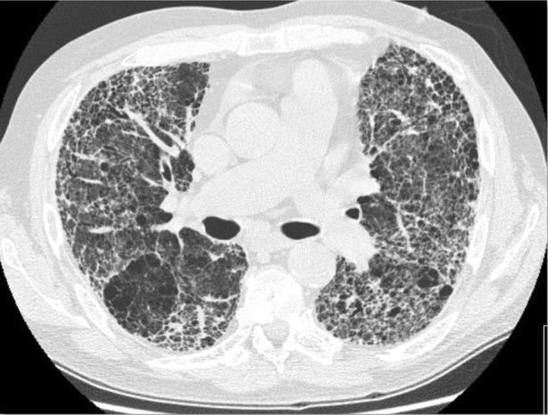
Axial non-contrast computed tomography (CT) of the chest demonstrating advanced interstitial lung disease. The imaging shows a classic Usual Interstitial Pneumonia (UIP) pattern characteristic of Idiopathic Pulmonary Fibrosis (IPF). Prominent bilateral, predominantly peripheral, and subpleural reticular opacities are present throughout the lung parenchyma. There is significant architectural distortion featuring extensive honeycombing, characterized by clusters of cystic airspaces with thickened walls. Associated findings include traction bronchiectasis, where secondary fibrotic changes cause pathological dilation of the bronchi, and thickened interlobular septa. Some ground-glass opacities are interspersed within the reticular network. The mediastinal structures and great vessels are visible centrally. This visual information is clinically relevant for the diagnosis of chronic fibrosing interstitial pneumonia and serves as a primary example of severe parenchymal destruction in elderly ex-smokers.
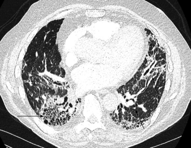
This diagnostic image is an axial chest CT scan showing a cross-section of the thorax. The image demonstrates a pattern of chronic interstitial lung disease with features consistent with Usual Interstitial Pneumonia (UIP). Prominent findings include bilateral, subpleural honeycombing, characterized by clustered, thick-walled cystic airspaces primarily located in the lower lobes. Also visible are subpleural reticular opacities and architectural distortion. Traction bronchiectasis is evident, marked by the abnormal dilation of the airways (indicated by arrows) within areas of surrounding fibrosis. These features signify irreversible lung parenchymal scarring and destruction. The distribution is predominantly peripheral and basilar, typical of advanced pulmonary fibrosis, which in this clinical context is associated with drug-induced lung injury. This scan is an essential educational example for identifying the hallmark radiologic features of interstitial fibrosis and traction bronchiectasis in a clinical setting.
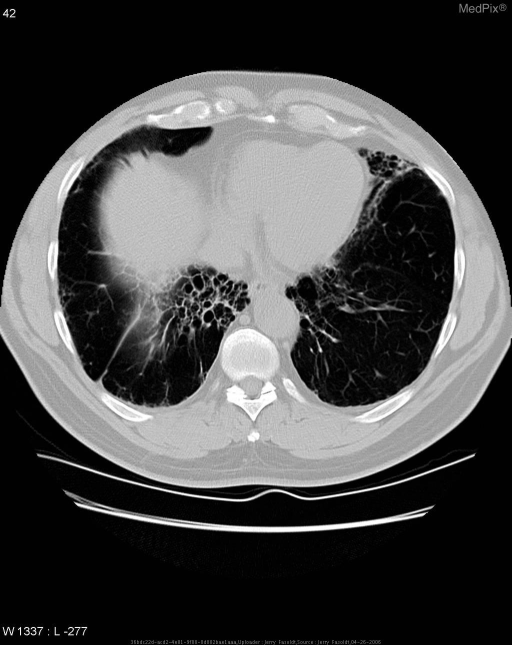
**Imaging Modality:** Axial Computed Tomography (CT) of the chest, lung window.
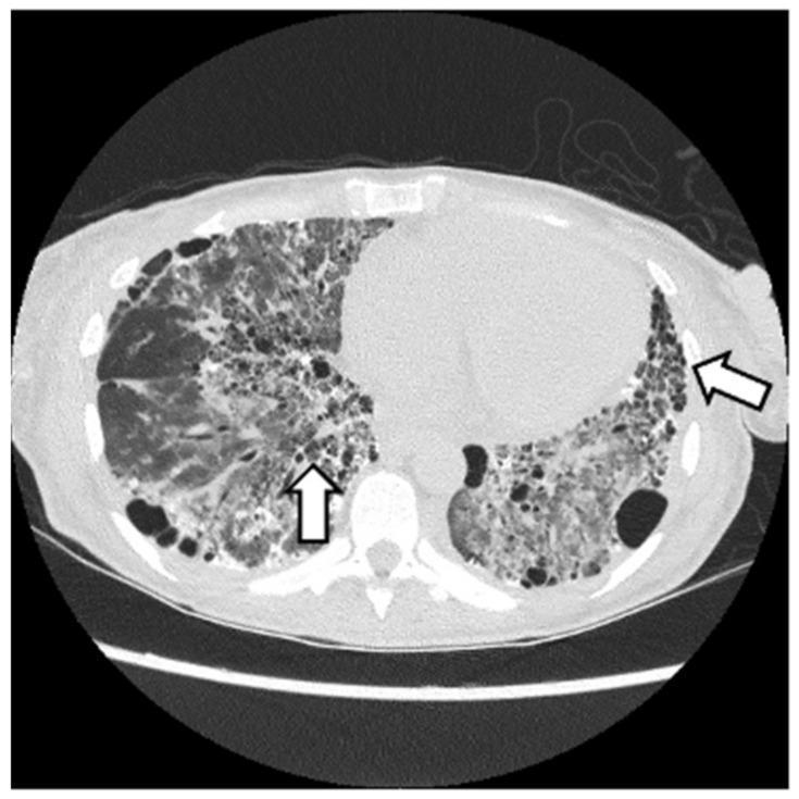
This diagnostic image is an axial computed tomography (CT) scan of the chest at the level of the lower lung fields, demonstrating advanced interstitial lung disease (ILD) with a pattern consistent with Usual Interstitial Pneumonia (UIP). Key visual features include extensive honeycombing, characterized by clusters of cystic airspaces with thick, well-defined walls, and coarse reticular opacities. The distribution is predominantly subpleural and basal, as indicated by the white arrows. Significant architectural distortion is present, with evidence of traction bronchiectasis (dilated airways within fibrotic regions). The lung parenchyma shows a heterogeneous density, with ground-glass opacities interspersed among areas of terminal fibrosis. These findings are clinically significant in the context of antisynthetase syndrome (ASyS) with anti-Jo-1 positivity, illustrating the severe fibrotic pulmonary manifestations that can occur in inflammatory myopathies. The image serves as an educational example of advanced pulmonary fibrosis and the UIP radiologic phenotype.
CT Chest Analysis - Lung Window
Key Imaging Findings
Pattern Recognition and Differential Diagnosis
| Feature | UIP (Usual Interstitial Pneumonia / IPF) | NSIP (Non-Specific Interstitial Pneumonia) |
|---|---|---|
| Distribution | Subpleural, basal-predominant | Subpleural, bilateral symmetric |
| Key finding | Honeycombing + traction bronchiectasis | GGO + reticular, minimal honeycombing |
| GGO extent | Minor | Major |
| Temporal pattern | Heterogeneous | Homogeneous |
| Common cause | Idiopathic (IPF) | CTD, drugs, idiopathic |
- Fibrotic NSIP - bilateral symmetric GGO + reticulation + traction bronchiectasis without dominant honeycombing; strongly associated with connective tissue diseases (RA, SSc, polymyositis/dermatomyositis)
- UIP / IPF - if honeycombing is present on other slices; typically more subpleural and basal
- Hypersensitivity Pneumonitis (chronic/fibrotic HP) - reticular + GGO + traction bronchiectasis; typically upper/mid-lobe sparing; need occupational/exposure history
- Combined Pulmonary Fibrosis and Emphysema (CPFE) - given the large right anterior bulla alongside bilateral fibrosis; classically in male smokers
- CTD-ILD (Rheumatoid arthritis, Scleroderma) - NSIP or UIP pattern in the setting of systemic disease
Reference CT Images for Comparison
Recommended Next Steps
| Investigation | Purpose |
|---|---|
| Full HRCT chest (thin-slice, prone views) | Characterize distribution precisely; detect honeycombing |
| PFTs (spirometry, DLCO, TLC) | Confirm restrictive pattern; assess severity |
| ANA, anti-CCP, RF, anti-Scl-70, anti-Jo-1, myositis panel | Screen for underlying CTD |
| Bronchoalveolar lavage (BAL) | Cell differential helps distinguish HP vs NSIP vs UIP |
| Multidisciplinary discussion (pulmonologist + radiologist + pathologist) | Gold standard for ILD classification |
| Surgical lung biopsy (if non-diagnostic) | Histologic confirmation if pattern remains uncertain |
Important Notes
- This is a single axial slice; full characterization requires the complete CT dataset with all levels (basal views are particularly important for UIP).
- Clinical context (age, sex, smoking, occupational exposure, autoimmune history, medications) is essential for narrowing the differential.
- The ATS/ERS/JRS/ALAT guidelines recommend multidisciplinary team diagnosis as the standard of care for ILD.