
Pain pathways
Reading File
Pain pathways involve peripheral nociceptors, spinal cord transmission via the dorsal horn, and ascending tracts (mainly spinothalamic) to the brain, where pain is perceived and modulated. There are also descending inhibitory pathways from the brainstem (periaqueductal gray, raphe nuclei) that gate pain via opioid, serotonergic, and noradrenergic mechanisms.
Searching sources...
Finding Sources
Finding Sources
Finding Sources
Reading File
Good - I have a rich section on pain pathways. Let me read more, plus fetch the key diagrams, and also grab the pain section from neuroscience.
Reading File
Loading Image
Loading Image
Now let me get more content - the nociceptors, spinal cord processing, and descending modulation sections:
Reading File
Reading File
Loading Image
Loading Image
Reading File
Loading Image
Finding Sources
Reading File
I now have comprehensive content from two authoritative neuroscience textbooks. Let me compile the full answer.
Pain Pathways
Pain (nociception) involves a sequence of events: transduction (at peripheral receptors), transmission (along sensory nerves to the spinal cord and brain), perception (in the cortex), and modulation (descending controls). Here is a complete account of each stage.
1. Peripheral Nociceptors
Nociceptors are free nerve endings that respond only to stimuli strong enough to threaten or damage tissue. They are found in skin, bone, muscle, most internal organs, blood vessels, and meninges - but notably absent from the substance of the brain itself.
Three main classes of nociceptive afferents:
| Fiber type | Myelination | Conduction | Pain quality |
|---|---|---|---|
| Aδ (Type I) | Thinly myelinated | ~5-30 m/s | Sharp, well-localised "first pain" |
| Aδ (Type II) | Thinly myelinated | ~5-30 m/s | Heat-sensitive |
| C fibers | Unmyelinated | <2 m/s | Dull, burning, aching "second pain" |
The cell bodies of these primary afferents sit in the dorsal root ganglia (DRG). A key channel for nociceptive action potential generation is Na$_v$1.7, a voltage-gated sodium channel encoded by SCN9A. Loss-of-function mutations in SCN9A cause congenital insensitivity to pain.
2. Peripheral Sensitisation and the Inflammatory Soup
When tissue is injured, a cocktail of chemicals - called the inflammatory soup - is released, sensitising nociceptors to produce hyperalgesia and allodynia:
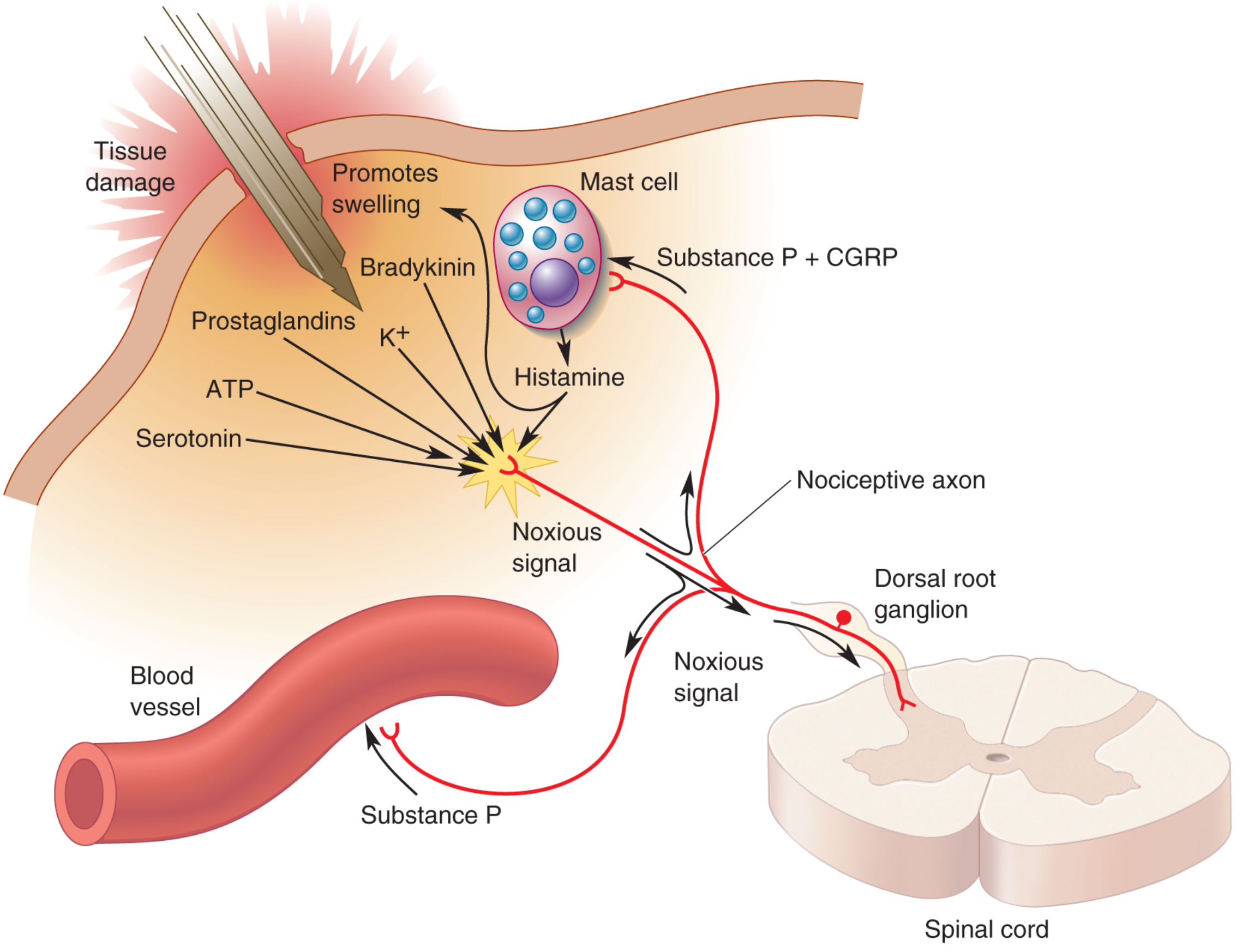
Peripheral chemical mediators of pain (Neuroscience: Exploring the Brain, 5th Ed.)
Constituents include: glutamate, serotonin, ATP, bradykinin, substance P, CGRP, prostaglandins, endocannabinoids, cytokines, K⁺, H⁺, and neurotrophins.
- Primary hyperalgesia - increased sensitivity at the site of injury (peripheral sensitisation)
- Secondary hyperalgesia - sensitisation of surrounding uninjured tissue (central sensitisation)
- Allodynia - pain from a normally non-painful stimulus
3. Spinal Cord Dorsal Horn (First Synapse)
The central branches of DRG neurons enter the spinal cord and terminate in the dorsal horn in a lamina-specific pattern (Rexed laminae):
| Lamina | Input | Function |
|---|---|---|
| I (Marginal zone) | Aδ and C fibers | Nociceptive-specific neurons; projects to midbrain and thalamus |
| II (Substantia gelatinosa) | C fibers | Inhibitory and excitatory interneurons; modulates nociceptive flow |
| III-IV | Aβ fibers | Light touch; some project supraspinally |
| V | Wide dynamic range neurons | Convergent noxious + innocuous input; projects to brain stem and thalamus |
Key neurotransmitters at the first synapse:
- Glutamate (fast, via AMPA receptors) - sharp pain
- Substance P (slow neuropeptide, via NK-1 receptors) - prolonged pain, central sensitisation
- NMDA receptor activation is central to wind-up and chronic pain development
4. Ascending Pain Pathways
The dominant pathway carrying pain and temperature is the anterolateral system (spinothalamic tract):
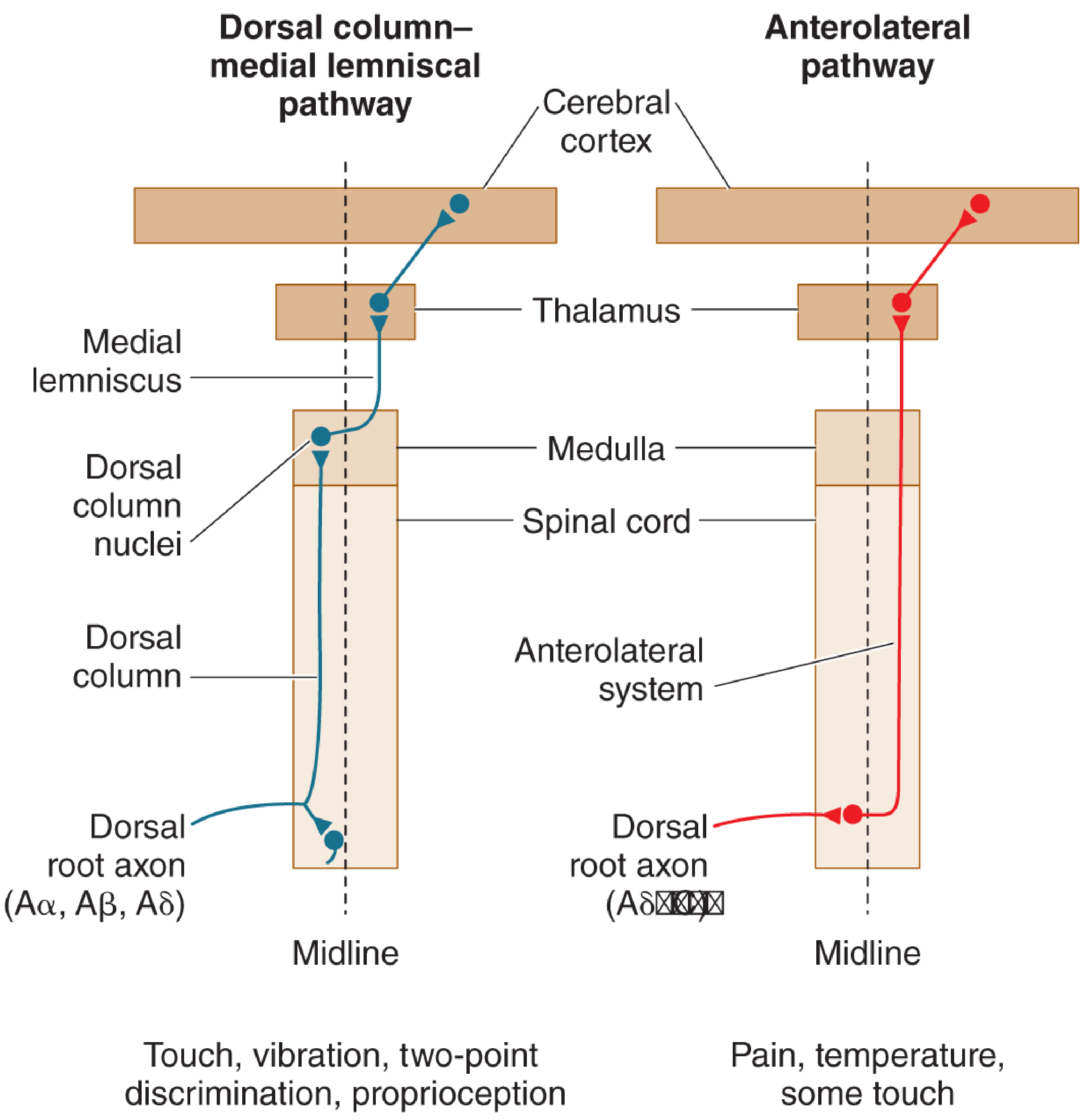
Two major ascending sensory pathways (Neuroscience: Exploring the Brain, 5th Ed.)
Spinothalamic tract route:
- First-order neuron: DRG → dorsal horn (laminae I, IV-V)
- Axons cross the midline (decussate) in the anterior white commissure at the same spinal level
- Ascend in the anterolateral column
- Terminate in the thalamus (VPL nucleus and intralaminar nuclei)
Contrast this with the dorsal column-medial lemniscal system (touch, vibration, proprioception), which ascends ipsilaterally and decussates at the level of the medulla.
5. Thalamus and Cortex
Nociceptive axons from the anterolateral system terminate across wider thalamic territory than touch fibers:
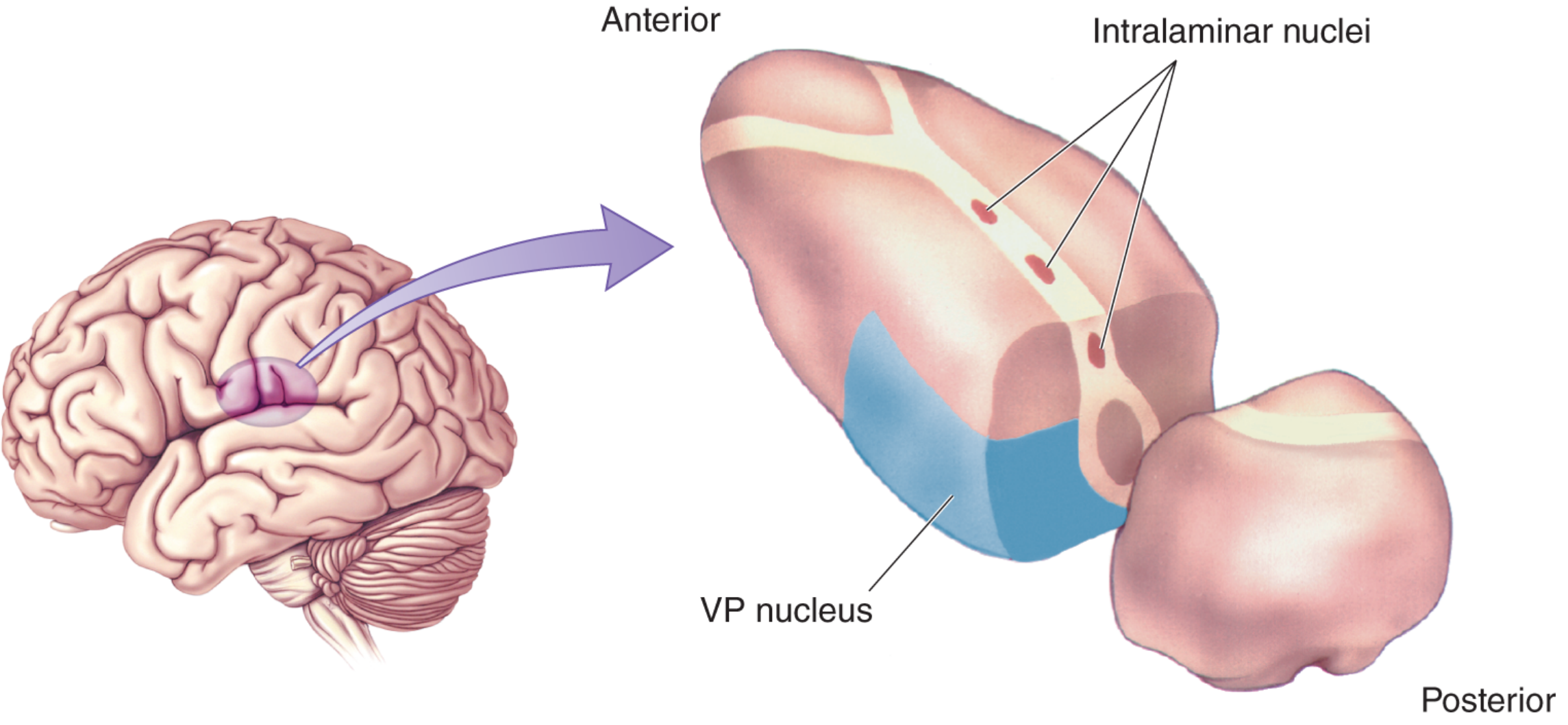
Thalamic relay nuclei for pain (Neuroscience: Exploring the Brain, 5th Ed.)
- VP (ventrobasal) nucleus - projects to primary somatosensory cortex (S1, S2): localisation and intensity coding
- Intralaminar nuclei - project diffusely to cortex and striatum: arousal, affective-motivational aspects
- The anterior cingulate cortex and insular cortex process the emotional unpleasantness of pain
- The prefrontal cortex mediates cognitive appraisal and descending control
This distributed cortical processing accounts for the two dimensions of pain: sensory-discriminative (where? how intense?) and affective-motivational (how unpleasant?).
6. Trigeminal Pain Pathway
Pain from the face and head uses an analogous but parallel route:
- Small-diameter Aδ and C fibers in the trigeminal nerve (CN V)
- Synapse on second-order neurons in the spinal trigeminal nucleus of the brainstem (equivalent to the substantia gelatinosa)
- Axons cross the midline and ascend in the trigeminal lemniscus
- Terminate in the VPM nucleus of the thalamus → somatosensory cortex
- Neuroscience: Exploring the Brain, 5th Ed.
7. Pain Modulation
Gate Control Theory (Melzack & Wall, 1965)
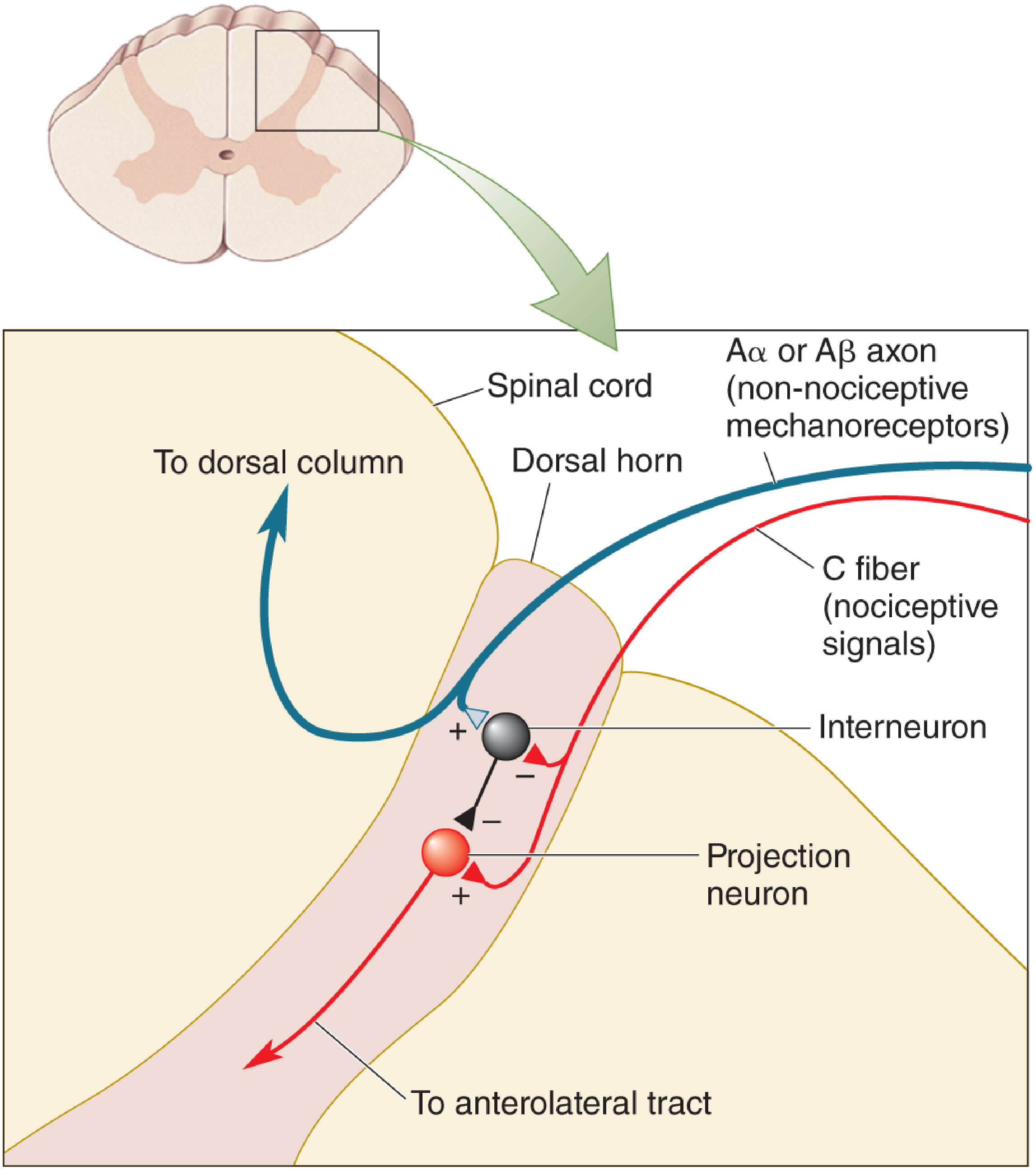
Gate control theory of pain (Neuroscience: Exploring the Brain, 5th Ed.)
Large-diameter Aβ touch fibers activate inhibitory interneurons in the dorsal horn that suppress nociceptive C-fiber signals. This explains why rubbing an injury reduces pain, and is the basis for TENS (transcutaneous electrical nerve stimulation).
Descending Inhibitory Pathway
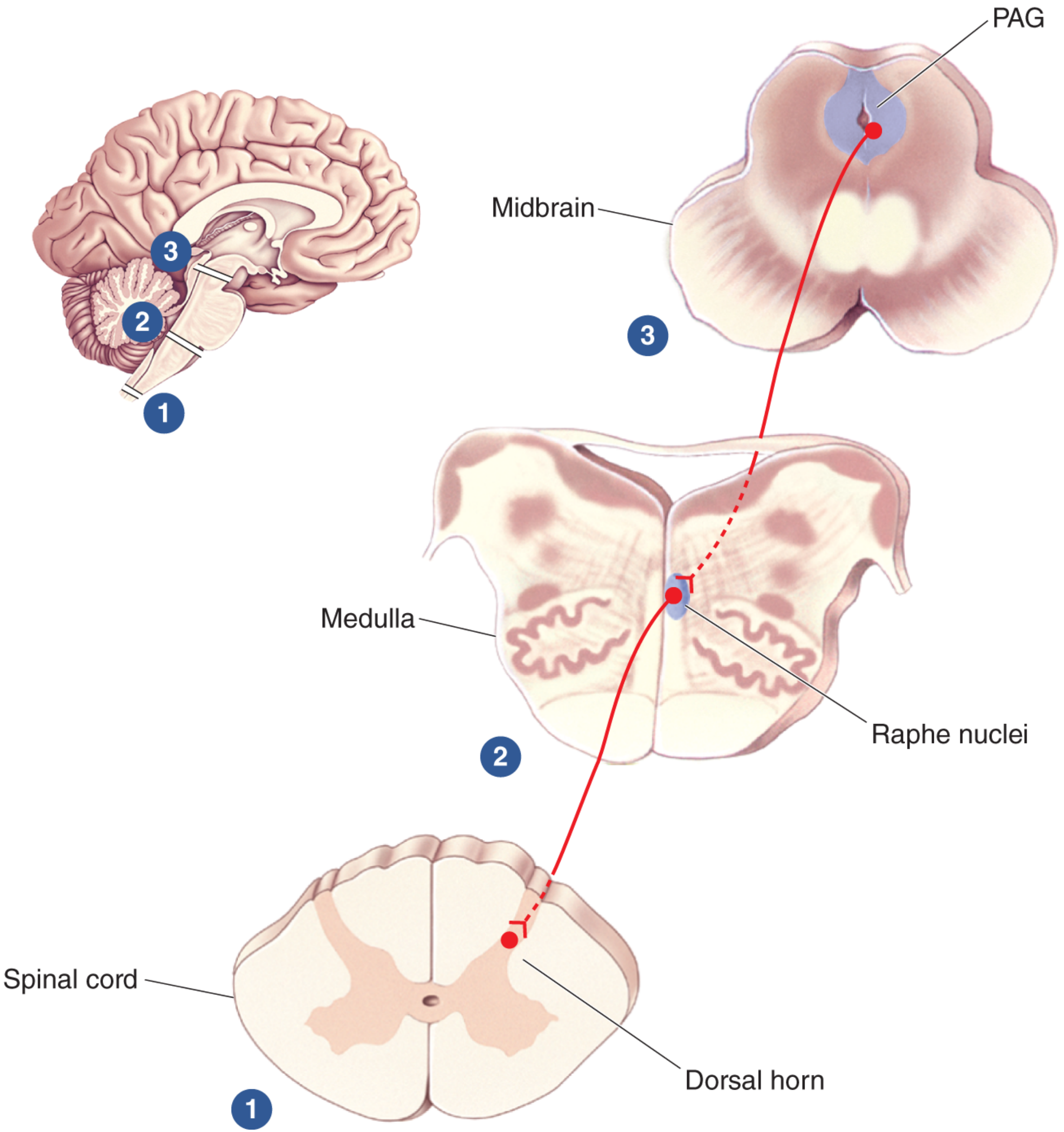
Descending pain-control pathway (Neuroscience: Exploring the Brain, 5th Ed.)
Key structures:
- Periaqueductal gray (PAG) - midbrain; receives emotional input from cortex, hypothalamus, and amygdala; electrical stimulation produces profound analgesia
- Raphe nuclei - medulla; serotonergic neurons project to dorsal horn
- Locus coeruleus - pons; noradrenergic neurons project to dorsal horn
- These brainstem neurons inhibit (or sometimes facilitate) nociceptive neurons in the dorsal horn
8. Endogenous Opioid System
The brain manufactures its own opioid-like peptides that bind to opioid receptors concentrated in pain-processing areas:
| Peptide family | Receptor |
|---|---|
| Endorphins | μ (mu) |
| Enkephalins | δ (delta) |
| Dynorphins | κ (kappa) |
Small injections of morphine or endorphins directly into the PAG produce powerful analgesia. Opioid receptors are found at multiple levels: peripheral nociceptors, dorsal horn, PAG, and the rostral ventromedial medulla. Naloxone (an opioid antagonist) blocks both exogenous opioid analgesia and stress-induced analgesia, confirming the role of endogenous opioids in pain modulation.
Summary Flowchart
Noxious stimulus
↓
Peripheral nociceptors (Aδ, C fibers) activated
↓
Inflammatory soup sensitises receptors → peripheral sensitisation
↓
DRG → Dorsal horn (Laminae I, II, V) - first synapse
Neurotransmitters: glutamate, substance P
Gate control modulation by Aβ fibers
↓
Anterolateral tract (spinothalamic) - crosses midline
↓
Thalamus (VPL/intralaminar nuclei)
↓
Cortex: S1, S2 (localisation), anterior cingulate + insula (affect)
↓
PAIN PERCEPTION
Descending modulation:
Cortex/hypothalamus/amygdala → PAG → Raphe/LC → Dorsal horn
(via serotonin, norepinephrine, endogenous opioids)
Sources: Neuroscience: Exploring the Brain, 5th Ed. | Kandel's Principles of Neural Science, 6th Ed.
This is a shared conversation. Sign in to Orris to start your own chat.