

image.png
muhs important micro 2 mbbs
Reading File
analyse_media
The image shows MUHS (Maharashtra University of Health Sciences) important microbiology topics for 2nd MBBS - Hepatobiliary section with Short Notes (SN) and Long Answer Questions (LAQ) on Hepatitis and Hydatid cyst. Let me now pull comprehensive content from the textbook library on all these topics simultaneously.
Searching sources...
Finding Sources
Finding Sources
Finding Sources
Finding Sources
Finding Sources
Finding Sources
Now let me read the key sections from Medical Microbiology and other sources on all these topics simultaneously.
Reading File
Reading File
Reading File
Excellent data - now let me read the Echinococcus section and more HBV content in parallel.
Reading File
Reading File
Searching Images
Echinococcus granulosus life cycle hydatid cyst diagram
Searching Images
Hepatitis B serological markers HBsAg HBeAg timeline
Reading File
Reading File
Loading Image
Loading Image
I now have excellent content. Let me also fetch the Echinococcus life cycle diagram and read more on HBV lab diagnosis and hepatitis classification.
Loading Image
Loading Image
Finding Sources
I now have all the core content I need. Let me read the hepatitis classification table from Harrison's and the HBV lab diagnosis section.
Reading File
Reading File
I now have comprehensive content for all MUHS topics. Let me compile a complete, exam-oriented answer.
MUHS 2nd MBBS Microbiology - Hepatobiliary (Important Topics)
📋 HEPATITIS - LAQ ⭐⭐⭐⭐⭐ (Most Important)
1. Classification of Hepatitis Viruses
| Virus | Genome | Size | Envelope | Family | Transmission | Chronicity |
|---|---|---|---|---|---|---|
| HAV | +ssRNA | 27 nm | No | Picornaviridae | Feco-oral | No |
| HBV | dsDNA (partial) | 42 nm (Dane particle) | Yes | Hepadnaviridae | Parenteral/sexual/perinatal | Yes (~5-10%) |
| HCV | +ssRNA | 55 nm | Yes | Flaviviridae | Parenteral | Yes (~80%) |
| HDV | -ssRNA (circular) | 35-37 nm | Yes (HBsAg coat) | Deltaviridae | Parenteral (needs HBV) | Yes |
| HEV | +ssRNA | 32-34 nm | No | Hepeviridae | Feco-oral | No (except immunocomp.) |
From Harrison's Principles of Internal Medicine 22E (2025)
2. Morphology of HBV ⭐
HBV is a 42-nm, double-shelled DNA virus - the "Dane particle" (discovered by Blumberg, 1963). Three morphological forms circulate in serum:
- 22-nm spherical particles - most numerous (non-infectious), represent excess HBsAg envelope protein
- Tubular/filamentous forms - varying length, also non-infectious
- 42-nm Dane particle - the complete, infectious virion (scolex + double shell)
Structure of Dane particle:
- Outer envelope - contains HBsAg (surface antigen = "Australia antigen")
- Inner nucleocapsid - contains HBcAg (core antigen), HBeAg, partially double-stranded circular DNA, DNA-dependent DNA polymerase (with reverse transcriptase activity)
Replication: HBV uses reverse transcription of pregenomic RNA to make minus-strand DNA - unique among DNA viruses. Entry via NTCP receptor (sodium taurocholate cotransporting polypeptide).
3. Pathogenesis of HBV ⭐
- Virus infects hepatocytes → binds NTCP receptor
- Does NOT directly kill liver cells - liver damage is immune-mediated (cytotoxic T-lymphocytes kill HBV-infected hepatocytes)
- CD8+ CTLs recognise HBcAg + MHC class I on hepatocytes → hepatocyte destruction
- Insufficient immune response → chronic infection and carrier state
- Chronic HBV → liver fibrosis → cirrhosis → hepatocellular carcinoma (HCC)
- HBV DNA integrates into host genome → oncogenesis
- 8-20% cumulative risk of cirrhosis over 5 years
4. Serological Markers of HBV ⭐⭐⭐⭐ (Exam Favourite)
Three antigens produce corresponding antibodies:
| Antigen/Antibody | Significance |
|---|---|
| HBsAg | First marker to appear (from ~2 months post-exposure); indicates active infection or carrier state; persists >6 months = chronic |
| Anti-HBs | Appears after clearance of HBsAg (indicates recovery + immunity); also seen after vaccination |
| HBcAg | NOT detectable in serum (stays inside hepatocytes) |
| IgM Anti-HBc | Early antibody; indicates acute HBV infection; best marker for acute HBV; fills "window period" |
| IgG Anti-HBc | Indicates past infection; persists for life |
| HBeAg | Indicates active viral replication + high infectivity; correlates with high HBV DNA |
| Anti-HBe | Appearance indicates declining replication; lower infectivity |
| HBV DNA / DNA polymerase | Most sensitive marker of active replication |
Window Period: HBsAg has cleared but Anti-HBs not yet detectable → only IgM anti-HBc is positive. This is the diagnostic window.
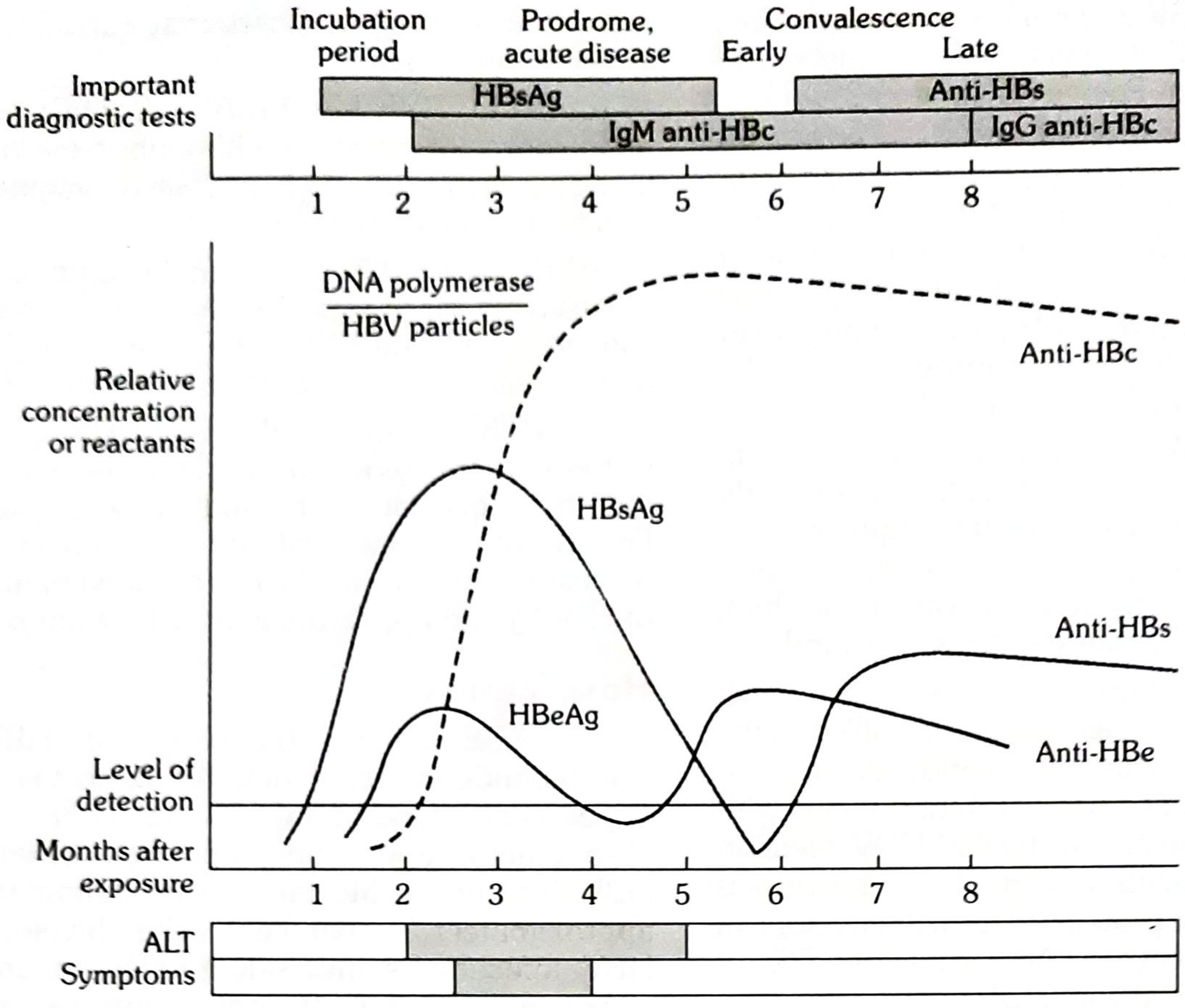
Interpretation table for MUHS:
| Scenario | HBsAg | Anti-HBs | IgM Anti-HBc | IgG Anti-HBc | HBeAg |
|---|---|---|---|---|---|
| Acute HBV | + | - | + | - | +/- |
| Window period | - | - | + | - | - |
| Recovered | - | + | - | + | - |
| Vaccinated | - | + | - | - | - |
| Chronic active | + | - | - | + | + |
| Chronic low replication | + | - | - | + | - |
5. Laboratory Diagnosis of Hepatitis B ⭐⭐⭐⭐
A. Serological Tests (most important for exams):
- ELISA / RIA - detect HBsAg, anti-HBs, anti-HBc (IgM + IgG), HBeAg, anti-HBe
- CLIA (Chemiluminescent immunoassay) - now standard in labs
- HBsAg detection = most widely used screening test
B. Molecular Tests:
- HBV DNA by PCR - most sensitive; quantifies viral load (IU/mL); used to monitor treatment
- HBV DNA polymerase assay - indicates active replication
C. Liver Function Tests:
- Raised serum ALT and AST (ALT > AST in viral hepatitis)
- Raised bilirubin (conjugated + unconjugated)
- Raised alkaline phosphatase
- Prolonged prothrombin time (severe disease)
D. Liver Biopsy:
- "Ground glass hepatocytes" - HBsAg-laden hepatocytes stain with Orcein/Victoria blue stain
- Knodell/Metavir scoring for fibrosis staging in chronic HBV
E. Imaging:
- USG abdomen - hepatomegaly, echogenicity, cirrhosis changes, portal hypertension
6. Modes of Transmission of HBV
- Parenteral route - blood transfusion, contaminated needles/syringes, dialysis, tattooing, ear piercing, acupuncture, shared razors
- Sexual transmission - sexual contact (semen, vaginal secretions, saliva); high risk in promiscuous individuals
- Perinatal/vertical - mother to baby at birth (most common in Asia); rarely transplacental in utero
- Horizontal transmission - child to child via skin contacts/cuts
HBV survives on surfaces for 7 days and is more infectious than HIV (100x).
7. Prophylaxis of HBV ⭐
Specific (Active) Immunization:
- Recombinant HBV vaccine (Engerix-B, Recombivax HB) - yeast-derived HBsAg
- Schedule: 0, 1, 6 months (3 doses); birth dose critical in endemic areas
- Anti-HBs titre >10 mIU/mL = protective
- Booster not routinely needed if initial response achieved
Passive Immunization:
- HBIG (Hepatitis B Immunoglobulin) - for post-exposure prophylaxis (needlestick, newborn of HBsAg+ mother)
- Given within 24-48 hours of exposure + vaccine simultaneously
Other measures:
- Screen blood donors for HBsAg
- Use disposable needles, safe sex practices
- Universal precautions in health care
SN Topics
SN 1: Hepatitis B - Laboratory Diagnosis ⭐
(Covered in detail above in section 5 - key points for short note format:)
Write about: Serology (HBsAg/anti-HBs/anti-HBc markers), PCR for HBV DNA, LFTs, liver biopsy (ground glass hepatocytes), imaging. Include the window period concept and ELISA as the standard screening test.
SN 2: Echinococcus granulosus - Life Cycle ⭐⭐
Causative organism: Echinococcus granulosus (a cestode/tapeworm) - causes Cystic Echinococcosis (Hydatid disease)
Hosts:
- Definitive host: Canines (dog, fox, wolf, coyote, jackal, dingo) - adult tapeworm in intestines
- Intermediate host: Herbivores (sheep, cattle, pigs) - larval cyst stage in viscera
- Accidental intermediate host: Humans (dead-end host)
Life Cycle:
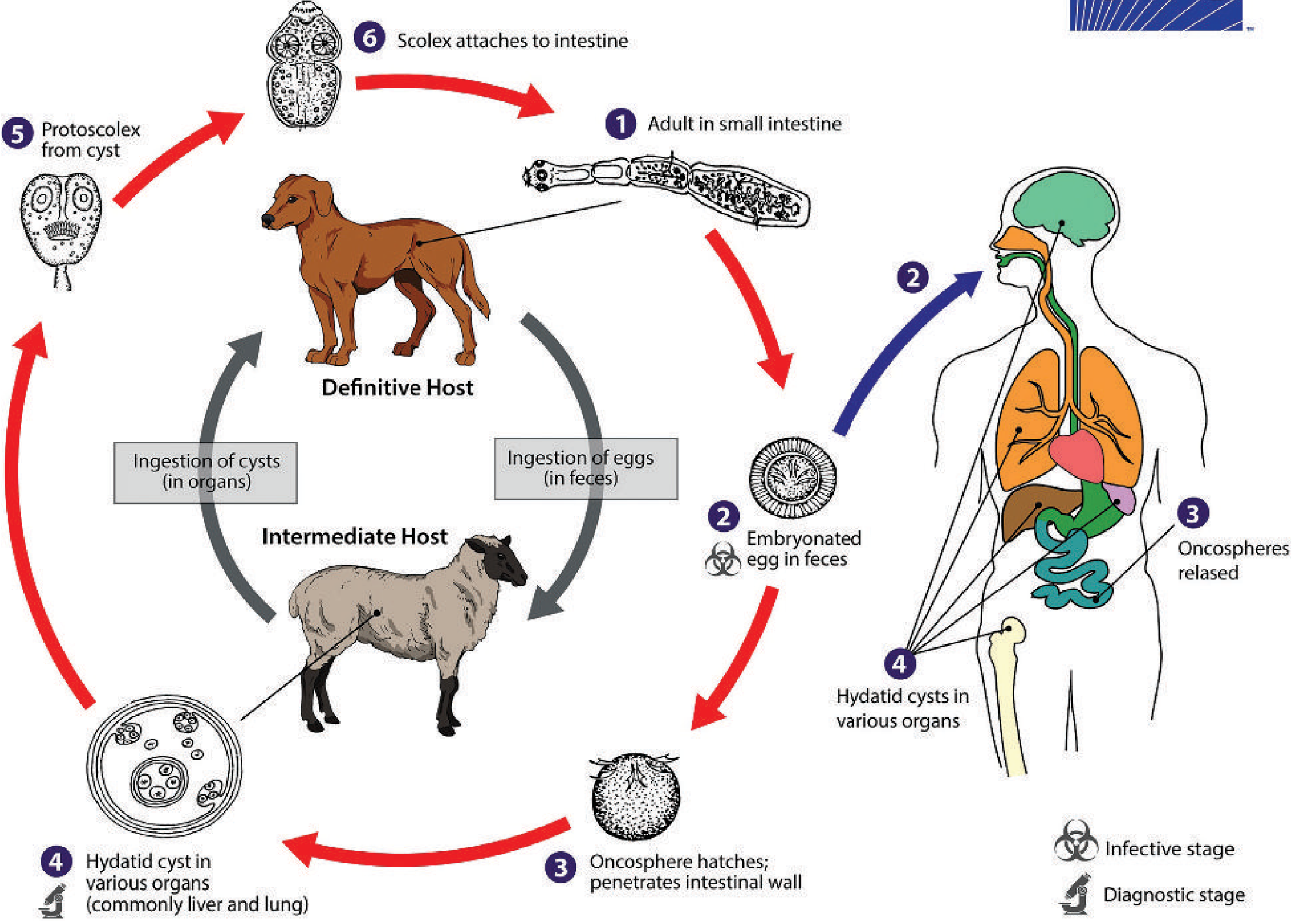
- Adult worm lives in small intestine of dog; 3 proglottids (immature, mature, gravid)
- Gravid proglottid releases embryonated eggs in dog feces → contaminate vegetation/water
- Eggs ingested by sheep (or humans) → oncosphere (6-hooked larva) hatches in intestine
- Oncosphere penetrates intestinal wall → enters portal circulation → reaches liver (first filter) or lungs (second filter) or other organs (bone, brain)
- In tissues → develops into unilocular hydatid cyst
- Dog eats infected sheep viscera → protoscolices from cyst → develop into adult worms in dog's intestine → cycle completes
Adult worm features:
- Taenia-like scolex with 4 sucking disks + double row of hooklets
- Strobila of only 3 proglottids (unusually small for a tapeworm)
SN 3: Hydatid Cyst ⭐⭐ - Causative Agent, Sites Affected, Cross-section Structure
Causative agent: Echinococcus granulosus (dog tapeworm, Cestoda class)
Sites affected (in order of frequency):
- Liver (~70%) - right lobe most common
- Lungs (~20%)
- Others: spleen, kidneys, brain, bone, peritoneum, heart
Cross-section structure of hydatid cyst (3 layers):
  ┌─────────────────────────────────┐
  │  PERICYST (Host layer)          │  ← Dense fibrous layer formed by host
  │  ┌───────────────────────────┐  │
  │  │  ECTOCYST (Laminated)     │  │  ← Acellular, white, laminated (parasite layer)
  │  │  ┌─────────────────────┐  │  │
  │  │  │  ENDOCYST            │  │  │  ← Inner germinal/germinative layer (parasite origin)
  │  │  │  (Germinal layer)    │  │  │     → produces brood capsules → protoscolices
  │  │  │  [Brood capsules]    │  │  │     → daughter cysts possible
  │  │  │  [Daughter cysts]    │  │  │
  │  │  │  [Hydatid fluid]     │  │  │
  │  │  └─────────────────────┘  │  │
  │  └───────────────────────────┘  │
  └─────────────────────────────────┘
Key contents:
- Hydatid fluid - clear, watery, antigenic (anaphylaxis if spilled); toxic
- Brood capsules - arise from germinal layer; contain protoscolices
- Protoscolices - future tapeworm heads
- Daughter cysts - develop within mother cyst from detached brood capsules
- Hydatid sand - degenerated brood capsules + free protoscolices + hooklets settled at bottom of cyst
Clinical features:
- Slow growing (5-20 years before symptoms)
- Liver: pain, biliary obstruction, jaundice
- Lungs: cough, dyspnea, chest pain
- Rupture: fever, urticaria, anaphylactic shock (potentially fatal)
- Bone: erosion of marrow cavity
- Brain: raised intracranial pressure
Diagnosis:
- USG / CT scan (imaging of choice) - cyst with daughter cysts is pathognomonic
- Serology (ELISA/IHA) - sensitive for liver, less for lung; false negative in 10-40%
- Casoni's skin test (historical, unreliable)
- Aspiration CONTRAINDICATED (risk of anaphylaxis and dissemination)
- "Hydatid sand" on aspiration (if done) = diagnostic
Treatment:
- PAIR (Puncture-Aspiration-Injection-Reaspiration) with hypertonic saline
- Surgical resection (treatment of choice)
- Albendazole (drug of choice for medical management; also pre/post-op)
Quick Revision Summary for MUHS
| Topic | Stars | Key Points |
|---|---|---|
| Hepatitis classification | ⭐⭐⭐⭐⭐ | HAV/HBV/HCV/HDV/HEV - genome, envelope, transmission, chronicity |
| HBV serological markers | ⭐⭐⭐⭐⭐ | HBsAg/anti-HBs/IgM anti-HBc/HBeAg - window period |
| Lab diagnosis HBV | ⭐⭐⭐⭐ | ELISA for serology + HBV DNA by PCR + LFTs + ground glass hepatocytes |
| HBV pathogenesis | ⭐ | Immune-mediated damage (CTLs), NTCP receptor, reverse transcriptase |
| HBV prophylaxis | included | Recombinant vaccine (0,1,6 months) + HBIG for post-exposure |
| Echinococcus life cycle | ⭐⭐ | Dog (definitive) → eggs in feces → human/sheep (intermediate) → hydatid cyst → dog eats viscera |
| Hydatid cyst structure | ⭐⭐ | 3 layers: pericyst (host) + ectocyst (laminated) + endocyst (germinal); brood capsules; hydatid sand |
Sources: Park's Textbook of Preventive & Social Medicine; Medical Microbiology 9e (Murray); Harrison's Principles of Internal Medicine 22E; Tietz Textbook of Laboratory Medicine
This is a shared conversation. Sign in to Orris to start your own chat.