
Describe deltoid muscle under attachments, relations, actions, nerve supply and applied aspects.
Reading File
Now let me search the medical library for deltoid muscle content in parallel with an image search.
Let me search more specifically for anatomy textbook content on the deltoid:
Excellent - I found the key anatomy sources. Let me now read the Gray's Anatomy for Students and THIEME Atlas sections in parallel:
Reading File
Reading File
Reading File
Excellent content found. Now let me read the THIEME section on the deltoid variants/relations and the Miller's orthopaedics section for applied aspects:
Reading File
Reading File
Finding Sources
Now let me read the Gray's axillary nerve section and quadrangular space syndrome section for applied aspects:
Reading File
Reading File
Finding Sources
Now let me fetch the key diagram images and search for more applied anatomy content on deltoid nerve injury:
Loading Image
Loading Image
Finding Sources
Let me fetch the main deltoid anatomy diagram from Gray's and also read more clinical details:
Loading Image
Reading File
Reading File
I now have comprehensive information from multiple authoritative textbooks. Let me compile the full answer.
Deltoid Muscle
The deltoid is a large, triangular, multi-pennate muscle forming the rounded contour of the shoulder. Its name derives from its resemblance to the inverted Greek letter delta (Δ). It is divided into three parts - clavicular (anterior), acromial (middle), and spinal (posterior) - each with distinct actions.
1. Attachments
Origin
The deltoid arises from a continuous U-shaped line that mirrors the adjacent insertion of the trapezius:
| Part | Origin |
|---|---|
| Clavicular (anterior) | Anterior border and superior surface of the lateral third of the clavicle |
| Acromial (middle) | Superior surface and middle third of the lateral margin of the acromion |
| Spinal (posterior) | Posterior third of the lateral acromion and the entire length of the spine of the scapula |
Insertion
All three parts converge into the deltoid tuberosity - a roughened, V-shaped impression on the lateral surface of the mid-shaft of the humerus. The muscle fibers of the acromial part are multipennate, giving it greater power-to-size ratio, while the clavicular and spinal fibers are more parallel (fusiform), giving greater range of movement.
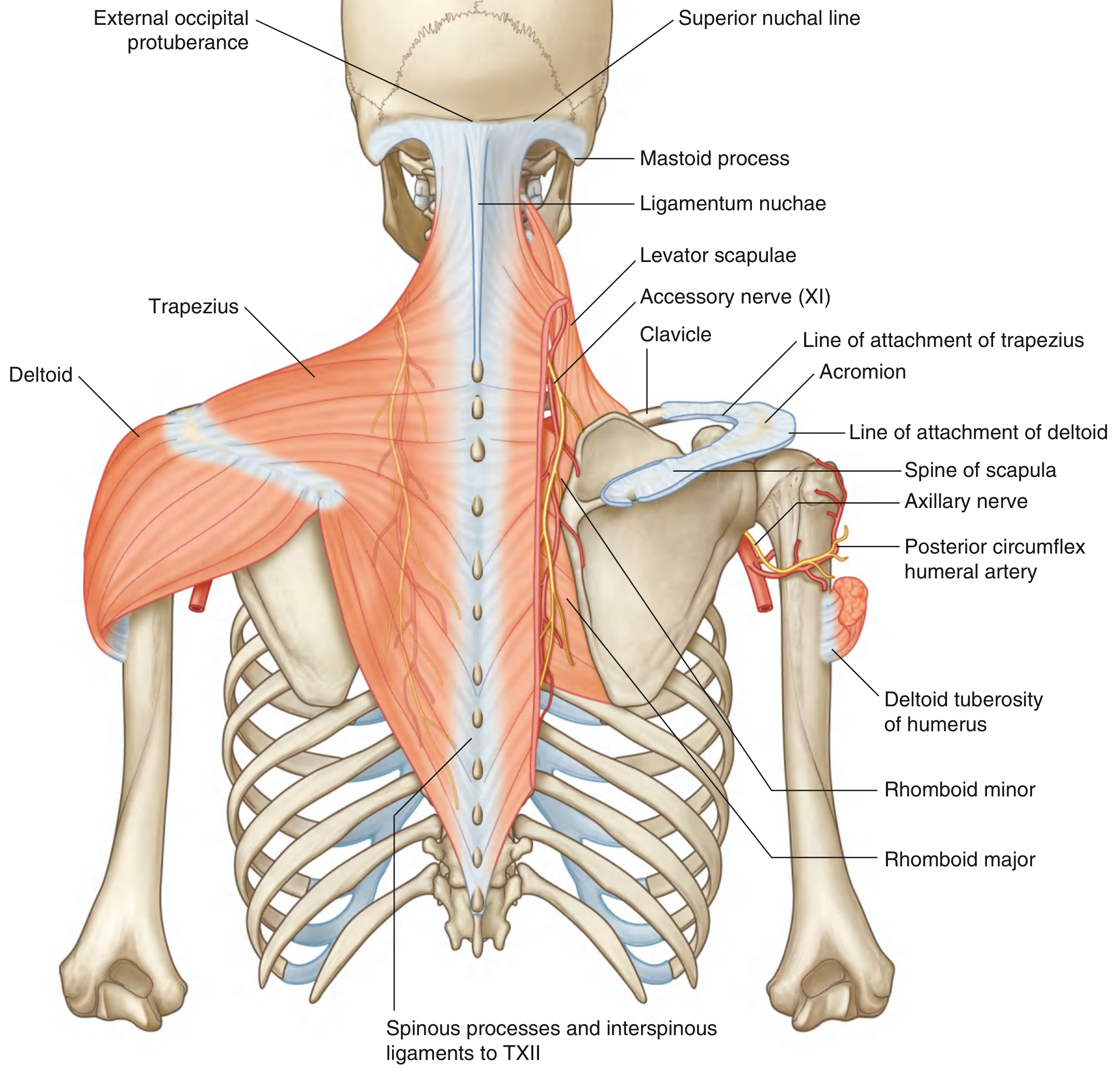
Attachments and neurovascular supply of the trapezius and deltoid muscles. (Gray's Anatomy for Students)
2. Relations
Superficially: Skin and superficial fascia. The superior lateral cutaneous nerve of the arm (sensory branch of the axillary nerve) pierces the deep fascia at the inferior border of the muscle.
Deep surface relations:
- The glenohumeral (shoulder) joint capsule lies deep to it, covered by the rotator cuff muscles (supraspinatus, infraspinatus, teres minor)
- The subacromial bursa (subdeltoid bursa) separates it from the supraspinatus tendon and greater tuberosity - this bursa does not normally communicate with the joint cavity
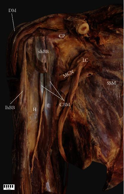
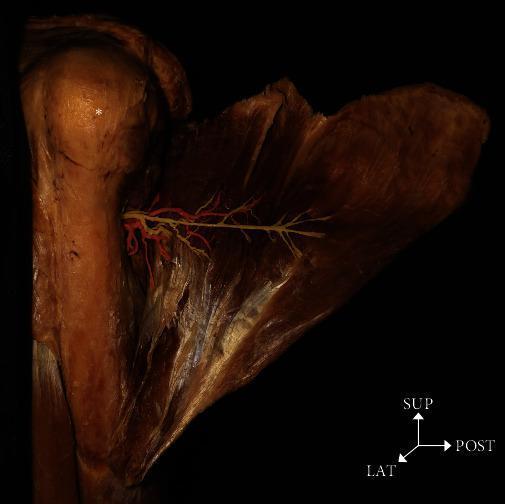
- The axillary nerve and posterior circumflex humeral artery run on its deep surface, entering the muscle by passing posteriorly around the surgical neck of the humerus through the quadrangular space
Medial border is contiguous with the pectoralis major anteriorly (forming the deltopectoral groove, through which the cephalic vein ascends) and with the infraspinatus and teres minor posteriorly.
The quadrangular (quadrilateral) space, through which the axillary nerve passes to reach the deltoid, is bounded by:
- Superiorly: teres minor and subscapularis
- Inferiorly: teres major
- Medially: long head of triceps
- Laterally: surgical neck of humerus
3. Actions
The actions of the three parts depend on their position relative to the axis of shoulder movement:
| Part | Primary Actions |
|---|---|
| Clavicular (anterior) | Flexion, medial (internal) rotation, adduction (below 60°), assists abduction above 60° |
| Acromial (middle) | Abduction (primary mover from 15° to 90°) |
| Spinal (posterior) | Extension, lateral (external) rotation, adduction (below 60°), assists abduction above 60° |
Key biomechanical point: Below 60° of abduction, the clavicular and spinal parts act as antagonists to the acromial part (they adduct). As the arm moves past ~60° of abduction, these parts cross the sagittal abduction/adduction axis and switch to acting as synergists, assisting in abduction. Thus all three parts contribute to abduction above 60°.
The deltoid cannot initiate abduction from 0° alone - the supraspinatus initiates the first 15° and depresses the humeral head, providing a fulcrum for the deltoid to work against.
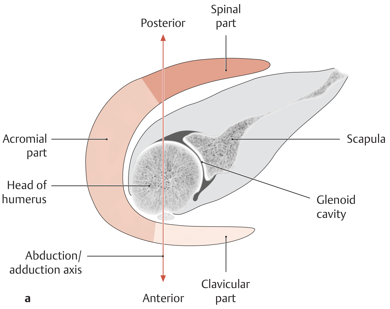
Schematic showing the three parts of the deltoid relative to the abduction/adduction axis. (THIEME Atlas of Anatomy)
4. Nerve Supply
- Motor nerve: Axillary nerve (C5, C6), a branch of the posterior cord of the brachial plexus
- Sensory (cutaneous): Superior lateral cutaneous nerve of the arm - the terminal cutaneous branch of the axillary nerve, supplying skin over the lower half of the deltoid ("regimental badge" area)
Course of the axillary nerve to the deltoid:
- Arises from the posterior cord at the lateral border of pectoralis minor
- Descends on the subscapularis muscle, behind the axillary artery
- Exits the axilla through the quadrangular space with the posterior circumflex humeral artery
- Winds around the posterior/lateral aspect of the surgical neck of the humerus
- Divides into anterior and posterior branches on the deep surface of the deltoid
- Also supplies teres minor and gives an articular branch to the glenohumeral joint capsule
The deltoid is also supplied by the posterior circumflex humeral artery (branch of the axillary artery), which accompanies the nerve through the quadrangular space.
5. Applied Aspects
A. Axillary Nerve Injury
The axillary nerve is vulnerable at the surgical neck of the humerus - a common site of fractures and dislocations. Injury results in:
- Paralysis of deltoid → loss of shoulder abduction (15°-90°)
- Paralysis of teres minor → loss of lateral rotation
- Sensory loss over the "regimental badge" area (skin over the inferior deltoid)
- Flattening of the shoulder contour (loss of deltoid bulk)
Causes of axillary nerve injury include:
- Fractures of the surgical neck of humerus
- Anterior shoulder dislocation (the nerve is stretched over the humeral head)
- Reduction of shoulder dislocation
- Intramuscular injections placed too high on the posterior shoulder
- Quadrangular space syndrome (see below)
- Crutch palsy (posterior cord injury)
- Neuralgic amyotrophy (Parsonage-Turner syndrome)
Important clinical note: The axillary nerve should always be tested before and after attempted reduction of a shoulder dislocation to establish baseline function and detect iatrogenic injury.
B. Quadrangular Space Syndrome
Hypertrophy or fibrosis of the muscles forming the quadrangular space can compress the axillary nerve within it. This typically produces atrophy of teres minor (more commonly than deltoid weakness), affecting rotator cuff control. It may present with point tenderness over the quadrangular space and pain on abduction/external rotation.
C. Intramuscular Injection Site
The deltoid is a commonly used IM injection site (vaccines, antipsychotic depots). The safe injection site is the upper outer quadrant of the deltoid, at least 2-3 finger-breadths below the acromion. Injections placed too high or too posteriorly risk injuring the axillary nerve.
D. Deltoid Rupture and Dehiscence
Complete rupture is uncommon and usually requires repair to bone. However, deltoid dehiscence (avulsion from the acromial origin) is a recognized complication of open rotator cuff repair and open acromioplasty. It is difficult to repair once established and causes significant loss of shoulder function.
E. Deltoid Fibrosis (Injection Fibrosis)
Repeated IM injections into the deltoid (especially antibiotics in children) can cause fibrous contracture of the muscle, resulting in restricted abduction and a characteristic "abduction jerk" gait pattern.
F. Deltoid-Splitting Surgical Approach
The deltoid can be split in line with its fibers for access to the shoulder (anterolateral approach). The axillary nerve limits the extent of this split - it must not extend more than 5 cm below the acromion to avoid damaging the nerve as it enters the deep surface of the muscle.
G. Shoulder Contour in Dislocation
In anterior shoulder dislocation, the humeral head moves anteroinferiorly, leaving a hollow under the acromion. The deltoid, which normally rounds the shoulder contour, now drapes over an empty space, producing the characteristic "sulcus sign" or flattened shoulder appearance.
Sources:
- Gray's Anatomy for Students - Deltoid, Axillary Nerve, Quadrangular Space sections
- THIEME Atlas of General Anatomy and Musculoskeletal System - Section 2.5 Deltoid
- Imaging Anatomy: Bones, Joints, Vessels and Nerves (Vol. 3) - Deltoid Muscle, p. 100
- Localization in Clinical Neurology, 8e - Axillary nerve anatomy and lesions
- Miller's Review of Orthopaedics, 9th Ed - Deltoid rupture and clinical management
- Campbell's Operative Orthopaedics, 15th Ed 2026 - Deltoid muscle dysfunction and surgical approaches
This is a shared conversation. Sign in to Orris to start your own chat.