
What is normal endometrial thockness is diff phases of menstrual cycle
The endometrial thickness varies across the menstrual cycle: proliferative phase (days 6-14) ~4-8 mm, periovulatory ~8-12 mm, secretory phase (days 15-28) ~7-14 mm, and menstrual phase ~2-4 mm.
Searching sources...
Reading File
Finding Sources
Searching the Web
normal endometrial thickness different phases menstrual cycle ultrasound
Reading File
Reading File
Loading Image
Reading File
Here is a complete summary of normal endometrial thickness across all phases of the menstrual cycle, drawn from authoritative medical textbooks:
Normal Endometrial Thickness Across the Menstrual Cycle
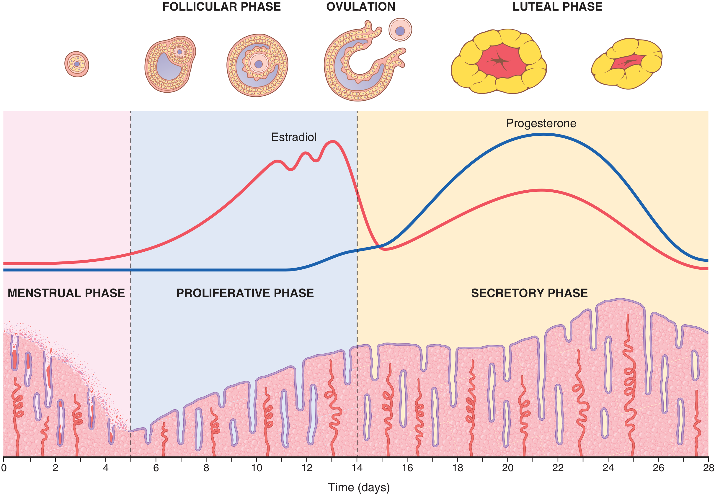
Phase-by-Phase Breakdown
| Phase | Days (28-day cycle) | Thickness | Hormone driver | Appearance on US |
|---|---|---|---|---|
| Menstrual | 1-5 | ~1-2 mm (thin stripe) | Estrogen + progesterone withdrawal | Thin echogenic line; minimal fluid |
| Early Proliferative | 5-9 | ~1-4 mm | Rising estrogen | Thin echogenic band |
| Late Proliferative (periovulatory) | 10-14 | ~3-8 mm (up to 11 mm) | Peak estrogen | Classic trilaminar (triple-layer) pattern |
| Secretory | 15-28 | ~5-6 mm (can reach 7-14 mm) | Progesterone dominant | Homogenous, echogenic, corkscrew glands |
Key Details by Phase
1. Menstrual Phase (Days 1-5)
- The corpus luteum involutes, estrogen and progesterone fall sharply
- The endometrium involutes to about 65% of its previous thickness
- Only the stratum basale (~1 mm) survives - this is the regenerative layer
- On ultrasound: thin, bright echogenic stripe with possible small amount of fluid
- Histology A Text and Atlas, p. 2245
2. Proliferative Phase (Days 5-14) - Estrogen-driven
- Driven by rising estradiol from developing follicles
- Glands are straight/tubular with narrow lumina and slightly wavy appearance
- At end of proliferative phase (day 14, just before ovulation): thickness reaches approximately 3-5 mm by histologic standards
- On ultrasound, thickness reaches up to 8-11 mm by the periovulatory period
- A distinctive trilaminar (triple-layer) appearance develops periovulatorily: a central echogenic line + hypoechoic functional layer + echogenic outer line
- Guyton and Hall, p. 1023; Histology A Text and Atlas, p. 2247
3. Secretory Phase (Days 15-28) - Progesterone-driven
- Progesterone from corpus luteum causes marked swelling and secretory changes
- Glands enlarge, become corkscrew-shaped and sacculated, fill with mucoid fluid rich in glycogen
- Endometrium becomes edematous
- At peak secretory phase (~day 21, 1 week after ovulation): thickness is 5-6 mm (histologic) or can reach 7-14 mm on ultrasound
- Spiral arteries lengthen and become more coiled, extending nearly to the surface
- On ultrasound: homogenous, uniformly echogenic appearance (progesterone effect)
- Guyton and Hall, p. 1023; Histology A Text and Atlas, p. 2247
Quick Reference Summary
| Source | Proliferative end | Secretory peak |
|---|---|---|
| Guyton & Hall (physiology) | 3-5 mm | 5-6 mm |
| Histology A Text and Atlas | ~3 mm | 5-6 mm |
| Radiopaedia / Ultrasound | up to 11 mm (periovulatory) | 7-14 mm |
Note on measurement discrepancy: The thickness values in physiology/histology textbooks reflect direct tissue measurements, while ultrasound (transvaginal) measurements are double-layer (measuring both the anterior and posterior endometrium together), which gives higher numbers. Ultrasound is the standard clinical measurement.
Postmenopausal Reference
- Normal: < 4-5 mm (no hormone replacement)
-
5 mm warrants investigation for endometrial pathology (e.g., carcinoma)
Clinical Pearl
The endometrium is best assessed by transvaginal ultrasound (TVS) in the sagittal plane, measuring the thickest echogenic area from one basal interface to the other. Because thickness varies so much with cycle phase, in premenopausal women with irregular bleeding, measurements are most reliable when timed to the early proliferative phase (days 5-10). As Radiopaedia notes, the significance of endometrial thickness in premenopausal women depends heavily on the cycle phase.
This is a shared conversation. Sign in to Orris to start your own chat.