

image.jpg
Answer these questions for bds 1st year biochemistry board exam of kathmandu university
analyse_media
Reading File
Now I have the questions. Let me search the medical library for all the topics simultaneously.
Finding Sources
Finding Sources
Finding Sources
Finding Sources
Finding Sources
Finding Sources
Finding Sources
Finding Sources
Good - I have Basic Medical Biochemistry (a perfect source for BDS 1st year). Let me read from that source plus Harrison's for porphyria.
Reading File
Reading File
Reading File
Good. I have enough from the library. Now let me also fetch the glycolysis diagram and do a web search for the specific BDS biochemistry topics to supplement my comprehensive answers.
Loading Image
Searching the Web
heme biosynthesis pathway regulation ALA synthase enzymes steps
I now have rich content from the textbooks and web sources. Let me compile comprehensive answers for all questions.Here are fully detailed answers to all the questions from your Haematopoietic section of BDS 1st Year Biochemistry:
HAEMATOPOIETIC BIOCHEMISTRY - BDS 1st Year Board Exam Answers
Q1. Define and classify porphyria. Explain the regulation of heme biosynthesis. [1+2+2=5]
Definition of Porphyria [1 mark]
Porphyrias are a group of inherited (rarely acquired) metabolic disorders caused by enzyme defects in the heme biosynthesis pathway, leading to accumulation of porphyrins or their precursors (ALA, PBG) in tissues, urine, and feces.
Classification of Porphyria [2 marks]
A. Based on site of enzyme defect:
| Type | Site | Examples |
|---|---|---|
| Hepatic porphyrias | Liver | Acute Intermittent Porphyria (AIP), Porphyria Cutanea Tarda (PCT), Hereditary Coproporphyria (HCP), Variegate Porphyria (VP) |
| Erythropoietic porphyrias | Bone marrow/RBCs | Congenital Erythropoietic Porphyria (CEP), Erythropoietic Protoporphyria (EPP) |
B. Based on clinical presentation:
- Acute porphyrias - present with neurovisceral attacks (acute abdominal pain, neuropsychiatric features, autonomic dysfunction). Examples: AIP, HCP, VP
- Cutaneous porphyrias - present with photosensitive skin lesions. Example: PCT
- Mixed (neurocutaneous) - both features. Example: VP, HCP
Regulation of Heme Biosynthesis [2 marks]
Rate-limiting step: The first step, catalyzed by ALA synthase (ALAS), is the principal regulatory point.
The 8-step heme synthesis pathway:
Mitochondria:
1. Glycine + Succinyl-CoA → ALA (enzyme: ALA synthase - rate-limiting, PLP-dependent)
Cytosol:
2. 2 ALA → Porphobilinogen (PBG) (enzyme: ALA dehydratase / PBG synthase)
3. 4 PBG → Hydroxymethylbilane (HMB) (enzyme: PBG deaminase / HMBS)
4. HMB → Uroporphyrinogen III (enzyme: Uroporphyrinogen III synthase)
5. Uroporphyrinogen III → Coproporphyrinogen III (enzyme: Uroporphyrinogen decarboxylase)
Mitochondria:
6. Coproporphyrinogen III → Protoporphyrinogen IX (enzyme: Coproporphyrinogen oxidase)
7. Protoporphyrinogen IX → Protoporphyrin IX (enzyme: Protoporphyrinogen oxidase)
8. Protoporphyrin IX + Fe²⁺ → HEME (enzyme: Ferrochelatase)
Regulation mechanisms:
-
Feedback inhibition by heme: Heme (the final product) inhibits ALAS-1 (liver isoform) at three levels:
- Inhibits ALAS transcription
- Inhibits import of ALAS precursor into mitochondria
- Directly inhibits ALAS enzyme activity
-
Two isoforms of ALAS:
- ALAS-1 (liver): regulated by heme feedback; induced by drugs, starvation, hormones
- ALAS-2 (erythroid cells): regulated by iron - erythropoietin increases its expression; its mRNA has an iron-responsive element (IRE) that responds to intracellular iron levels
-
Drug-induced regulation: Drugs metabolized by cytochrome P450 (e.g., barbiturates, griseofulvin) deplete heme, removing the feedback inhibition and causing massive ALAS-1 induction - this can precipitate acute porphyria attacks
-
Ferrochelatase inhibition: Lead inhibits both ALA dehydratase (step 2) and ferrochelatase (step 8), causing porphyrin accumulation in lead poisoning
Q2. Write Short Notes
a) Structure and Function of Hemoglobin
Structure:
- Hemoglobin (Hb) is a tetrameric globular protein - 4 polypeptide subunits, each carrying one heme group
- HbA (adult): 2alpha + 2beta chains (most abundant, ~97%)
- HbA2: 2alpha + 2delta chains (~2.5%)
- HbF (fetal): 2alpha + 2gamma chains (higher O2 affinity - beneficial in fetus)
- Each subunit has a heme prosthetic group: protoporphyrin IX ring + Fe²⁺ (ferrous iron)
- Fe²⁺ can bind one O2 molecule, so each Hb can carry 4 molecules of O2
- The protein exists in two states:
- T (tense/deoxy) state: low O2 affinity; stabilized by 2,3-BPG, H⁺, CO2
- R (relaxed/oxy) state: high O2 affinity
Functions:
- O2 transport: Picks up O2 in lungs (high pO2) and releases in tissues (low pO2)
- CO2 transport: ~23% of CO2 transported as carbaminohemoglobin (Hb-NHCOO⁻)
- Buffering: Histidine residues act as buffers, helping maintain blood pH
- Bohr Effect: H⁺ and CO2 decrease O2 affinity of Hb, facilitating O2 release in tissues
- 2,3-BPG effect: Binds beta chains in T state, stabilizing deoxy form and reducing O2 affinity (important in adaptation to altitude/anemia)
b) G6PD Deficiency
Definition: G6PD (Glucose-6-phosphate dehydrogenase) deficiency is the most common enzyme deficiency worldwide, affecting ~400 million people. It is X-linked recessive.
Biochemistry:
- G6PD catalyzes the first step of the HMP (Hexose Monophosphate) shunt / Pentose Phosphate Pathway:
- Glucose-6-phosphate + NADP⁺ → 6-Phosphogluconolactone + NADPH
- NADPH is essential for regenerating glutathione (GSH) via glutathione reductase
- GSH neutralizes oxidative radicals (H2O2) via glutathione peroxidase
- RBCs lack mitochondria - they depend entirely on the HMP shunt for NADPH
Clinical consequences of G6PD deficiency:
- Without sufficient NADPH, GSH cannot be maintained
- Oxidative stress (drugs, infections, fava beans) causes:
- Oxidation of Hb → Heinz bodies (denatured Hb precipitates)
- RBC membrane damage → Hemolytic anemia
- Triggers: Primaquine, dapsone, nitrofurantoin, aspirin, infection, fava beans
- Labs: Heinz body stain positive, decreased G6PD enzyme assay, normocytic hemolytic anemia
Why it is protective against malaria: Infected RBCs lyse more readily in G6PD-deficient individuals, limiting parasite proliferation.
c) Sickle Cell Haemoglobin and Its Laboratory Diagnosis
Molecular defect:
- Autosomal recessive point mutation in the beta-globin gene (chromosome 11)
- Glutamate (Glu) → Valine (Val) at position 6 of the beta chain
- This creates HbS (alpha2 beta2S) instead of normal HbA
Pathophysiology:
- Under deoxygenated conditions, HbS polymerizes (forms long, rod-like fibers)
- These fibers distort RBCs into a sickle (crescent) shape
- Sickled cells:
- Are rigid and non-deformable - obstruct small vessels (vaso-occlusive crisis)
- Have shortened lifespan (~20 days vs normal 120 days) - hemolytic anemia
- Cause ischemia, pain crises, acute chest syndrome, stroke
HbS properties:
- Reduced O2 affinity compared to HbA
- HbS is insoluble when deoxygenated - this is the basis of the sickling test
Laboratory Diagnosis:
| Test | Principle | Result in SCA |
|---|---|---|
| Peripheral blood smear | Morphology | Sickle cells, target cells, Howell-Jolly bodies |
| Sickling test (Na2S2O4) | Sodium metabisulfite/dithionite deoxygenates blood; HbS sickles | Positive |
| Hb solubility test | HbS precipitates in high-phosphate buffer | Turbid (positive) |
| Hb electrophoresis | HbS migrates differently than HbA at alkaline pH | HbS band at distinct position; confirms and distinguishes HbSS, HbSC, HbS-beta-thal |
| HPLC | Gold standard for Hb variant identification | Identifies and quantifies HbS accurately |
| Osmotic fragility test | Decreased fragility in SCA (target cells) | Decreased |
| DNA analysis | Detects beta-globin mutation | Definitive; used for prenatal diagnosis |
d) Glycolytic Products in RBCs
RBCs are unique in that they lack mitochondria, nuclei, and ribosomes. They rely entirely on glycolysis (anaerobic) for energy.
Key glycolytic products in RBCs:
-
ATP (2 mol per mol glucose):
- Powers Na⁺/K⁺-ATPase - maintains RBC shape and volume
- Powers Ca²⁺-ATPase
- Maintains membrane flexibility
-
NADH:
- Reduces methemoglobin (Fe³⁺) back to functional hemoglobin (Fe²⁺) via methemoglobin reductase
-
Pyruvate → Lactate:
- In RBCs, pyruvate is converted to lactate (anaerobic end product)
- Regenerates NAD⁺ to keep glycolysis running
-
2,3-Bisphosphoglycerate (2,3-BPG):
- Unique to RBCs via the Rapoport-Luebering shunt (bypasses the ATP-generating step)
- Produced from 1,3-BPG by bisphosphoglycerate mutase
- Key allosteric regulator of Hb-O2 affinity: binds to deoxyhemoglobin between beta chains, stabilizes T-state, reduces O2 affinity, facilitating O2 delivery to tissues
- Increased in altitude, anemia, chronic lung disease
-
NADPH (via HMP shunt):
- Maintains glutathione in reduced form (GSH) - protects against oxidative damage
e) Regulation of Heme Synthesis
(See Q1 detailed answer above)
Key points summary:
- Main regulator: ALA synthase (ALAS) - rate-limiting enzyme
- ALAS-1 (hepatic): Inhibited by heme (end-product feedback); induced by drugs (P450 inducers)
- ALAS-2 (erythroid): Regulated by iron via IRE in mRNA; induced by erythropoietin
- Lead poisoning: Inhibits ALA dehydratase and ferrochelatase - causes accumulation of ALA and Zn-protoporphyrin
f) Porphyria
(See Q1 definition and classification above)
Additional clinical points for short note:
Acute Intermittent Porphyria (AIP) - most tested:
- Enzyme defect: PBG deaminase (HMBS) deficiency (step 3)
- Autosomal dominant
- Hepatic type; NO skin manifestations
- Clinical triad:
- Acute abdominal pain (colicky)
- Neuropsychiatric symptoms (anxiety, hallucinations, seizures)
- Peripheral neuropathy (ascending motor neuropathy)
- Urine turns dark reddish-brown on standing (oxidation of PBG to porphyrins)
- Diagnosis: Elevated urine ALA + PBG during acute attacks
- Precipitating factors: Drugs (barbiturates, sulfonamides, OCP), fasting, alcohol, infection, stress, hormones
- Treatment: IV glucose (10-20 g/hr), IV hematin; Givosiran (siRNA) for recurrent attacks
Porphyria Cutanea Tarda (PCT):
- Most common porphyria
- Enzyme defect: Uroporphyrinogen decarboxylase (step 5)
- Photosensitive skin blisters on sun-exposed areas
- Associated with alcohol, estrogen, hepatitis C, HIV, iron overload
- Treatment: Phlebotomy, low-dose hydroxychloroquine
Q3. Discuss diagrammatically the absorption of non-heme iron, its transport and storage. How does hepcidin regulate iron absorption? [4+2=6]
Non-Heme Iron Absorption [Diagrammatic]
LUMEN (duodenum/upper jejunum)
|
Non-heme iron (Fe³⁺)
|
Duodenal cytochrome b (DCYTB) ← reduces Fe³⁺ → Fe²⁺
|
Fe²⁺ enters enterocyte via
DMT-1 (Divalent Metal Transporter-1)
|
Inside enterocyte:
- Small pool stored as FERRITIN
- Rest transferred across basolateral membrane
|
FERROPORTIN (FPN-1) exports Fe²⁺ from enterocyte → portal blood
|
HEPHAESTIN (copper-containing ferroxidase) oxidizes Fe²⁺ → Fe³⁺
|
Fe³⁺ binds TRANSFERRIN (each molecule binds 2 Fe³⁺)
|
Transported via BLOOD to tissues
Factors enhancing non-heme iron absorption:
- Vitamin C (ascorbic acid) - reduces Fe³⁺ to Fe²⁺
- Acidic pH (gastric HCl)
- Animal protein (MFP factor)
- Iron deficiency state
- Pregnancy, erythropoiesis
Factors reducing non-heme iron absorption:
- Phytates (cereals), oxalates (spinach), polyphenols (tea, coffee)
- Alkaline pH, antacids
- Calcium, zinc (compete with DMT-1)
- Iron overload (hepcidin upregulation)
Heme iron (from meat): absorbed as intact heme by HCP1 receptor; heme oxygenase releases Fe²⁺ inside enterocyte.
Iron Transport and Storage
Transport in blood:
- Transferrin: Major iron transport protein; a beta-1 globulin glycoprotein made in liver; carries Fe³⁺; normally ~30% saturated (Transferrin Saturation)
- Transferrin receptor (TfR1): On all cells, especially erythroid precursors and liver; binds transferrin-Fe complex; endocytosed; iron released in endosome (acidification)
Storage:
- Ferritin: Cytosolic storage protein; spherical shell of 24 apoferritin subunits; stores up to 4,500 iron atoms as Fe³⁺-phosphate complex; major storage in liver, spleen, bone marrow, muscle
- Hemosiderin: Aggregated, partially degraded ferritin; insoluble; mobilized slowly; seen in iron overload (Golden-brown granules with Prussian Blue staining)
Release from macrophages:
- Macrophages phagocytose old RBCs, break down heme via heme oxygenase, release iron via ferroportin back into circulation
Hepcidin Regulation of Iron Absorption [2 marks]
Hepcidin:
- A 25 amino acid antimicrobial peptide hormone produced by the liver
- The master regulator of systemic iron homeostasis
Mechanism of action:
- Hepcidin binds to Ferroportin (FPN-1) on enterocytes, macrophages, and hepatocytes
- This binding causes internalization and degradation of ferroportin
- Without ferroportin, iron is trapped inside enterocytes and macrophages
- Result: reduced iron absorption from gut and reduced iron release from stores
When is hepcidin upregulated (iron blocked)?
- Iron overload (high transferrin saturation, high ferritin)
- Inflammation/infection (IL-6 induces hepcidin) - this causes anemia of chronic disease
- BMP-SMAD signaling pathway activated by BMP6 (liver) when iron stores are high
When is hepcidin downregulated (iron absorption allowed)?
- Iron deficiency
- Hypoxia (via HIF-2alpha signaling)
- Increased erythropoiesis (erythropoietin, growth differentiation factor 15/ERFE)
- Anemia
Clinical relevance:
- Hemochromatosis (type 1): HFE gene mutation → hepcidin not produced → unregulated iron absorption → iron overload (liver, heart, pancreas, joints)
- Anemia of chronic disease: High hepcidin (from inflammation) → iron trapped → functional iron deficiency despite adequate stores
- Hepcidin mimetics are being developed to treat iron overload disorders
Q4. Write down the absorption, transport, storage and release of iron in our body [5]
(This overlaps significantly with Q3 - see above for full detail)
Summary table:
| Stage | Site | Key Molecules |
|---|---|---|
| Absorption | Duodenum/jejunum | DCYTB, DMT-1, Ferroportin, Hephaestin |
| Transport (blood) | Plasma | Transferrin (Fe³⁺) |
| Uptake by cells | Liver, bone marrow | Transferrin receptor 1 (TfR1) |
| Storage | Liver, spleen, BM | Ferritin, Hemosiderin |
| Release from stores | Macrophages, hepatocytes | Ferroportin (regulated by hepcidin) |
Daily iron cycle:
- Total body iron: ~3-4 g in men, ~2-3 g in women
- Daily dietary requirement: 8-10 mg/day (men), 18 mg/day (women)
- Daily absorption: ~1-2 mg (equal to daily loss)
- Most iron (~25 mg/day) is recycled from senescent RBCs by macrophages
Q5. Importance of HMP Shunt Pathway
Definition: The Hexose Monophosphate (HMP) shunt, also called the Pentose Phosphate Pathway (PPP) or Phosphogluconate pathway, is an alternative pathway of glucose oxidation that operates parallel to glycolysis.
Occurs in: Cytosol of cells (active in liver, RBCs, adrenal cortex, mammary gland, testes)
Two phases:
-
Oxidative (irreversible) phase: Glucose-6-P → Ribulose-5-P
- G6PD catalyzes: G-6-P + NADP⁺ → 6-Phosphogluconolactone + NADPH + H⁺ (rate-limiting)
- 6-Phosphogluconate dehydrogenase: 6-PG + NADP⁺ → Ribulose-5-P + NADPH + CO2
- Yield: 2 NADPH per glucose
-
Non-oxidative (reversible) phase: Interconverts pentose phosphates → fructose-6-P and glyceraldehyde-3-P (enters glycolysis)
Why the HMP shunt is important - 5 key roles:
-
Production of NADPH:
- Maintains glutathione (GSH) in reduced form - protects RBCs and other cells from oxidative damage
- Required for fatty acid synthesis (liver, mammary gland, adipose)
- Required for steroid/cholesterol synthesis (adrenal cortex, gonads)
- Required for cytochrome P450 reactions (liver detoxification)
- Phagocytic killing in neutrophils (NADPH oxidase generates superoxide)
-
Production of Ribose-5-phosphate:
- Precursor for nucleotide and nucleic acid synthesis (ATP, GTP, NAD, FAD, RNA, DNA)
- Essential for proliferating cells
-
Critical for RBCs:
- Only source of NADPH in RBCs (no mitochondria)
- Protects hemoglobin from oxidation (met-Hb formation)
- G6PD deficiency disrupts this - causes hemolytic anemia with oxidative drugs
-
Provides erythrose-4-phosphate:
- For synthesis of aromatic amino acids (via shikimate pathway in bacteria - basis of antibiotic targets)
-
Reductive biosynthesis in tissues:
- NADPH used in synthesis of fatty acids, bile acids, neurotransmitters, and nitric oxide (by NO synthase)
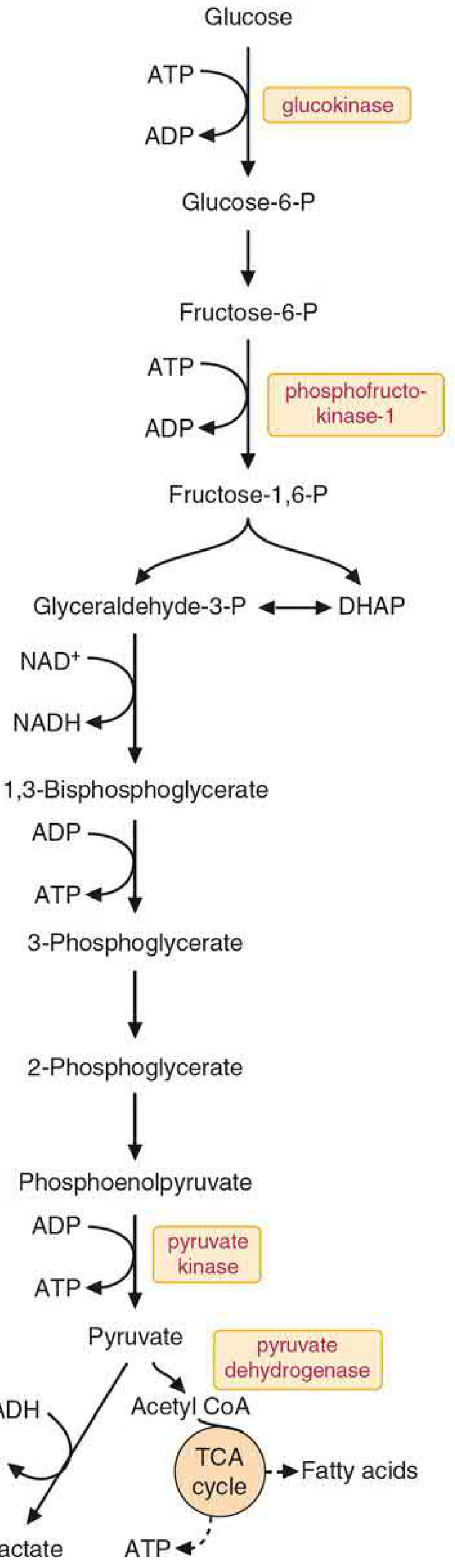
Q6. Reactions of Glycolysis with Rate-Limiting Enzymes
Glycolysis: The breakdown of one glucose molecule into two pyruvate molecules, occurring in the cytosol of all cells.
Net yield: 2 ATP, 2 NADH, 2 pyruvate (aerobic); 2 ATP, 2 lactate (anaerobic)
The 10 Steps of Glycolysis:
Phase I - Preparatory/Investment Phase (ATP used):
| Step | Reaction | Enzyme | Notes |
|---|---|---|---|
| 1 | Glucose → Glucose-6-P | Hexokinase (liver: Glucokinase) | ATP used; IRREVERSIBLE; regulated |
| 2 | Glucose-6-P → Fructose-6-P | Phosphoglucose isomerase | Reversible |
| 3 | Fructose-6-P → Fructose-1,6-bisP | Phosphofructokinase-1 (PFK-1) | ATP used; MOST IMPORTANT rate-limiting step; IRREVERSIBLE |
| 4 | Fructose-1,6-bisP → DHAP + Glyceraldehyde-3-P | Aldolase | Reversible |
| 5 | DHAP → Glyceraldehyde-3-P | Triose phosphate isomerase | Reversible |
Phase II - Pay-off Phase (ATP generated):
| Step | Reaction | Enzyme | Notes |
|---|---|---|---|
| 6 | Glyceraldehyde-3-P + NAD⁺ + Pi → 1,3-BPG + NADH | Glyceraldehyde-3-P dehydrogenase | Inhibited by iodoacetate |
| 7 | 1,3-BPG + ADP → 3-Phosphoglycerate + ATP | Phosphoglycerate kinase | Substrate-level phosphorylation |
| 8 | 3-Phosphoglycerate → 2-Phosphoglycerate | Phosphoglycerate mutase | Reversible |
| 9 | 2-Phosphoglycerate → PEP + H2O | Enolase | Inhibited by fluoride |
| 10 | PEP + ADP → Pyruvate + ATP | Pyruvate kinase | IRREVERSIBLE; regulated; third rate-limiting enzyme |
Three Rate-Limiting (Irreversible) Enzymes:
-
Hexokinase / Glucokinase (Step 1)
- Inhibited by: Glucose-6-P (product inhibition) - hexokinase
- Glucokinase (liver/pancreas): NOT inhibited by G6P; induced by insulin; low affinity (high Km) - acts as glucose sensor
-
Phosphofructokinase-1 - PFK-1 (Step 3) - THE most important
- Inhibited by: High ATP, citrate, H⁺ (acidosis)
- Activated by: AMP, ADP, Fructose-2,6-bisphosphate (most potent activator), Pi
- Fructose-2,6-bisP is made by PFK-2 (activated by insulin, inhibited by glucagon)
-
Pyruvate Kinase (Step 10)
- Inhibited by: ATP, alanine, glucagon (via phosphorylation)
- Activated by: Fructose-1,6-bisP (feedforward activation), AMP, insulin
- Pyruvate kinase deficiency causes hemolytic anemia (important in RBC)
Glycolysis in RBCs - Special Features:
- Anaerobic only (no mitochondria)
- ATP produced: 2 mol per mol glucose (net)
- Pyruvate is converted to lactate (by lactate dehydrogenase) to regenerate NAD⁺
- Rapoport-Luebering shunt: 1,3-BPG → 2,3-BPG (bypasses ATP production at step 7)
- 2,3-BPG produced is crucial for O2 delivery regulation
- Pyruvate kinase deficiency in RBCs: most common glycolytic enzyme deficiency causing hemolytic anemia; buildup of 2,3-BPG; right-shifted O2 dissociation curve (cells tolerate anemia better)
Q7. Give an Account of Acute Intermittent Porphyria (AIP)
Definition: AIP is the most common acute hepatic porphyria, caused by a partial deficiency (~50%) of porphobilinogen deaminase (PBGD/HMBS), the third enzyme in heme biosynthesis.
Genetics:
- Autosomal dominant, chromosome 11q23
- Penetrance is low - most carriers never develop attacks
- More common in women (female hormones trigger ALAS-1 induction)
Pathogenesis:
- When demand for heme increases (drugs, fasting, hormones) → ALAS-1 is induced
- Due to PBGD deficiency, ALA and PBG accumulate
- ALA is neurotoxic - interferes with GABA receptors and inhibitory neurons
- This causes the neurovisceral symptoms
Clinical Features - "Rule of 4Ps":
- Pain - severe, acute abdominal pain (most common presenting symptom)
- Polyneuropathy - peripheral and autonomic neuropathy
- Psychiatric - anxiety, confusion, hallucinations, psychosis
- Port-wine urine - reddish-brown urine on standing (PBG oxidizes to porphyrins)
Other features: Tachycardia, hypertension, constipation, urinary retention, SIADH (hyponatremia), seizures
Precipitants: "FACTS"
- Fasting / low carbohydrate diet
- Alcohol
- Chemicals/drugs (barbiturates, sulfonamides, rifampicin, OCP, griseofulvin, anticonvulsants)
- Toxins
- Stress / infection / surgery / menstrual cycle (luteal phase)
Diagnosis:
- Urine: elevated ALA and PBG (Watson-Schwartz test - urine turns pink-red with Ehrlich reagent for PBG)
- Red fluorescence of urine under UV light
- Erythrocyte PBGD activity reduced (~50%)
- No photosensitivity (unlike other porphyrias)
- Normal during remission - diagnosis missed unless tested during attack
Treatment:
- Remove precipitating factors (stop offending drug)
- IV Glucose (300-400 g/day) - reduces ALAS-1 induction (glucose effect)
- IV Hematin (3-4 mg/kg/day x 4 days) - provides exogenous heme, inhibits ALAS-1
- Supportive: IV fluids, pain management (opioids), beta-blockers for tachycardia/hypertension, benzodiazepines for seizures (NOT barbiturates or phenytoin - these are UNSAFE)
- Givosiran (siRNA) - monthly subcutaneous injection for recurrent attacks; neutralizes ALA mRNA in hepatocytes (approved 2019)
Complications of recurrent attacks:
- Chronic renal failure
- Hepatocellular carcinoma (liver)
- Chronic neuropathy
*Sources: Harrison's Principles of Internal Medicine 22E; Basic Medical Biochemistry (A Clinical Approach) 6E; Heme biosynthesis pathway*Here is the Glycolysis pathway diagram from the textbook for reference:
Here's a quick-reference summary of all questions answered:
| # | Topic | Key Point to Remember |
|---|---|---|
| Q1 | Porphyria + Heme regulation | 2 types (hepatic/erythropoietic); ALAS is rate-limiting; feedback by heme |
| Q2a | Hemoglobin structure | Tetramer; 4 heme groups; T & R states; 2,3-BPG |
| Q2b | G6PD deficiency | X-linked; no NADPH → no GSH → oxidative hemolysis; Heinz bodies |
| Q2c | Sickle cell Hb | Glu→Val mutation; HbS polymerizes when deoxygenated; diagnose by electrophoresis |
| Q2d | Glycolytic products in RBCs | ATP, NADH, 2,3-BPG, Lactate - no mitochondria |
| Q2f | Porphyria (short note) | AIP: PBG deaminase deficiency; port-wine urine; neurovisceral attacks |
| Q3/Q4 | Iron absorption/hepcidin | DCYTB→DMT-1→Ferroportin; Hepcidin degrades ferroportin |
| Q5 | HMP shunt importance | NADPH (protection, synthesis); Ribose-5-P (nucleotides) |
| Q6 | Glycolysis + enzymes | 3 rate-limiting: Hexokinase, PFK-1 (most important), Pyruvate kinase |
| Q7 | Acute Intermittent Porphyria | PBGD deficiency; ALA+PBG accumulate; Rx: glucose + hematin |
This is a shared conversation. Sign in to Orris to start your own chat.