
Why thyroid cancer causes respiratory problems
thyroid cancer respiratory problems mechanisms tracheal compression invasion
thyroid cancer tracheal compression airway invasion anatomy
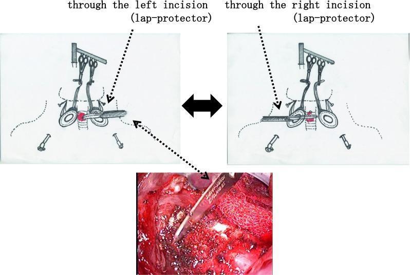
This composite educational resource illustrates the surgical 'shaving' technique for tracheal invasion during thyroid cancer surgery. The figure consists of two anatomical diagrams (top) and one intraoperative photograph (bottom). The diagrams depict a bilateral surgical approach using small supraclavicular incisions fitted with lap protectors. Visible instruments include retractors secured to a cradle and various clamps/ligating tools providing exposure to the tracheal cartilage. The bilateral view emphasizes that the invasive lesion can be accessed through either the left or right incision ports. The lower clinical photograph provides a close-up, intraoperative view of the procedure, showing a scalpel performing a tangential dissection (shaving) of tumor tissue from the tracheal wall while attempting to preserve the integrity of the airway. This technique is typically employed in thyroid surgery when complete resection is possible without necessitating a full tracheal resection and reconstruction.
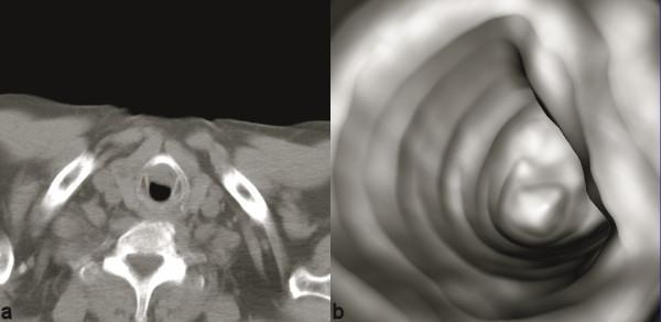
This composite diagnostic image features an axial computed tomography (CT) scan (left) and a corresponding reformatted virtual endoscopy (right) demonstrating severe tracheal stenosis secondary to thyroid cancer invasion. The axial CT at the level of the neck shows significant parenchymal irregularity and asymmetrical enlargement of the right thyroid lobe. There is clear evidence of the thyroid mass eroding the right lateral tracheal wall and extending intraluminally, causing a high-grade reduction in the airway diameter. The virtual endoscopy provides a 3-dimensional perspective of the same region, highlighting the eccentric nature of the tracheal narrowing. This volume-rendered view allows for the assessment of the intraluminal surface and the longitudinal extent of the stricture, which is critical for surgical planning. The educational focus is on the integration of 2D cross-sectional imaging with 3D reconstructive techniques to evaluate airway patency and tissue invasion in thoracic and head-and-neck oncology.
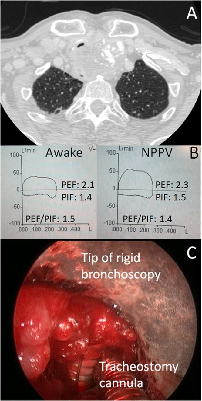
This composite educational image illustrates a case of extrinsic tracheal compression due to thyroid cancer and its subsequent management. Panel A is an axial CT scan of the upper chest showing a large, heterogeneous soft tissue mass anterior and lateral to the trachea, causing significant luminal narrowing and posterior displacement. Panel B presents Pulmonary Function Test (PFT) flow-volume loops comparing 'Awake' and 'Non-Invasive Positive Pressure Ventilation' (NPPV) states. The curves demonstrate a classic 'box' shape characteristic of fixed upper airway obstruction, with minimal change in Peak Expiratory Flow (PEF) and Peak Inspiratory Flow (PIF) despite positive pressure, indicating a firm, non-dynamic stenosis. Panel C is an intraoperative endoscopic photograph showing a rigid bronchoscopy-guided percutaneous dilatational tracheostomy (PDT). Visible landmarks include the tip of the rigid bronchoscope and a tracheostomy cannula positioned within an inflamed, stenotic tracheal lumen. This multi-modal presentation highlights the diagnostic pathway from radiological imaging and physiological assessment to surgical intervention for malignant airway obstruction.
How Thyroid Cancer Causes Respiratory Problems
1. Tracheal Compression (External)
- Dyspnea (shortness of breath, especially on exertion)
- Stridor (a high-pitched, noisy breathing sound from turbulent flow through a narrowed airway)
- Wheezing or sense of tightening in the throat
- A characteristic "box-shaped" fixed upper airway obstruction pattern on pulmonary function testing
2. Direct Tracheal Invasion (Intraluminal)
- Severe, potentially life-threatening airway narrowing
- Hemoptysis (coughing up blood) from erosion of mucosal vessels
- Asphyxia in extreme cases
- Post-obstructive pneumonia distal to the blockage
3. Recurrent Laryngeal Nerve (RLN) Invasion or Compression
- A breathy, barely audible voice
- Inefficient breathing (poor airway protection, air wasted with speech)
- Aspiration risk (food/liquid entering the airway) - can cause aspiration pneumonia
- Bilateral RLN involvement (rare but serious) can cause near-complete airway obstruction
4. Compression of Adjacent Structures
- Dyspnea
- Dysphagia (swallowing difficulty, which can cause aspiration and secondary respiratory issues)
- Horner syndrome (ptosis, miosis, anhidrosis from sympathetic chain compression)
- Superior vena cava syndrome (from mediastinal involvement causing venous congestion that can indirectly impair breathing)
- Pericardial or pleural effusions (which compress the lungs)
5. Distant Metastases to the Lungs
- Progressive dyspnea from reduced lung parenchyma
- Pleural effusion (fluid around the lungs, compressing them)
- Lymphangitic spread causing diffuse infiltrates
Summary Table
| Mechanism | Respiratory Effect | Cancer Type Most Commonly Involved |
|---|---|---|
| Tracheal compression | Stridor, dyspnea, fixed upper airway obstruction | Any large thyroid mass |
| Tracheal wall invasion/intraluminal tumor | Severe obstruction, hemoptysis, asphyxia | Anaplastic, advanced papillary |
| Recurrent laryngeal nerve paralysis | Vocal cord palsy, aspiration, weak cough | Any locally invasive cancer |
| Mediastinal/SVC compression | Venous congestion, pleural effusion | Large/mediastinal extension |
| Pulmonary metastases | Progressive dyspnea, effusion | Papillary, follicular (well-differentiated) |