
Equipment name with two definition 1.Medical others name (old to all A2Z name)give 123.... 2.physiology with principal of equipment 2 line 3.indication 4.contraindication 5.complication and don't use pt 6.picture equipment with parts 7.parts name and use and function of equipment 8.equipment with each part placement of anatomy lande mark 9.types of equipment and size adult and paediatric and male and female 10.if applicable colour code 11.sterlization of equipment 12. Procedure use equipment with vedio link youtube Reference Refers book Code blue manual guide and international trauma life support and tinnalae 9th editions and atls procedures latha ganti latest editions and Reichman’s Emergency Medicine Procedures and Miller book of anaesthetic and ROBERTS & HEDGES CLINICAL PROCEDURES in Emergency Medicine and UNDERSTANDING ANESTHETIC EQUIPMENT & PROCEDURES A PRACTICAL APPROACH Editors Dwarkadas K Baheti Et tube Vandana V Laheri
endotracheal tube parts cuff pilot balloon Murphy eye connector
"Intubation, Intratracheal"[MeSH Terms] AND "Equipment Design"[MeSH Terms]
endotracheal intubation procedure video YouTube NEJM OR Medcram OR Laerdal
Endotracheal (ET) Tube: Equipment Note
Definitions
- Definition 1 (structural): A flexible, curved, usually PVC tube with a 15 mm connector at one end and a beveled tip at the other. It is passed through the mouth or nose and between the vocal cords into the trachea.
- Definition 2 (functional): A cuffed or uncuffed airway device that gives a sealed, open channel between the outside and the trachea. It allows positive-pressure ventilation, oxygen delivery, suction, and protection from aspiration.
1. Other names (A to Z)
- Armoured tube (spiral-wire reinforced)
- Breathing tube
- Cole tube (neonatal, shouldered, uncuffed)
- Cuffed tube
- Double-lumen tube (DLT, a related specialised tube)
- Endotracheal tube (ETT, ET tube)
- Flexometallic tube
- Laser-resistant tube
- Magill tube (no Murphy eye)
- Microlaryngeal tube (MLT)
- Murphy tube (with Murphy eye)
- Nasotracheal tube
- Oral or nasal RAE tube (preformed)
- Orotracheal tube
- Oxford tube (L-shaped)
- Portex or Mallinckrodt tube (brand names)
- Reinforced tube
- Tracheal tube
- Uncuffed (plain) tube
2. Physiology and principle
- The tube bypasses the pharynx and larynx, so air moves directly between the machine or bag and the trachea. The inflated cuff seals against the tracheal wall. This allows positive-pressure ventilation and reduces the chance of aspiration.
- Resistance to flow depends mainly on tube diameter, and also on tube length and curvature. The choice of size is always a compromise between larger flow and less airway trauma. - Morgan and Mikhail's Clinical Anesthesiology 7e, p. 588
3. Indications
- Failure of oxygenation or ventilation.
- Inability to maintain or protect the airway (low GCS, risk of aspiration).
- Anticipated clinical deterioration (for example burns, expanding neck haematoma, severe sepsis, shock).
- General anaesthesia where a secure airway is needed (surgery on the head, neck, or abdomen; prone or lateral position; long cases).
- Cardiac arrest and resuscitation.
- Need for pulmonary toilet or mechanical ventilation.
4. Contraindications
- Complete laryngeal or tracheal disruption, or a laryngeal fracture. Use a surgical airway or a controlled approach.
- Complete upper airway obstruction that prevents tube passage. Go to a front-of-neck access (cricothyroidotomy).
- Unstable cervical spine. This is not a bar, but it requires in-line stabilisation.
- Acute epiglottitis in a child. Intubate in a controlled setting, with a surgeon ready.
- Nasal route only: basal skull fracture, severe midface fracture, coagulopathy or anticoagulation, nasal obstruction, sinusitis.
5. Complications
- Dental, lip, or tongue injury.
- Pharyngeal or laryngeal injury, vocal cord damage, arytenoid dislocation.
- Oesophageal intubation.
- Right main bronchus (endobronchial) intubation.
- Hypoxia, aspiration, laryngospasm, bronchospasm.
- Hypertension, tachycardia, arrhythmia, raised intracranial pressure.
- Epistaxis (nasal route).
- Tracheal perforation.
- Cuff over-pressure causing mucosal ischaemia.
- Tube blockage, kinking, or biting.
- Accidental extubation.
- Sore throat and hoarseness.
- Ventilator-associated pneumonia.
- Sinusitis (nasal tubes).
- Post-extubation stridor.
- Subglottic or tracheal stenosis and tracheomalacia.
- Vocal cord granuloma or paralysis.
- Tracheo-oesophageal fistula.
6. Picture with parts
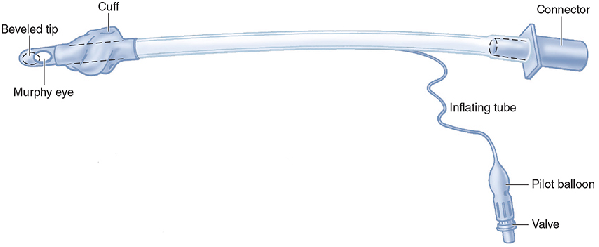
7. Parts, use, function, and anatomical landmark (adult oral tube)
| Part | Function | Where it sits |
|---|---|---|
| Beveled tip (patient end) | The bevel helps the tube pass between the vocal cords. It faces left in a standard tube. | Mid-trachea, about 3 to 5 cm above the carina. The carina is at T4-T5, level with the sternal angle. |
| Murphy eye | A side hole opposite the bevel. It keeps gas flowing if the tip abuts the carina or tracheal wall. | Just beyond the cuff, in the trachea. |
| Cuff | A high-volume, low-pressure cuff seals the trachea. It permits positive-pressure ventilation and reduces aspiration. | Upper trachea, below the cricoid cartilage (C6). Its upper edge is at least 1.5 to 2 cm below the vocal cords. |
| Vocal cord guide (black marker line, some brands) | Shows the correct depth at the cords. | At the glottis. |
| Tube shaft (PVC, with centimetre markings and a radio-opaque line) | Carries gas. The ID and cm depth are printed on it, and the line shows on chest X-ray. | From the lips (or nostril) to the trachea. |
| Inflating tube | A channel inside the tube wall that links the valve to the cuff. | Runs along the shaft and exits near the lips. |
| Pilot balloon | A rough indicator that the cuff is inflated. | Outside the mouth, at the cheek. |
| Valve | Stops air leaking from the cuff after inflation. A syringe fits here. | Outside, at the end of the pilot line. |
| 15 mm connector | Connects to the breathing circuit or bag. | Outside the mouth, at the lips. |
- Cuff inflation:
- Inflate until no leak is heard during bag ventilation, usually 5 to 8 mL.
- In an emergency, 10 mL is acceptable, then adjust once the patient is stable.
- For long-term use, keep cuff pressure at 20 to 25 mm Hg.
- Capillary flow in the mucosa is compromised above 30 mm Hg. - Roberts and Hedges' Clinical Procedures, Ch 4, p. 118
- Depth at the teeth: about 21 cm for adult females and 23 cm for adult males (from my own knowledge). Morgan gives a cut length of 24 cm. For a nasal tube, add about 2 to 3 cm.
- Related equipment: laryngoscope (handle, blade, bulb or light, battery), stylet or bougie, 10 mL syringe, Magill forceps, suction, bag-valve-mask, and capnography. - Morgan and Mikhail 7e, p. 590
8. Types and sizes
- Plain or Magill (no eye) and Murphy (with eye).
- Cuffed and uncuffed.
- Oral and nasal.
- Preformed (RAE).
- Armoured (spiral-wound, wire-reinforced).
- Microlaryngeal.
- Double-lumen.
- Tubes with bronchial blockers.
- Laser-resistant (metal).
- Nerve integrity monitor (NIM) tubes. - Morgan and Mikhail 7e, p. 589-590
| Group | Cuffed ID (mm) | Notes |
|---|---|---|
| Adult male | 7.5 to 9.0 | A 7.5 mm tube is enough for almost all patients. |
| Adult female | 7.0 to 8.0 | Morgan gives 7.0 to 7.5. |
| Preterm | 2.5 | |
| Term | 3.0 | |
| 6 months | 3.0 to 3.5 | |
| 1 to 2 years | 4.0 | |
| 3 to 4 years | 4.5 | |
| 5 to 6 years | 5.0 | |
| 10 years | 6.0 | |
| 12 years | 7.0 | |
| 14 years and above | Use adult sizing |
- Children over 2 years: uncuffed ID = (age/4) + 4. Cuffed ID = (age/4) + 3. - Roberts and Hedges, p. 118
- Tintinalli gives cuffed ID = (age/4) + 4 for children over 1 year, so sources differ. The AHA formula is (age/4) + 3.5. A cuffed tube is usually 0.5 mm smaller than an uncuffed one.
- The width of the little fingernail is a quick guide to size.
- In a child, the narrowest part of the airway is the cricoid ring, not the cords. If the tube passes the cords but not farther, use the next smaller size.
- Cuffed tubes are now increasingly favoured in children. - Morgan and Mikhail 7e, p. 589
- Oral depth in a child (from my own knowledge): about 3 times the ID, or (age/2) + 12 cm.
- Nasal tubes are 0.5 to 1.0 mm smaller than oral tubes.
- A cuffed tube is the better choice in a patient with a burn, because swelling may prevent a later tube change.
9. Colour code
- There is no universal colour code for adult ET tubes. The tube is clear, and the ID is printed on it. Some brands colour the connector or pilot balloon by size, so read the printed size.
- In children, colour comes from the length-based tape (Broselow). The tape colour gives the tube size, blade, and other equipment (from my own knowledge, so check your tape version):
| Colour | Cuffed ET tube ID (mm) |
|---|---|
| Grey | 3.5 |
| Pink | 3.5 |
| Red | 3.5 |
| Purple | 4.0 |
| Yellow | 4.5 |
| White | 5.0 |
| Blue | 5.5 |
| Orange | 6.0 |
| Green | 6.5 |
10. Sterilisation and reuse (from my own knowledge, not from the retrieved textbook text)
- The ET tube is single-use. The manufacturer sterilises it, usually with ethylene oxide. Do not resterilise or reuse it. Check that the pack is intact and in date.
- Laryngoscope blades:
- Disposable blades are preferred.
- Reusable metal blades need cleaning, then autoclaving or high-level disinfection, per the manufacturer.
- Wipe the handle with a low-level disinfectant.
- Stylets and bougies are single-use where possible. Reusable ones need cleaning and autoclaving.
- Reusable Magill forceps are autoclaved.
11. Procedure and videos
- Prepare equipment, check the cuff and light, and plan a back-up airway.
- Position the patient (sniffing position) and preoxygenate.
- Give induction and paralysis drugs, as needed.
- Open the mouth and insert the blade. Lift to see the cords.
- Pass the tube through the cords until the cuff is just beyond them.
- Remove the stylet, inflate the cuff, and ventilate.
- Confirm placement with waveform capnography, chest rise, and bilateral breath sounds. Secure the tube and obtain a chest X-ray.
Recent evidence check
Next opa
oropharyngeal airway Guedel parts flange bite block
"oropharyngeal airway" AND "airway management"
oropharyngeal airway insertion video YouTube demonstration sizing
Oropharyngeal Airway (OPA): Equipment Note
Definitions
- Structural: A rigid, curved plastic device with a flat flange at one end. It is placed over the tongue, through the mouth, to the back of the throat.
- Functional: A temporary airway adjunct that holds the tongue away from the back of the throat, keeping an air channel open in an unconscious patient. It is not a definitive airway.
1. Other names (A to Z)
- Berman airway (side-channelled, open design)
- Brook airway (mouth-to-mouth resuscitation type)
- Guedel airway (the commonest name)
- Mayo airway (older, tubular type)
- Oral airway
- Ovassapian airway (fibreoptic intubating airway, a related type)
- Oropharyngeal airway (OPA)
- Patil-Syracuse airway (related intubating type)
- Pharyngeal airway
- Safar airway (a pharyngeal tube for mouth-to-mouth)
- Williams airway (related intubating type)
2. Physiology and principle
- In a deeply unconscious patient, muscle tone falls and the tongue and soft tissue drop back against the posterior pharyngeal wall. This blocks the airway at the hypopharynx.
- The curved body of the OPA follows the tongue and lifts its base forward. Air can then pass through and around the device. It does this without any airway stimulation reflex control, so it is tolerated only when the gag reflex is absent. - Tintinalli's Emergency Medicine, Ch 28
3. Indications
- Comatose or deeply obtunded patient with tongue-base obstruction and no gag reflex. - Tintinalli's, Ch 28
- Adjunct to bag-mask ventilation when the airway is obstructed. - Miller's Anesthesia 10e, Mask Ventilation
- Cardiac arrest and resuscitation.
- Bite block during oral intubation or oral endoscopy, so the patient cannot bite the tube (own knowledge).
- A step in the apnoea sequence: reposition, jaw thrust, oral airway, nasal airway, supraglottic device, intubation, surgical airway.
4. Contraindications
- Intact gag reflex or a conscious or semi-conscious patient. The device can trigger vomiting and laryngospasm.
- Severe oral or dental injury, loose teeth, or recent oral surgery (relative, own knowledge).
- Airway obstruction that is not caused by the tongue, for example a foreign body, swelling, or a mass.
- Small child: do not rotate the airway. Use a tongue depressor instead (see section 10).
5. Complications
- Gagging, vomiting, aspiration, and laryngospasm in a patient who is not deeply obtunded.
- Injury to teeth, lips, tongue, gums, or palate.
- A wrongly sized airway can worsen obstruction. If too small, it pushes the tongue back. If too large, it can press on the epiglottis or larynx. - Miller's 10e
- Artificial airways may cause significant injuries. - Fishman's Pulmonary Diseases and Disorders
- Pressure injury to the tongue or lips with prolonged use (own knowledge).
- Bleeding, and pinching of the lips between teeth and device.
6. Pictures
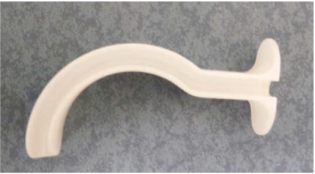
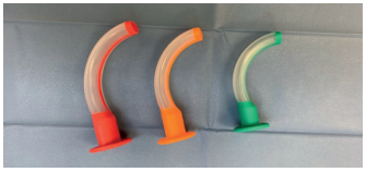
7. Parts, use, function, and anatomical landmark (Guedel type; own knowledge)
| Part | Function | Where it sits |
|---|---|---|
| Flange (proximal end) | A flat rim that stops the airway sliding into the mouth. It also gives a grip for insertion and removal. Its colour shows the size. | Outside, resting on the lips and incisors. |
| Bite block | A reinforced, rigid section so the patient cannot bite and close the channel. | Between the upper and lower teeth. |
| Curved body (channel) | A hollow curved shaft that follows the tongue. Air passes through it, and a suction catheter can go down it. | Over the dorsum of the tongue, under the hard palate. |
| Distal tip | Holds the base of the tongue off the posterior pharyngeal wall. | In the oropharynx or hypopharynx, above the epiglottis. |
8. Types and sizes
- The correct size extends from the corner of the mouth to the earlobe or the angle of the mandible. - Roberts and Hedges' Clinical Procedures, Fig 3.5
- Tintinalli's and Miller's use the corner of the mouth to the angle of the mandible. Current Surgical Therapy 14e uses the midpoint of the incisors to the angle of the mandible.
| Size | Length | Usual patient |
|---|---|---|
| 000 | 40 mm | Premature or neonate |
| 00 | 50 mm | Infant |
| 0 | 60 mm | Small infant |
| 1 | 70 mm | Young child |
| 2 | 80 mm | Older child or small adult |
| 3 | 90 mm | Medium adult, usually female |
| 4 | 100 mm | Large adult, usually male |
| 5 | 110 mm | Very large adult |
9. Colour code
- Colour codes are not universal and differ between manufacturers. The size number is usually printed on the flange as well.
- A commonly used European (own knowledge) scheme: size 2 green, size 3 yellow, size 4 red. The smaller sizes vary by brand. The Bailey and Love photo shows orange and green bases.
- Read the printed size on the device and check the pack.
10. Sterilisation (own knowledge)
- Most OPAs are single-use disposable plastic, supplied clean. Discard after use and do not reprocess.
- Reusable (rubber or autoclavable plastic) ones need washing and then autoclaving or high-level disinfection, per the manufacturer.
11. Procedure
- Measure along the side of the face (mouth corner to earlobe or angle of the jaw).
- Open the mouth with the thumb and index finger.
- Insert the airway inverted (concave side up), along the hard palate.
- When well into the mouth, rotate it 180 degrees so the tip lies in the hypopharynx. It may help to pull the jaw forward during passage.
- Alternative: use a tongue blade to push the tongue down, and advance the airway in its natural curve. No rotation is needed.
- Tintinalli's also describes inserting with the concave side horizontal and rotating 90 degrees.