
Pars flaccida and pars densa
Reading File
The pars flaccida and pars tensa are the two parts of the tympanic membrane (eardrum). The pars flaccida (Shrapnell's membrane) is the smaller, upper portion above the lateral malleolar folds, while the pars tensa forms the larger, taut main body of the eardrum.
Searching sources...
Finding Sources
Finding Sources
Reading File
Reading File
Reading File
Reading File
Loading Image
Loading Image
Here is a comprehensive overview of the pars flaccida and pars tensa - the two parts of the tympanic membrane (TM).
Tympanic Membrane: Pars Flaccida vs. Pars Tensa
Endoscopic view of the right tympanic membrane (normal):
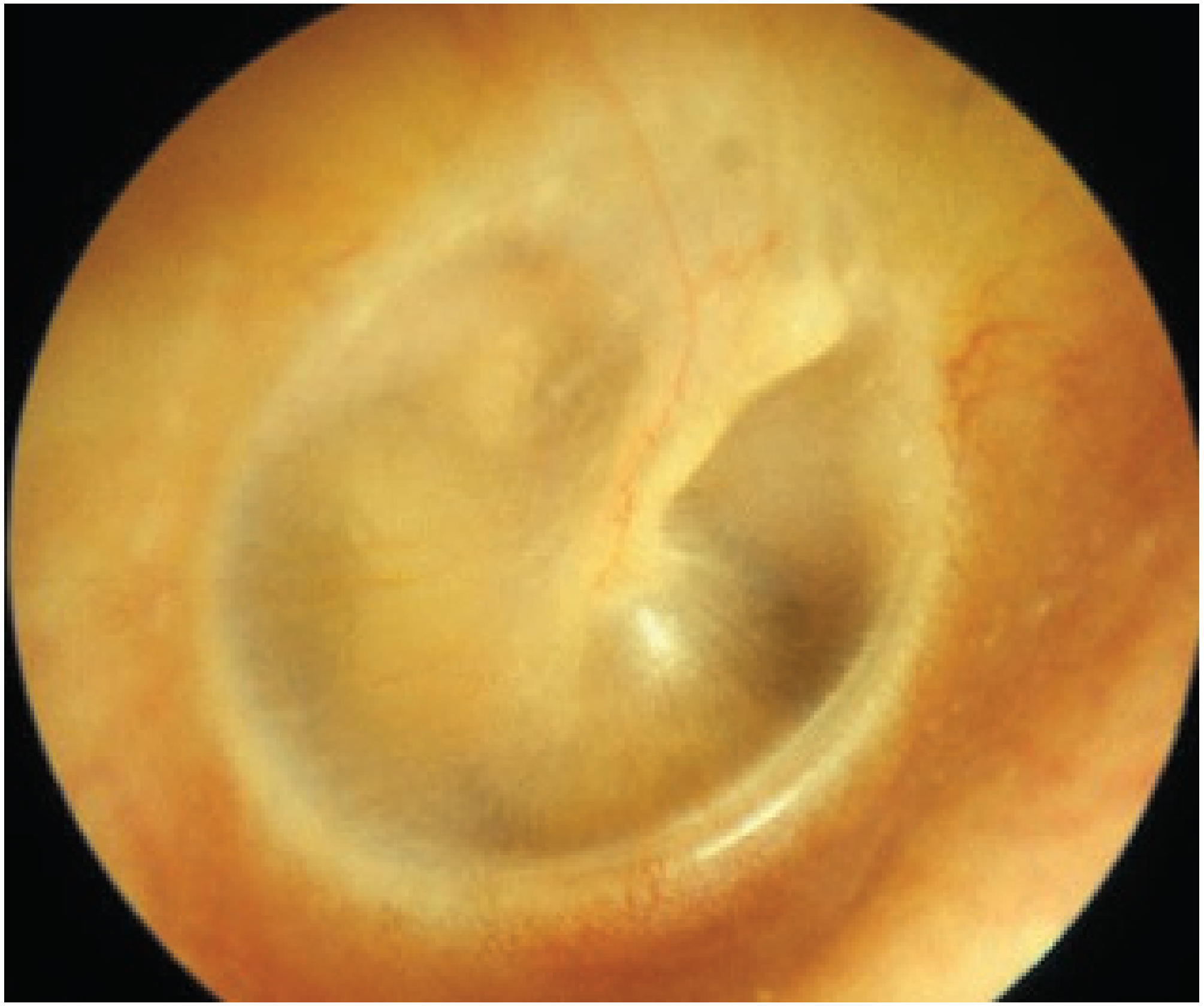
Normal tympanic membrane (labelled structures - short process of malleus, umbo, chorda tympani, long process of incus):
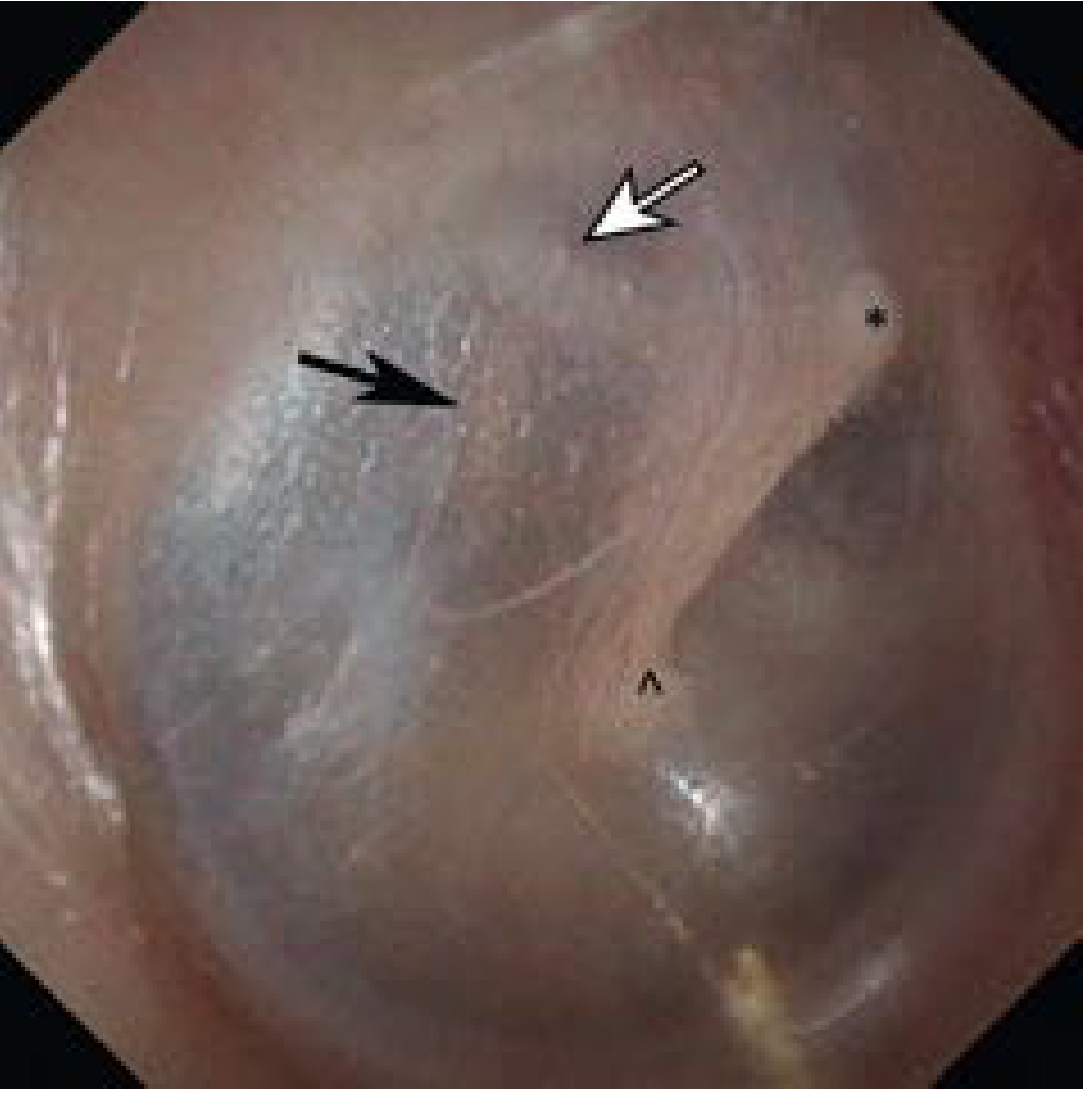
Overview
The TM lies at the medial end of the external auditory canal (EAC). It is slightly oval, ~9-10 mm in its longest diameter (posterosuperior to anteroinferior), and is oriented at ~55° with the floor of the meatus. It is divided into two distinct portions by the anterior and posterior malleolar folds (also called alveolar folds):
| Feature | Pars Tensa | Pars Flaccida (Shrapnell's Membrane) |
|---|---|---|
| Location | Majority of the TM (lower 4/5ths) | Small, superior triangular area above malleolar folds |
| Bony support | Attached to fibrous annulus in tympanic sulcus | Sits in the notch of Rivinus - no bony sulcus/annulus |
| Layers | Three layers (epithelium + lamina propria + mucosa) | Three layers (same) |
| Lamina propria | Well-organized: radial (outer) + circular/parabolic/transverse (inner) fibres; abundant type II collagen | Less marked, loosely and randomly arranged collagen; mainly type I collagen; more elastic |
| Rigidity | Firm and taut | Flaccid, more compliant |
| Thickness | Thinner, strong | Actually thicker than pars tensa, but with less-organized, looser collagen |
| Medial relation | Tympanic cavity (mesotympanum) | Prussak's space (epitympanum/attic) |
| Collagen density | Dense, well-organized | Sparse, loosely arranged in a vascular lamina propria |
Anatomy of the Pars Flaccida
- It occupies the notch of Rivinus, a region where the bony tympanic ring is deficient (formed by the squama of the temporal bone rather than tympanic bone)
- Bounded above by the notch, and below by the anterior and posterior malleolar folds which run to the lateral process of the malleus
- Medially it overlies Prussak's space - the most common site of primary acquired cholesteatoma formation
- Because it lacks the fibrous annulus and has disorganized collagen, it is inherently more prone to retraction under negative middle ear pressure
Anatomy of the Pars Tensa
- Forms the bulk of the TM
- Attached peripherally by the fibrocartilaginous tympanic annulus within the tympanic sulcus
- Concave toward the EAC; the tip of the malleus handle attaches centrally at the umbo
- The posterosuperior quadrant of the pars tensa is a weak point - it has thinner, more sparsely distributed collagen and increased vascularity, making it susceptible to retraction pockets
- Perforations of the pars tensa are always described as "central" (by definition, since they are surrounded by the annulus) - indicative of tubotympanic (safe/mucosal) disease
Clinical Relevance
Chronic Otitis Media (COM) Classification
| Type | TM Involvement |
|---|---|
| Tubotympanic (mucosal/safe) COM | Retraction/perforation of pars tensa only |
| Atticoantral (squamous/unsafe) COM | Retraction/involvement of pars flaccida (attic) |
- Pars flaccida retractions involve the attic and lead to atticoantral disease
- Pars tensa retractions are confined to the mesotympanum
Cholesteatoma
- The most susceptible area for retraction and cholesteatoma is the pars flaccida
- Retraction of the pars flaccida into Prussak's space forms primary acquired cholesteatoma
- Pars tensa retractions, especially posterosuperiorly, can also lead to cholesteatoma (secondary acquired)
Retraction Pockets
The pars flaccida and posterosuperior pars tensa are vulnerable because:
- Eustachian tube dysfunction generates negative middle ear pressure
- The pars flaccida has thicker but loosely organized, weakly supported collagen
- Inflammation/infection further damages collagenous support
- The result is progressive retraction, atelectasis, and potential cholesteatoma
Retraction of the pars tensa is graded by the Sade classification; retraction of the pars flaccida is graded by the Tos classification.
Innervation (both portions)
- Lateral surface: auriculotemporal nerve (Vc) posteroinferiorly; auricular branch of vagus (Arnold's nerve) anterosuperiorly; additional VII nerve fibres
- Medial surface: tympanic branch of glossopharyngeal nerve (Jacobson's nerve)
Sources: Shambaugh Surgery of the Ear; Scott-Brown's Otorhinolaryngology Head & Neck Surgery Vol. 2; Cummings Otolaryngology Head and Neck Surgery
This is a shared conversation. Sign in to Orris to start your own chat.