
Lasik surgery procedure
Reading File
LASIK (Laser-Assisted In Situ Keratomileusis) is a refractive surgical procedure that reshapes the cornea to correct myopia, hyperopia, and astigmatism using an excimer laser.
Searching sources...
Finding Sources
Searching the Web
LASIK surgery procedure steps technique 2024
Finding Sources
Reading Web Page
https://www.ncbi.nlm.nih.gov/books/NBK555970
Reading File
Reading File
Reading File
Reading File
Loading Image
Here is a thorough overview of the LASIK procedure, drawn from Kanski's Clinical Ophthalmology (10th ed.), The Wills Eye Manual, and StatPearls (NIH).
LASIK (Laser-Assisted In Situ Keratomileusis)
What It Is
LASIK is one of the most commonly performed refractive surgical procedures worldwide. It uses an excimer laser to reshape the corneal stroma exposed beneath a thin hinged flap, permanently altering the cornea's refractive power to correct myopia, hypermetropia, and astigmatism.
How It Corrects Vision
| Refractive Error | Laser Action |
|---|---|
| Myopia | Central ablative flattening reduces curvature |
| Hypermetropia | Mid-peripheral ablation steepens the central cornea |
| Astigmatism | Asymmetric ablation normalizes corneal contour |
Treatable range:
- Myopia: up to -6 to -8 D
- Hypermetropia: up to +3 to +4 D
- Astigmatism: up to 5 D (depending on corneal thickness)
Preoperative Evaluation
Before surgery, the following assessments are mandatory:
- Complete ocular examination (refraction must be stable for at least 1 year)
- Tear film osmolarity - to exclude dry eye syndrome
- Corneal pachymetry - to measure thickness and detect mild keratoconus
- Corneal topography - to exclude irregular astigmatism, keratoconus, and forme fruste keratoconus
- Corneal OCT - to produce an epithelial map
- Wavefront aberrometry (corneal and whole eye)
- Contact lens wearers: soft lenses discontinued for 2 weeks, rigid/gas-permeable lenses for at least 3 weeks before examination
Surgical Technique (Step by Step)
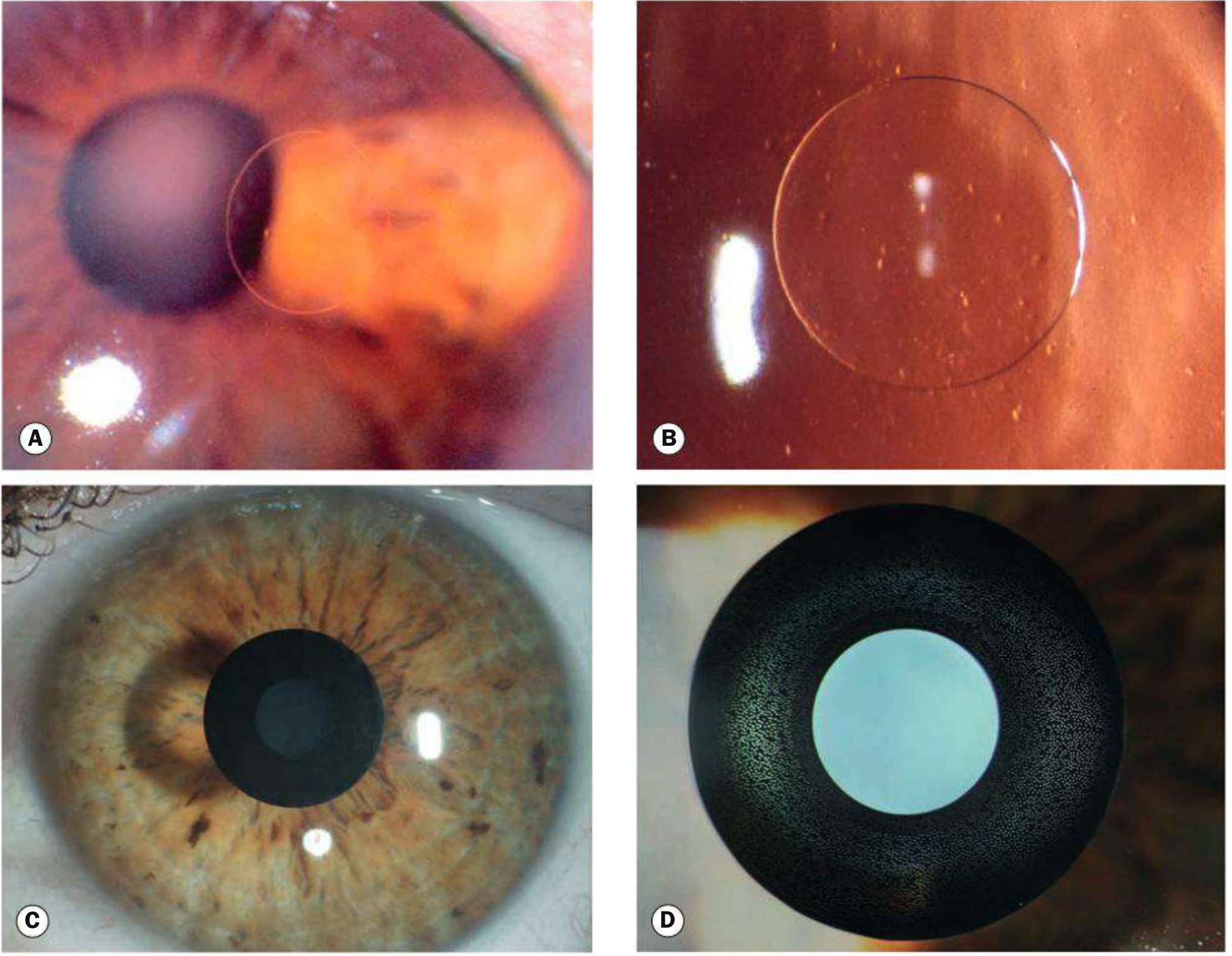
- Patient positioning - Supine position; fellow eye taped closed; operative eye held open with a speculum.
- Anesthesia - Topical anesthetic eye drops applied.
- Suction ring application - A suction ring is centered on the cornea, substantially raising intraocular pressure. This stabilizes the eye and provides a guide track.
- Flap creation - A thin corneal flap (including epithelium, Bowman's membrane, and superficial stroma) is created in one of two ways:
- Mechanical microkeratome - An oscillating blade advances across the cornea
- Femtosecond laser (now preferred) - Infrared laser pulses create microcavitation bubbles in a precise cleavage plane, producing a flap of uniform thickness (100-120 µm). Significantly reduces flap-related intraoperative complications.
- Flap reflection - The hinged flap is folded back to expose the underlying stromal bed.
- Excimer laser ablation - The stroma is reshaped by the excimer laser (wavelength 193 nm, UV range) with negligible disruption to surrounding tissue.
- Flap repositioning - The flap is carefully replaced over the stromal bed. It adheres naturally - no sutures required.
- Post-op assessment - The flap interface and centration are confirmed.
Variations
- Wavefront-guided LASIK - Excimer laser uses a custom ablation pattern based on whole-eye wavefront aberrometry measurements to reduce post-op higher-order aberrations.
- "Optimised" LASIK - Uses a specific ablation algorithm to create larger optical zones and better corneal asphericity, reducing night glare.
- LASIK Plus (LASIK + Cross-linking) - Riboflavin is applied to the stromal bed before flap replacement, followed by UV light exposure after repositioning. Considered for patients at risk of ectasia or refractive regression (usually >-7 D).
Contraindications
Absolute:
- Refractive instability (>0.5 D change in the last year)
- Corneal ectasia or keratoconus
- Residual corneal bed < 250 µm after planned ablation
- Uncontrolled diabetes mellitus
- Pregnancy/nursing
Relative:
- Severe dry eye
- Thin corneas
- High refractive errors outside treatable range (intraocular procedure preferred instead)
- Occupations/activities with high eye trauma risk (flap dislocation risk)
Advantages Over Surface Ablation (PRK)
- Greater postoperative comfort
- Faster visual rehabilitation
- More rapid stabilization of refraction
- Milder stromal haze
- Can treat a wider range of refractive errors
Intraoperative Complications
- Buttonholing - Microkeratome penetrates through the flap (rare with femtosecond laser)
- Flap amputation - Incomplete hinge (rare)
- Irregular flap creation
- Anterior chamber penetration (very rare)
Postoperative Complications
Early (within 2 weeks)
| Complication | Notes |
|---|---|
| Dry eye / neurotrophic keratopathy | Most common early complication; occurs in 85-98% at 1 week, ~60% at 1 month. Due to severed corneal nerves. Treated with artificial tears; resolves as nerves regenerate (3-12 months). |
| Flap dislocation/wrinkling | Requires prompt repositioning |
| Diffuse lamellar keratitis (DLK) | "Sands of the Sahara" - granular interface deposits, occurs days 1-7. Treated with intensive topical antibiotic + steroid. |
| Epithelial defect | Usually self-limited |
| Central toxic keratopathy (CTK) | Corneal thinning and flattening; usually improves over 6-12 months |
| Infection | Rare; must culture organism; treat with targeted topical antibiotic |
Late (weeks to months)
| Complication | Notes |
|---|---|
| Dry eye / tear film instability | Most common overall; up to 70%; usually self-resolves |
| Transient light sensitivity syndrome | 2-8 weeks post-op; more common after hyperopic LASIK (9%). Responds to topical steroids. |
| Subepithelial haze | Causes glare, haloes, night starbursts; settles in 3-6 months |
| Epithelial ingrowth | Under the flap (1-2%); may need flap lift and scraping if vision affected |
| Regression / under- or over-correction | Refractive stability takes up to 3 months |
| Corneal ectasia | Most serious late complication (0.2-0.6%). Progressive thinning/steepening. Risk factors: abnormal topography, thin cornea, high myopia, low residual stromal bed, young age. |
| Pressure-induced stromal keratitis (PISK) | Fluid cleft from elevated IOP due to steroid use; presents as under-correction |
| Irregular astigmatism | Decentered ablation, central island, or flap irregularity |
Postoperative Care
- Topical antibiotics and steroids for the first 1-4 weeks
- Artificial tears for dry eye management
- Avoid eye rubbing (flap disruption risk)
- Follow-up at day 1, week 1, 1 month, and 3 months
- Final refraction usually stable by 3 months
Comparison: LASIK vs. PRK vs. SMILE
| Feature | LASIK | PRK | SMILE |
|---|---|---|---|
| Flap | Yes | No | No |
| Recovery speed | Fast | Slow | Fast |
| Pain | Minimal | More painful | Minimal |
| Dry eye risk | High | Moderate | Lower |
| Ectasia risk | Low-moderate | Lower | Lower |
| Range of correction | Wider | More limited | Similar to LASIK |
Sources: Kanski's Clinical Ophthalmology 10th ed., pp. 301-303 | The Wills Eye Manual | StatPearls - LASIK (NIH)
This is a shared conversation. Sign in to Orris to start your own chat.