
Typhoid
typhoid fever rose spots rash clinical
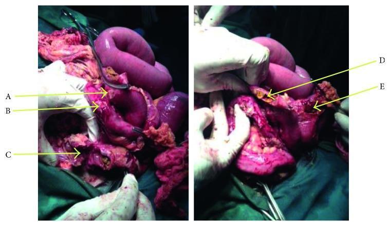
This clinical photograph provides an intraoperative view from an emergency exploratory laparotomy, demonstrating multiple ileal perforations secondary to typhoid fever. The image shows distended loops of the small bowel (ileum) with significant pathological changes. Marked at locations A through E are several distinct intestinal perforations, characterized by ragged, irregular edges and disrupted serosal surfaces. The intestinal wall appears inflamed, edematous, and discolored, with evident denudation of the serosa in multiple spots. Yellowish-green fibrinous exudate and fecal material are visible at the sites of perforation, indicating active fecal peritonitis. Gloved hands and surgical retractors are shown manipulating the bowel to expose the extent of the damage. This visual illustrates the severe surgical complications of typhoid enteritis, specifically typhoid intestinal perforation (TIP), which typically occurs in the distal ileum and requires segmental resection and peritoneal lavage.
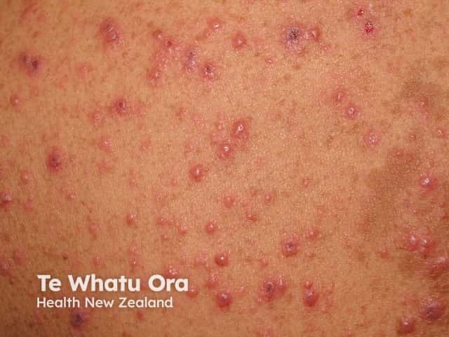
This clinical photograph shows a vesicular-papular rash on an erythematous base, distributed predominantly on the trunk with face involvement, consistent with varicella-zoster virus infection (chickenpox). The lesions appear at multiple stages, with small macules evolving to clear fluid-filled vesicles and some crusted plaques, giving the classic dew drop on a rose petal appearance. The skin is mildly erythematous around vesicles; lesions are superficial, non-palpable. No mucosal involvement is evident in this image. The distribution is generalized but has centripetal predominance. This presentation is highly suggestive of primary varicella, particularly in unvaccinated or susceptible individuals, and is contagious through vesicle fluid and respiratory secretions. Differential considerations include disseminated herpes simplex infection, disseminated herpes zoster in immunocompromised patients, or pityriasis rosea with vesicular components, though the latter is less typical. Clinically, the image supports diagnosis when accompanied by fever, malaise, pruritus, and a compatible exposure history. Reassurance, antiviral therapy consideration (e.g., acyclovir in appropriate age groups), and supportive care are relevant. The photograph is useful for medical education, telemedicine assessment, and dermatology training in recognizing staged varicella lesions, public health documentation, and patient counseling on contagion risk. This image supports clinical triage, outbreak documentation, and patient education on contagious rash in practice.
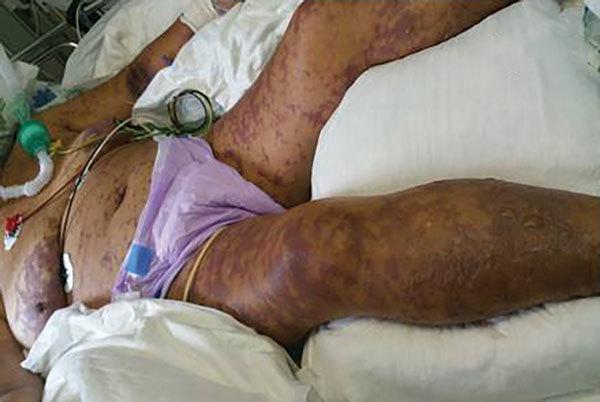
Clinical photograph of a patient in an intensive care unit exhibiting dermatological manifestations of Israeli spotted fever and purpura fulminans. The image shows a diffuse, widespread purpuric rash with a characteristic fern-leaf (arborescent) pattern. The morphology consists of non-blanching reddish-purple spots and larger ecchymotic patches across the abdomen and lower extremities. Progressed lesions on the lower right leg demonstrate the formation of bullae filled with serous fluid, suggesting tissue necrosis. The clinical context is reinforced by life-support equipment, including an endotracheal tube with a green connector for mechanical ventilation, ECG monitoring leads on the chest, and multiple intravenous lines. This visual record illustrates the severe cutaneous evolution of rickettsial diseases, specifically the transition into disseminated intravascular coagulation (DIC) and purpura fulminans in a critical care setting.
typhoid fever management
Typhoid Fever (Enteric Fever)
Definition & Causative Organism
Epidemiology
- Affects up to 30 million people worldwide each year, with ~200,000 deaths.
- Endemic in regions with poor sanitation and unsafe drinking water - Africa, the Americas, Southeast Asia, and the Western Pacific.
- In the United States, ~400 travel-associated cases are diagnosed annually.
- Children and adolescents bear the highest burden in endemic areas.
- A 2025 systematic review and meta-analysis (PMID 41125003) confirmed real-world effectiveness of typhoid conjugate vaccine in children and adolescents.
Transmission & Pathogenesis
- S. Typhi is ingested and taken up by M cells overlying Peyer patches in the terminal ileum.
- Bacteria are engulfed by mononuclear phagocytes in the underlying lymphoid tissue.
- Peyer patches enlarge into plateau-like elevations up to 8 cm in diameter; mucosal shedding creates oval ulcers oriented along the long axis of the ileum.
- Unlike nontyphoidal Salmonella, S. Typhi disseminates via lymphatics and bloodstream, causing reactive hyperplasia of mesenteric lymph nodes.
- The red pulp of the spleen expands from phagocyte hyperplasia.
- Typhoid nodules - small foci of parenchymal necrosis with macrophage aggregates - appear in the liver, bone marrow, and lymph nodes.
- Gallbladder colonization may result in a chronic carrier state.
Clinical Features
Classic Stepwise Progression
| Week | Features |
|---|---|
| Week 1 | Fever (gradually rising), headache, malaise, dry cough, anorexia, myalgias |
| Week 2 | Sustained high fever (39-40°C), relative bradycardia, abdominal distension, splenomegaly, hepatomegaly, rose spots |
| Week 3 | Risk of complications: intestinal perforation, hemorrhage; some patients improve |
| Week 4 | Recovery or deterioration depending on complications |
Key Clinical Signs
- Fever: Prolonged, hectic, step-ladder pattern in classic cases
- Relative bradycardia (Faget sign): Pulse-temperature dissociation - classic but may be absent
- Rose spots: Pale red (salmon-colored) maculopapular lesions, 2-4 mm, appearing in crops on the chest and abdomen in fair-skinned individuals; seen in ~30% of patients
- Splenomegaly and hepatomegaly
- Abdominal distension and tenderness (may mimic appendicitis)
- Constipation in 30% of patients (diarrhea can also occur, especially in epidemic settings)
- Leukopenia and elevated liver enzymes (nonspecific)
- "Pea-soup" diarrhea in the second week in some cases
Complications
- Intestinal perforation (most feared; occurs in distal ileum at Peyer patch ulcers) - see image below
- Intestinal hemorrhage
- Disseminated intravascular coagulation (DIC)
- Encephalopathy, meningitis, seizures, psychosis, ataxia
- Myocarditis, endocarditis
- Pneumonia
- Cholecystitis
- Septic arthritis, mycotic aneurysm
- Renal failure
- Deafness (as a sequela)
Diagnosis
- Blood culture: Gold standard in the first week; positive in ~80-90% during the febrile bacteremic phase
- Bone marrow culture: Most sensitive overall; remains positive even after antibiotics have been started
- Stool/urine culture: More useful from the second week onward
- Widal test: Measures agglutinating antibodies to O and H antigens; limited specificity, many false positives in endemic areas - largely superseded
- Rapid antigen tests / typhidot: Used in resource-limited settings
- Leukopenia, elevated transaminases, elevated CRP are supportive but nonspecific
Treatment
Antibiotic Therapy (Sleisenger & Fordtran, Tintinalli's Emergency Medicine)
| Antibiotic | Dose | Notes |
|---|---|---|
| Ciprofloxacin (fluoroquinolone) | 500 mg twice daily × 7-10 days | First-line; resistance increasing from South/Southeast Asia |
| Ceftriaxone | 2 g IV once or twice daily × 10-14 days | Preferred for severe disease, IV route |
| Azithromycin | 1 g daily × 5 days | Preferred in areas of high fluoroquinolone resistance |
| Cefixime | Oral option | For non-severe disease |
- Ampicillin, TMP-SMX, and chloramphenicol are now unreliable due to widespread resistance (multidrug-resistant/MDR strains).
- For severe typhoid with meningitis/encephalopathy or shock: add dexamethasone to antibiotics.
- Supportive care: IV rehydration, blood transfusion if significant GI blood loss.
- Relapses occur in 5-10% of patients after apparent clinical recovery.
- Untreated mortality: 10-20%, mainly in young children.
Chronic Carriers
- Defined as excretion of S. Typhi in stool for >1 year.
- Associated with gallbladder colonization/gallstones.
- Treated with prolonged fluoroquinolones; cholecystectomy may be required in refractory cases.
Prevention & Vaccination
- Ty21a - oral live-attenuated vaccine (3-4 doses); 51-76% efficacy; requires revaccination every 5 years.
- Vi polysaccharide (ViPS) - single IM injection; ~72% efficacy; revaccination every 2 years.
- Typhoid Conjugate Vaccine (TCV - Vi-TT): Newer single-dose IM vaccine; superior immunogenicity and longer-lasting; now preferred by the WHO for children >6 months. A 2025 meta-analysis (PMID 41125003) confirmed strong real-world effectiveness in children and adolescents.
Summary Table
| Feature | Details |
|---|---|
| Organism | S. Typhi, S. Paratyphi A/B/C |
| Reservoir | Humans only |
| Transmission | Fecal-oral (contaminated food/water) |
| Incubation | 1-3 weeks |
| Classic sign | Step-ladder fever, rose spots, relative bradycardia |
| Pathology | Peyer patch hypertrophy, oval ileal ulcers, typhoid nodules |
| Diagnosis | Blood culture (Week 1), bone marrow culture (most sensitive) |
| Treatment | Fluoroquinolones / ceftriaxone / azithromycin |
| Key complication | Intestinal perforation (distal ileum) |
| Mortality (untreated) | 10-20% |