
Types of dialysis
Reading File
Dialysis is a renal replacement therapy that removes waste products and excess fluid when the kidneys fail. The two main types are hemodialysis and peritoneal dialysis.
Searching sources...
Finding Sources
Searching PubMed
"dialysis" AND "hemodialysis OR peritoneal dialysis"
Reading File
Reading File
Finding Sources
Reading File
Finding Sources
Finding Sources
Reading File
Reading File
Reading File
Reading File
Finding Sources
Finding Sources
Reading File
Reading File
Reading File
Reading File
Loading Image
Now I have rich, comprehensive content from authoritative nephrology textbooks. Let me compile the full answer.
Types of Dialysis
Dialysis is a form of renal replacement therapy (RRT) that artificially removes waste products, excess solutes, and fluid from the blood when the kidneys can no longer do so adequately. There are two broad categories: Hemodialysis (HD) and Peritoneal Dialysis (PD). Each contains several subtypes. A third category, Continuous Renal Replacement Therapy (CRRT), is reserved primarily for critically ill patients.
1. Hemodialysis (HD)
Blood is pumped out of the body through a dialyzer (artificial membrane), where waste products and excess fluid are removed by diffusion and convection against a dialysate fluid, then returned to the body.
Access types used: arteriovenous (AV) fistula (preferred), AV graft, or central venous catheter.
Subtypes
| Subtype | Description |
|---|---|
| In-Center / Facility HD | Performed at a dialysis unit under nursing supervision; standard is 3-4 hours per session, three times weekly |
| Home HD | Performed by the patient at home; associated with better flexibility and potentially better outcomes |
| Conventional / Intermittent HD (IHD) | The standard modality; 3-5 hr sessions, thrice weekly. Dose measured as Kt/V (target ≥1.3 per session) |
| Daily HD | More frequent sessions (5-6x/week); reduces interdialytic fluid and solute swings; used in some home HD programs |
| Nocturnal HD | Long sessions (6-8 hr) overnight, typically 3-6 nights/week; excellent solute clearance and BP control |
| High-flux HD | Uses high-flux membranes that clear larger middle molecules (e.g., beta-2 microglobulin) more efficiently |
| Hemodiafiltration (HDF) | Combines diffusion (as in standard HD) with convection (using replacement fluid); superior middle-molecule clearance |
- Brenner and Rector's The Kidney, regarding standard IHD: "Patients typically undergo dialysis treatments for 3 to 5 hours on a thrice-weekly, alternate-day, or daily schedule, depending on catabolic demands, electrolyte disturbances, and volume status."
In Australia (~12% of HD patients receive home HD, second only to New Zealand at 15.6%), and in Japan (>85% in-center HD with >4 hr treatment times in ~80% of patients), practice patterns vary significantly worldwide. - Brenner and Rector's The Kidney
2. Peritoneal Dialysis (PD)
The peritoneal membrane (lining of the abdominal cavity) serves as the natural dialysis membrane. Dialysate fluid is infused into the peritoneal cavity via a permanent catheter (usually the double-cuff, swan-neck Tenckhoff catheter), dwells for a period, and is then drained. Solutes diffuse down their concentration gradient, while ultrafiltration is driven by osmotic pressure (usually from glucose in the dialysate). - Fischer's Mastery of Surgery, 8th ed.
PD better preserves residual kidney function compared with HD, partly because it avoids acute intravascular volume depletion. - Fischer's Mastery of Surgery
Subtypes
| Type | Description |
|---|---|
| CAPD (Continuous Ambulatory Peritoneal Dialysis) | Manual instillation and drainage of dialysate several times per day (typically 4 exchanges/day); no machine needed; continuous treatment |
| APD (Automated Peritoneal Dialysis) | Uses a cycler machine to instill and drain fluid automatically |
| CCPD (Continuous Cycling Peritoneal Dialysis) | APD performed at night by a cycler, plus a final "fill" that remains in the peritoneal cavity during the day; an extra daytime exchange may be added |
| NIPD (Nocturnal Intermittent Peritoneal Dialysis) | APD at night only - no fluid left in the cavity during the day; used when daytime fill causes discomfort |
| Tidal PD | A variation of APD where only part of the dialysate is drained each cycle, leaving a "tidal" volume to reduce discomfort and maintain contact with the membrane |
| Incremental PD | Lower dialysis dose at the start, adjusted as residual kidney function declines |
- Comprehensive Clinical Nephrology, 7th ed.: "PD can be prescribed up to 24 hours/day and 7 days/wk to patients in the form of CAPD or CCPD. In CCPD, patients receive treatment with an automated PD (APD) cycler at night plus a final 'fill' that remains in the peritoneal cavity during the day."
Osmotic agents used in PD dialysate:
- Glucose (1.5-4.25 g/dL) - most common; risk of hyperglycemia and glucose degradation products
- Icodextrin - non-glucose; better ultrafiltration, preserves residual kidney function, avoids hyperglycemia; note: can falsely elevate glucose readings on glucometers using glucose dehydrogenase strips. - Comprehensive Clinical Nephrology, 7th ed.
3. Continuous Renal Replacement Therapy (CRRT)
Used primarily in critically ill, hemodynamically unstable patients in the ICU, where the rapid fluid shifts of IHD would be poorly tolerated. CRRT is slower, gentler, and runs continuously over 24 hours.
| Subtype | Mechanism |
|---|---|
| CVVHD (Continuous Venovenous Hemodialysis) | Diffusion-based; blood flows counter-currently to dialysate; no replacement fluid |
| CVVH (Continuous Venovenous Hemofiltration) | Convection-based; large volume of replacement fluid is used; no dialysate |
| CVVHDF (Continuous Venovenous Hemodiafiltration) | Combines diffusion (dialysate) + convection (replacement fluid) for superior solute clearance |
| SLED / EDD (Sustained Low-Efficiency Dialysis / Extended Duration Dialysis) | Hybrid approach: intermediate duration (8-12 hr); combines some advantages of IHD and CRRT; allows off-machine time for procedures/therapy |
- Washington Manual of Medical Therapeutics: "CRRT can be used in specialized circumstances, particularly when the patient's hemodynamic status would not tolerate the rapid fluid shifts of IHD... The slower blood flows may necessitate anticoagulation (with either systemic heparin or regional citrate) to prevent the filter from clotting."
- Brenner and Rector's The Kidney (KDIGO guideline recommendation): "CRRT and PIRRT [be used] in hemodynamically unstable patients, and CRRT be used for patients with acute brain injury or other causes of increased intracranial pressure or generalized brain edema."
Key Comparison: HD vs. PD
| Feature | Hemodialysis | Peritoneal Dialysis |
|---|---|---|
| Frequency | 3x/week (in-center) | Daily (continuous) |
| Access | AV fistula, graft, catheter | Peritoneal (Tenckhoff) catheter |
| Setting | Hospital/clinic or home | Home (mainly) |
| Hemodynamic impact | Greater fluid shifts | Gentler; better cardiovascular tolerance |
| Residual kidney function | Less well preserved | Better preserved |
| Infection risk | AV access infection, bloodstream | Peritonitis, exit-site infection |
| Survival | No consistent difference vs. PD | No consistent survival advantage confirmed by RCT |
| Quality of life | Clinic-bound (in-center) | Greater freedom; home-based |
- Comprehensive Clinical Nephrology, 7th ed.: "Retrospective and prospective nonrandomized comparative studies have failed to indicate a consistent survival advantage for either modality... PD is associated with superior health-related quality of life."
Modality Selection Principles
- Choice is guided largely by patient preference, lifestyle, residual kidney function, and clinical factors. - Comprehensive Clinical Nephrology
- For AKI with hemodynamic instability: prefer CRRT or SLED
- For AKI with increased intracranial pressure: CRRT preferred (avoids cerebral edema worsening)
- For diabetes + ESKD: no evidence that one modality is superior; glucose management differs with PD (use icodextrin when possible). - Comprehensive Clinical Nephrology
- PD is contraindicated (relative) in: recent intra-abdominal surgery, large abdominal hernia, severe respiratory disease, inability to perform exchanges, active abdominal infection
Sources: Brenner and Rector's The Kidney (2-Volume Set); Comprehensive Clinical Nephrology, 7th ed.; Fischer's Mastery of Surgery, 8th ed.; Washington Manual of Medical Therapeutics; National Kidney Foundation Primer on Kidney Diseases, 8th ed.
Complete hemodialysis procedure
Reading File
Hemodialysis is a well-defined procedure involving patient assessment, vascular access, machine priming, the dialysis run itself, and termination with monitoring throughout.
Searching sources...
Finding Sources
Finding Sources
Finding Sources
Finding Sources
Finding Sources
Finding Sources
Reading File
Reading File
Finding Sources
Finding Sources
Reading File
Reading File
Reading File
Reading File
Reading File
Finding Sources
Finding Sources
Reading File
Reading File
Loading Image
Reading File
Reading File
Reading File
Complete Hemodialysis Procedure
Overview and Principle
Hemodialysis (HD) substitutes an artificial filter (the dialyzer) for the glomerulus to produce an ultrafiltrate of plasma. Blood is withdrawn from the patient, circulated through the dialyzer where waste products and excess fluid are removed, and returned to the patient. The core mechanism relies on:
- Diffusion - solutes move down a concentration gradient across a semipermeable membrane (removes urea, potassium, creatinine, phosphate)
- Ultrafiltration (UF) - fluid removal driven by a controlled hydrostatic pressure gradient across the membrane
- Convection (in high-flux/hemodiafiltration) - solute drag carried with water movement, improving middle-molecule clearance
Blood and dialysate flow countercurrently on opposite sides of the membrane to maximize the concentration gradient throughout the entire length of the dialyzer. - Brenner and Rector's The Kidney
Phase 1: Pre-Dialysis Assessment
Patient Evaluation Before Each Session
Before connecting the patient, the following are assessed:
| Parameter | Details |
|---|---|
| Dry weight (target weight) | Estimated weight with no excess fluid; guides total UF volume |
| Interdialytic weight gain | Compared to last session's post-dialysis weight; excess = fluid to remove |
| Blood pressure and heart rate | Pre-dialysis BP documented; guides UF rate |
| Temperature | Fever may indicate access infection |
| Access site inspection | AV fistula/graft: auscultate for bruit, palpate for thrill; catheter: check exit site for redness/discharge |
| Symptoms | Dyspnea, chest pain, nausea, neurological changes |
| Recent labs | Pre-dialysis BUN, potassium, bicarbonate, phosphate, hemoglobin; reviewed to adjust prescription |
The HD prescription must be individualized for each patient based on these clinical parameters. - Brenner and Rector's The Kidney
Phase 2: Vascular Access
Three types of access are used, in order of preference:
1. Arteriovenous (AV) Fistula (Preferred)
- Surgically created connection between an artery and vein (usually radial artery + cephalic vein at the wrist, or brachial artery + cephalic/basilic vein)
- Takes 6-12 weeks to "mature" (dilate and thicken) before use
- Cannulation technique: two 15-gauge large-bore needles inserted - one arterial (blood withdrawal, upstream) and one venous (blood return, downstream); placed 5-8 cm apart using the rope-ladder or buttonhole technique
- Preferred because of lowest infection rate, longest longevity, and best outcomes
2. AV Graft
- A synthetic (polytetrafluoroethylene, PTFE) or biological tube bridging artery to vein
- Can be used within 2-4 weeks of creation
- Cannulated with the same 15-gauge needle technique as a fistula
- Higher complication rate than native fistula (thrombosis, infection, stenosis)
3. Tunneled-Cuffed Central Venous Catheter (Last resort)
Used when AV access is not available or not yet mature.
Insertion procedure (as described in Brenner and Rector's The Kidney):
- Strict sterile technique and topical 1% Xylocaine; conscious sedation often used
- Access to the right internal jugular vein under real-time ultrasound guidance using a 21-gauge needle
- A guide wire is passed down to the inferior vena cava under fluoroscopy
- A skin pocket (~1 cm) is created at the insertion site
- A subcutaneous tunnel is created laterally and inferiorly, under the clavicle, approximately 5-7 cm from the needle insertion point
- The catheter tip is fed to the junction of the superior vena cava and right atrium
- The cuff is positioned 2 cm from the exit site
- Final X-ray confirms position and checks for kinks
Catheter sizes: 14.5 or 16 French; lengths: 24 cm (right IJ), 28 cm (left IJ), 36-42 cm (femoral)
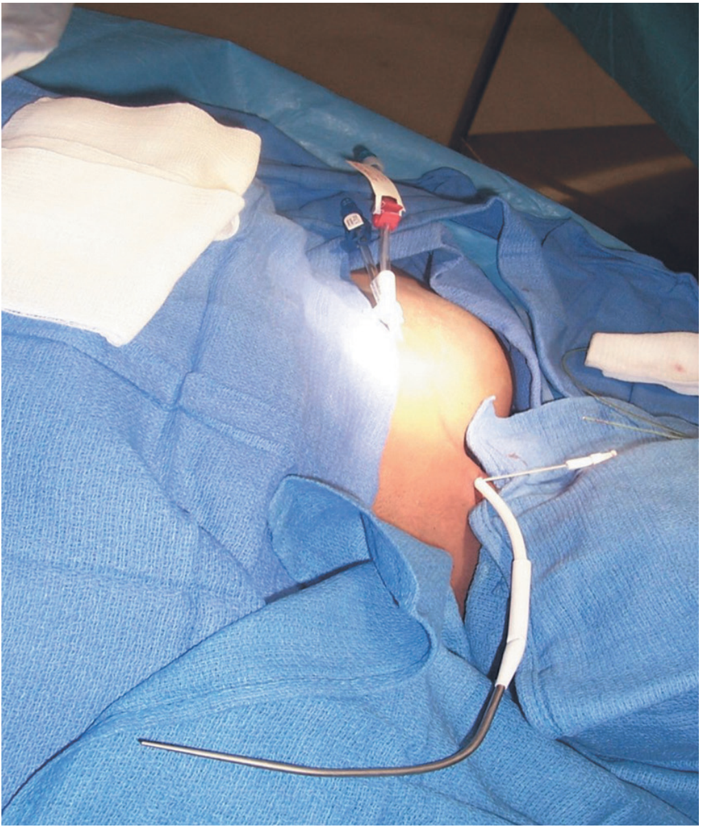
Tunneled hemodialysis catheter placement - the catheter tunneled from upper chest to the internal jugular entry site. - Brenner and Rector's The Kidney
Phase 3: The Dialysis Prescription
The complete dialysis prescription consists of the following individualized components (Box 63.1, Brenner and Rector's The Kidney):
| Prescription Component | Standard Values |
|---|---|
| Duration | 3-4 hours per session (US average ~3.5 hr) |
| Frequency | 3 times per week (Monday/Wednesday/Friday or Tuesday/Thursday/Saturday) |
| Vascular access type | AV fistula > AV graft > tunneled catheter |
| Dialyzer | Selected by membrane type, surface area, flux (low vs. high), biocompatibility |
| Blood flow rate (Qb) | 300-500 mL/min |
| Dialysate flow rate (Qd) | 500-800 mL/min (countercurrent to blood) |
| Ultrafiltration rate (UFR) | Calculated from weight gain ÷ session time; typically 0.5-1 L/hr |
| Dialysate composition | Tailored (see table below) |
| Anticoagulation | Heparin (most common) |
| Dialysate temperature | 35-37°C (cooling can reduce intradialytic hypotension) |
| Intradialytic medications | ESAs, IV iron, vitamin D analogs as needed |
Dialysate Composition
| Solute | Typical Concentration | Purpose |
|---|---|---|
| Sodium | 135-145 mEq/L | Fluid balance, BP control |
| Potassium | 1-4 mEq/L | Removes excess potassium |
| Bicarbonate | 33-40 mEq/L | Corrects metabolic acidosis |
| Calcium | 2.5-3.5 mEq/L | Bone disease management |
| Magnesium | 0.5-1.0 mEq/L | Prevents hypomagnesemia |
| Glucose | 100-200 mg/dL | Prevents hypoglycemia |
Dialysate concentrations mirror those normally maintained by the native kidneys. - Brenner and Rector's The Kidney
Phase 4: Anticoagulation
Anticoagulation prevents clotting within the extracorporeal circuit.
Standard Protocol - Unfractionated Heparin (UFH):
- Small amounts of IV heparin (1,000-2,000 units) prevent thrombosis at the vascular access site - Tintinalli's Emergency Medicine
- Loading dose: ~40 U/kg IV bolus at the start
- Continuous infusion: ~20 U/kg/hour, adjusted to maintain adequate circuit anticoagulation
- Target: activated clotting time (ACT) 200-250 seconds within the circuit
Alternatives when heparin is contraindicated (e.g., heparin-induced thrombocytopenia):
- Regional citrate anticoagulation - citrate infused pre-filter creates regional hypocalcemia in the circuit; calcium replaced post-filter
- Hirudin/lepirudin - direct thrombin inhibitors
- Heparin-free dialysis - frequent saline flushes of circuit; used in active bleeding
Phase 5: Machine Setup and Circuit Priming
Before connecting the patient:
- Dialyzer inspection - check for integrity, correct membrane type, expiry
- Blood tubing set - arterial and venous lines connected to dialyzer
- Circuit priming - the extracorporeal circuit is flushed with ~500-1000 mL normal saline to remove air and manufacturing preservatives (e.g., glycerine), and to check for leaks
- Dialysate preparation - machine mixes concentrated dialysate with purified water to achieve the prescribed ionic composition; online conductivity monitoring ensures correct preparation
- Water treatment - incoming water undergoes softening, activated carbon filtration, and reverse osmosis to remove ions, organic compounds, and endotoxins; standards for water purity are strict because microbe-generated impurities such as endotoxins can cross the dialyzer membrane and cause inflammation - Brenner and Rector's The Kidney
- Alarms set - arterial pressure, venous pressure, TMP (transmembrane pressure), air detector, conductivity, temperature, and blood leak detector alarms confirmed
Phase 6: Initiating Dialysis (Patient Connection)
For AV Fistula/Graft:
- Skin cleaned with antiseptic; sterile drape placed
- Local anesthetic (optional) applied over needle sites
- Arterial needle (16G or 15G) inserted first, bevel up, at 25-45° angle, in the direction of blood flow upstream
- Venous needle inserted downstream, 5-8 cm from the arterial needle
- Needles secured with tape/dressing
- Blood lines connected; clamps released
- Blood pump started at a slow initial rate (~100-150 mL/min), then gradually increased to target (300-500 mL/min) over several minutes
For Catheter:
- Sterile technique; mask and gloves mandatory
- Catheter caps removed; lumens aspirated to check blood return
- Locked anticoagulant solution (heparin lock) aspirated and discarded
- Blood lines connected to arterial (red) and venous (blue) ports
- Blood pump started
Once connected:
- Heparin loading dose administered
- Heparin infusion started
- UF rate set per prescription
- Dialysate circuit opened
Phase 7: During the Dialysis Session (Monitoring)
The patient is monitored continuously throughout the 3-4 hour session:
| Parameter | Frequency | Action if Abnormal |
|---|---|---|
| Blood pressure | Every 30 min (or continuous) | If hypotensive: reduce UFR, give normal saline bolus, recline patient |
| Heart rate | Every 30 min | |
| Symptoms | Continuous nursing observation | |
| Arterial/venous pressures | Continuous (machine alarm) | Access dysfunction or needle dislodgement |
| Transmembrane pressure (TMP) | Continuous | Clotting or filter fouling |
| Blood leak detector | Continuous | Dialyzer membrane rupture |
| Air detector (venous trap) | Continuous | Air embolism prevention |
| Conductivity and pH | Continuous | Incorrect dialysate preparation |
| Temperature | Continuous | |
| Blood glucose (diabetics) | Each session |
Common Intradialytic Complications and Management
| Complication | Frequency | Cause | Management |
|---|---|---|---|
| Hypotension | Most common (~25% of sessions) | Excessive/rapid fluid removal, low serum osmolality | Reduce UF rate, saline bolus 100-200 mL, trendelenburg position, cool dialysate |
| Muscle cramps | Common | Rapid fluid removal, low sodium dialysate | Reduce UF rate, saline bolus, hypertonic saline or glucose |
| Nausea and vomiting | Common | Hypotension, dialysis disequilibrium | Treat hypotension; antiemetics if needed |
| Headache | Common | Dialysis disequilibrium syndrome | Reduce blood flow rate, shorten session |
| Fever/chills | Occasional | Access infection, endotoxin contamination | Blood cultures, IV antibiotics; evaluate water quality |
| Chest pain | Occasional | Angina, air embolism, hemolysis | Stop dialysis; ECG, oxygen, evaluate cause |
| Air embolism | Rare | Air in circuit | Clamp lines, left lateral decubitus position, 100% oxygen |
| Hemolysis | Rare | Overheated dialysate, incorrect osmolality, kinking | Stop dialysis; do NOT return blood |
| Arrhythmia | Occasional | Electrolyte shifts (K+), myocardial disease | ECG monitoring; adjust dialysate K+ |
Dialysis Disequilibrium Syndrome - occurs especially in first sessions or after long gaps; caused by rapid removal of urea creating transient cerebral edema from osmotic shift. Prevented by using lower blood flow rates and shorter first sessions.
Phase 8: Terminating Dialysis (Takedown)
At end of session:
- Blood pump slowed to ~100 mL/min
- Blood lines rinsed with ~200-300 mL normal saline to return all blood to the patient (saline rinse-back)
- Blood pump stopped
- Lines clamped
- Needles removed from AV fistula/graft; firm pressure held for 5-10 minutes until hemostasis achieved
- For catheter: each lumen flushed with saline, then locked with heparin solution (1000-5000 U/mL per lumen volume); caps replaced with strict sterile technique
- Access site inspected and dressed
Post-Dialysis Assessment
- Post-dialysis weight recorded and compared to target dry weight
- Blood pressure and heart rate (supine and sitting) - watch for orthostatic hypotension
- Post-dialysis symptoms: fatigue, lightheadedness, dyspnea
- Blood samples drawn for post-dialysis BUN (to calculate Kt/V)
- Patient monitored for 10-15 minutes before discharge from the unit
Phase 9: Assessing Dialysis Adequacy (Kt/V)
Adequacy is measured by urea kinetic modeling, specifically Kt/V:
K = urea clearance (mL/min) t = dialysis time (min) V = volume of distribution of urea = total body water (liters)
Calculation (simplified):
From pre- and post-dialysis BUN:
URR (Urea Reduction Ratio) = [(Pre-BUN - Post-BUN) / Pre-BUN] × 100
A URR ≥65% is considered adequate. - Washington Manual of Medical Therapeutics
For more accurate Kt/V, formal urea kinetic modeling accounts for:
- Fluid removal during the session (which reduces V)
- Urea generation during treatment
- Post-dialysis urea rebound (the "double-pool effect" - urea rebounds ~30 min after dialysis ends as intracellular urea equilibrates with the blood)
Adequacy Targets (KDOQI Guidelines):
- Single-pool Kt/V ≥ 1.4 per session (minimum 1.2)
- Standard Kt/V ≥ 2.1/week for thrice-weekly dialysis
- For AKI: Kt/V ≥ 3.9/week (KDIGO recommendation) - Brenner and Rector's The Kidney
Residual kidney function (Ku) contributes additional continuous clearance between sessions and should be incorporated into the overall adequacy calculation - it confers a survival advantage far in excess of an equivalent dialyzer clearance. - Brenner and Rector's The Kidney
Phase 10: Machine Disinfection After Session
After each patient:
- Dialyzer (if for reuse program) rinsed, tested for volume/clearance, and chemically disinfected
- Blood tubing discarded as biohazardous waste
- Machine exterior wiped with appropriate disinfectant
- Dialysate pathways disinfected (heat or chemical)
- Reuse of dialyzers is prohibited by law in some countries (e.g., Egypt) and not practiced in others (Saudi Arabia, Libya, Iran) - Brenner and Rector's The Kidney
Summary: Complete HD Session Timeline
BEFORE SESSION
├── Patient assessment (weight, BP, labs, access inspection)
├── Prescription review & adjustment
└── Machine setup, priming, dialysate preparation

INITIATING DIALYSIS
├── Vascular access cannulation (AV needles) or catheter connection
├── Heparin loading dose
└── Blood pump started slow → ramp to target Qb

DURING SESSION (3-4 hours)
├── Continuous machine monitoring (pressures, alarms)
├── Vital signs every 30 min
├── UFR adjusted per fluid removal target
└── Intradialytic complications managed as they arise

TERMINATING DIALYSIS
├── Saline rinse-back of extracorporeal blood
├── Needles/catheter disconnected with sterile technique
├── Hemostasis (needles) or heparin lock (catheter)
└── Post-dialysis weight, BP, symptom check

POST-SESSION
├── Post-dialysis BUN for Kt/V calculation
├── Machine disinfection
└── Patient education and next appointment
Sources: Brenner and Rector's The Kidney, 2-Volume Set; Tintinalli's Emergency Medicine, A Comprehensive Study; Washington Manual of Medical Therapeutics; Comprehensive Clinical Nephrology, 7th ed.; Brenner and Rector's The Kidney (KDIGO AKI guidelines chapter)
What is wet weight in dialysis
In dialysis, "wet weight" (also called pre-dialysis weight) is the patient's weight when they arrive for dialysis, carrying excess fluid accumulated since the last session. It contrasts with "dry weight" (target weight), which is the ideal weight with no fluid overload.
Searching sources...
Finding Sources
Finding Sources
Finding Sources
Finding Sources
Reading File
Finding Sources
Reading File
Reading File
Reading File
Finding Sources
Finding Sources
Reading File
I now have comprehensive textbook content on this topic. Here is the full answer:
Wet Weight in Dialysis
Definition
Wet weight (also called pre-dialysis weight or pre-treatment weight) is the patient's actual body weight measured at the beginning of each dialysis session - after the interdialytic period (the days between sessions) during which the patient has accumulated excess fluid from food, beverages, and any residual kidney failure.
It represents the patient's weight while they are volume-overloaded - carrying water and sodium retained because their failed kidneys cannot excrete them.
The Key Weight Concepts in Dialysis
Understanding wet weight requires knowing all three weight terms used in practice:
| Term | Definition |
|---|---|
| Wet weight | Pre-dialysis weight; patient's weight on arrival, carrying excess fluid |
| Dry weight (target weight) | The lowest tolerated post-dialysis weight at which the patient has minimal signs and symptoms of either fluid overload or fluid depletion - the euvolemic ideal |
| Post-dialysis weight | Weight immediately after the session ends; should equal the dry weight if the session achieved the fluid removal target |
"Dry weight reflects the lowest tolerated post-dialysis weight at which the patient experiences minimal signs and symptoms of either hypovolemia or hypervolemia. According to this definition, the management of dry weight is based on an iterative process of gentle and gradual intensification of ultrafiltration guided by the patient's symptoms." - Comprehensive Clinical Nephrology, 7th ed. (Sinha and Agarwal, 2009)
How Wet Weight is Used
Calculating How Much Fluid to Remove
The difference between wet weight and dry weight gives the ultrafiltration (UF) volume target for the session:
UF target = Wet weight - Dry weight
For example: if a patient's wet weight is 73 kg and their dry weight is 71 kg, the machine must remove 2 liters of fluid during the session.
The Interdialytic Weight Gain (IDWG) is the wet weight minus the previous session's post-dialysis weight. An IDWG of 1-2 kg between sessions is generally acceptable; gains >3-4 kg are considered excessive and increase the risk of:
- Pulmonary edema
- Hypertension
- Excessive rapid fluid removal (with hypotension, cramping, cardiac stress)
What Drives Fluid Accumulation Between Sessions (Wet Weight Rise)
Patients with kidney failure cannot excrete water or sodium. Between sessions, weight increases primarily from:
- Drinking fluids
- Water content of food
- Sodium intake (stimulates thirst and water retention)
- IV fluids or medications received
- Residual urine output (if any)
Clinical Significance of Wet Weight
1. Guides the Ultrafiltration Prescription
The UFR (ultrafiltration rate) for the session is calculated as:
UFR = (Wet weight - Dry weight) ÷ Session time
Removing >13 mL/kg/hour is considered high-risk for intradialytic hypotension and cardiac injury ("myocardial stunning"). This is why fluid restriction between sessions is essential - the more a patient gains, the more aggressively fluid must be pulled, with more side effects.
2. Assesses Volume Status Trend
A consistently high wet weight relative to dry weight signals:
- Poor dietary fluid/sodium restriction
- Incorrect (too high) dry weight target
- Volume overload - a leading cause of hypertension in dialysis patients
3. Reflects Success of Previous Session
If the post-dialysis weight from last session was on target but wet weight today is high, the patient has gained too much between sessions.
Dry Weight Assessment: Not as Simple as It Looks
The dry weight is not a fixed number - it must be reassessed regularly because:
- Muscle mass or fat mass can change (illness, nutrition changes)
- A patient who appears clinically euvolemic may actually be covertly volume-overloaded
"Among hypertensive dialysis patients, the management of dry weight should not be based on the presence or absence of clinically overt hypervolemia. In such patients, volume excess is more often covert. The discriminatory power of physical examination in ruling in or out volume overload is low." - Comprehensive Clinical Nephrology, 7th ed.
Importantly: High IDWG does not always equal volume overload, and low IDWG does not rule it out. Studies showed that dialysis patients with low IDWG were often volume overloaded. - Comprehensive Clinical Nephrology, 7th ed.
Clinical Signs of Volume Excess (suggesting dry weight needs to be lowered):
- Shortness of breath / signs of congestive heart failure
- Uncontrolled hypertension confirmed on home BP monitoring
- Use of multiple antihypertensive medications
- Low IDWG (patient drinking little but still hypertensive - suggests chronic volume excess, not acute gain)
- Persistent BP elevation during dialysis (intradialytic hypertension)
Objective Tools to Assess Dry Weight
| Tool | Method | Notes |
|---|---|---|
| Clinical assessment | Symptoms, BP, lung exam, edema | Poor specificity; covert volume excess is common |
| Bioimpedance spectroscopy (BIS) | Electrical impedance estimates fluid compartments | Increasingly used; several RCTs tested BIS-guided vs. clinical dry weight adjustment |
| Relative plasma volume (RPV) monitoring | Continuous hematocrit monitoring during dialysis to estimate plasma volume contraction | Used in research (DRIP trial) |
| Lung ultrasound | Counts B-lines (comet tails) as marker of pulmonary congestion | Emerging tool; rapid bedside assessment |
| Chest X-ray, NT-proBNP, inferior vena cava diameter (echo) | Indirect markers of volume status | Used supplementarily |
Clinical Example
| Value | |
|---|---|
| Patient's dry weight | 70.0 kg |
| Wet weight today (pre-dialysis) | 72.8 kg |
| Fluid to remove | 2.8 L |
| Session length | 4 hours |
| UFR required | 2800 mL ÷ 240 min = ~11.7 mL/min = ~700 mL/hr |
| Risk assessment | Within acceptable range (<13 mL/kg/hr for a 70 kg patient = 910 mL/hr max) |
Summary
- Wet weight = pre-dialysis weight = patient's weight arriving with accumulated excess fluid
- Dry weight = target weight = the euvolemic post-dialysis weight goal
- The difference between the two = fluid to be removed by ultrafiltration
- Accurate dry weight assessment is one of the most important and challenging aspects of dialysis management
- Subclinical volume overload (low wet weight gain but chronically elevated dry weight) is a major, underappreciated cause of dialysis hypertension
Sources: Comprehensive Clinical Nephrology, 7th ed.; Brenner and Rector's The Kidney, 2-Volume Set
This is a shared conversation. Sign in to Orris to start your own chat.