
Frontal mucocoele, frontal recess stenosis and management of chronic refractory frontal sinusitis in detail according to standard ENT textbook in simplified manner all three answers separately
Reading File
Now let me search the medical library for all three topics in parallel.
Finding Sources
Finding Sources
Finding Sources
Finding Sources
Reading File
Reading File
Reading File
Reading File
Reading File
Reading File
Reading File
Reading File
Reading File
Reading File
Reading File
Reading File
Reading File
Reading File
Reading File
Reading File
I now have comprehensive data from Scott-Brown's and Cummings. Let me compile the full structured answer.
Searching Images
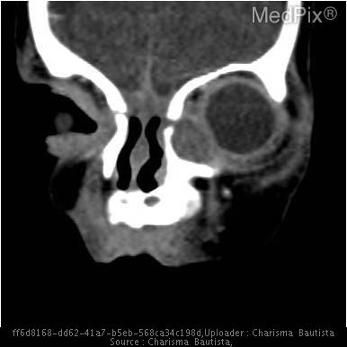
frontal sinus mucocele CT scan expansion
Searching Images
Draf frontal sinus surgery classification endoscopic
I have enough information to write a comprehensive, well-organized answer. Here is the full response based primarily on Scott-Brown's Otorhinolaryngology and Cummings Otolaryngology.
Topic 1: Frontal Mucocoele
Definition
A mucocoele is an expanding, epithelial-lined sac filled with inspissated mucus that completely fills and distends a paranasal sinus. It develops secondary to obstruction of the sinus outflow tract combined with an ongoing inflammatory process within the sinus. The lining is pseudostratified or low-columnar epithelium (not a distinct separate membrane as in a cyst). Where bony erosion occurs, the mucocoele epithelium often becomes fused with the dura or orbital periosteum.
(Scott-Brown's Otorhinolaryngology, Chapter 100)
Site of Development
Frontal sinuses are the most commonly affected site, followed by ethmoid, maxillary, and sphenoid sinuses (in descending order). This is because the frontal sinus has the most complex and narrow drainage pathway, making obstruction likely. Mucocoeles can also form in any aerated structure such as a concha bullosa.
Causes / Aetiology
Prior sinus surgery is now the leading cause (66-86% of cases across large series). The breakdown:
| Cause | Approximate incidence |
|---|---|
| Prior sinus surgery | 66-87% |
| Prior trauma | 10-14% |
| Spontaneous | 15-17% |
| Chronic rhinosinusitis (CRS) without surgery | 2-7% |
| Benign/malignant tumours | 1-5% |
Surgery in the frontal recess can cause circumferential injury to the frontal sinus outflow tract, leading to stenosis and mucocoele formation. Trauma-related mucocoeles have a mean latency of 9 years from injury to formation.
Pathogenesis
Two factors are considered essential for mucocoele formation:
- Obstruction of the sinus outflow tract
- Ongoing inflammatory process within the sinus
Osteolytic cytokines - interleukin-1 (IL-1) and tumour necrosis factor (TNF) - are present within the mucocoele lining. These are thought to be responsible for the progressive bony erosion and expansion that characterizes the lesion.
Clinical Features
Symptoms depend on the direction of expansion. Key features include:
- Forehead mass: A visible swelling of the forehead (most dramatic sign of a large frontal mucocoele)
- Ophthalmic symptoms (more common than rhinological/neurological): periorbital swelling, pain, exophthalmos, diplopia, limited ocular mobility, visual disturbance
- Optic neuropathy in up to 18% of patients with ophthalmic involvement (from compression in posterior ethmoid/sphenoid mucocoeles)
- Nasal obstruction (if arising in a concha bullosa)
- Epiphora or cystic medial canthal swelling (dacryocele)
- A pyocoele (infected mucocoele) presents with fever, rapid expansion, intense pain, and can cause orbital cellulitis
Radiology
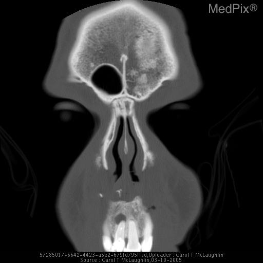
CT scan (primary investigation):
- Homogeneous, isodense lesion filling and expanding the sinus
- Bony remodelling and thinning of the sinus walls (not destruction)
- Bony defect of the lamina papyracea and/or superomedial orbital rim in fronto-ethmoidal mucocoeles
- Globe displaced laterally/inferiorly with proptosis on axial cuts
- Erosion of the posterior table (intracranial extension)
- Contrast enhancement occurs ONLY with a pyocoele (not a simple mucocoele)
MRI (supplementary):
- Indicated when there is significant bony erosion of the posterior table or lamina papyracea to delineate mucocoele from brain/soft tissue
- Mucocoeles generally have high water content - hyperintense on T1 and T2
- Pyocoeles have higher protein content and show variable signal intensity on both T1 and T2 - MRI can be misleading due to variability of contents
Differential Diagnosis
- Benign or malignant sinonasal tumours
- Allergic fungal sinusitis / other fungal disease
- Cholesterol granuloma (rare)
Surgical Management
The goal is wide marsupialization of the sac to provide permanent ventilation and drainage, and to relieve pressure on adjacent structures. Cosmetic deformities (forehead swelling) often resolve with time as bony remodelling occurs.
Fronto-ethmoidal mucocoeles:
- Endoscopic marsupialization is the first-line treatment (preferred method per Cummings)
- A Modified Endoscopic Lothrop Procedure (MELP / Draf III) is often required for complex frontal sinus mucocoeles, especially when there is loss of lateral support due to bony erosion/removal of the superior lamina papyracea
- Good results with endoscopic techniques: recurrence rates of 0 to 2.5%
- The most common complication is restenosis of the frontal recess - this is more common after Draf IIb (23%) than Draf IIa (3.6%) because more mucosal stripping occurs with extensive drilling
- Sometimes the mucocoele creates an "auto-Draf IIb" by itself - it erodes the medial wall of the frontal recess, creating a wide opening
Laterally located / complex mucocoeles:
- Combined approaches (endoscopic + external) are needed when thick bony septations prevent endoscopic-only access
- Lynch-Howarth external approach can assist with drilling of lateral bony septations, but carries a risk of long-term frontal outflow obstruction
- Osteoplastic frontal flap + MELP for far laterally placed mucocoeles
- Transorbital Neuroendoscopic Surgery (TONES) via the superior eyelid approach is a newer, less invasive alternative for lateral frontal sinus pathology
Stenting:
- Remains controversial; most authors do NOT advocate routine stenting, except for smaller unilateral marsupializations
- If a stent is placed, it must be loose-fitting to avoid circumferential pressure necrosis leading to stenosis
Obliteration:
- Has a 93% success rate, but carries a major complication rate >20% and has largely fallen out of favour
- Contraindicated when there is extensive posterior table erosion (unable to remove all respiratory epithelium from dura)
(Scott-Brown's, Chapter 100; Cummings, Chapter 46)
Topic 2: Frontal Recess Stenosis
Anatomy of the Frontal Recess
The frontal recess is the drainage pathway of the frontal sinus, not a distinct anatomical structure. Its boundaries are:
| Boundary | Structure |
|---|---|
| Anterior | Agger nasi cell, frontal process of maxilla (frontal beak) |
| Medial | Superior attachment of middle turbinate, lateral lamella of cribriform plate |
| Lateral | Lamina papyracea |
| Posterior | Anterior face of the ethmoid bulla (bulla lamella) |
| Superior | Communicates with frontal sinus |
- If the bulla lamella is absent superiorly, the suprabulla space communicates directly with the frontal recess - the anterior ethmoidal artery is then unprotected and at risk during dissection
- The agger nasi cell is present in over 90% of patients and is the key anterior landmark
- The uncinate process attaches superiorly to the lamina papyracea (33%), skull base (10%), or middle turbinate/combination (57%)
Causes of Frontal Recess Stenosis
Stenosis is the most feared complication of frontal sinus surgery. Causes include:
1. Mucosal Circumferential Injury (Post-surgical)
Any surgery in the frontal recess that causes circumferential injury to the mucosa of the outflow tract leads to scar formation, stenosis, and potentially mucocoele formation. The principle is that mucosa regenerating over bare bone lacks effective cilia, perpetuating disease.
2. Incomplete Dissection with Retained Cells
- A retained uncinate process or residual frontal recess cells (agger nasi, supra-agger cells, frontal bulla cells, etc.) can impede drainage and perpetuate mucosal inflammation
3. Lateralization of the Middle Turbinate
- Incidence of lateralized middle turbinates in revision surgery is 36-78%
- Causes medial wall obstruction of the frontal recess
- The frontal sinus rescue procedure was described for this: mucosa is dissected from the turbinate stump, medial mucosa discarded, and lateral sinus mucosa draped over the denuded stump
4. Osteoneogenesis
- Failure to preserve mucosa during surgery leads to new bone formation (osteoneogenesis)
- Incidence is higher after prior surgery
- Driven by persistent mucosal inflammation, ongoing infection, and surgical trauma
- The osteitis promotes further mucosal oedema, creating a vicious cycle of frontal recess stenosis
5. Scarring and Synechia
- Common in revision frontal sinus surgery but may not always be symptomatic
- Meticulous technique minimizes this risk
6. Anatomical Cell Variants
- Suprabulla cells, supraorbital ethmoid cells, frontal bulla cells, and recessus terminalis have been significantly associated with frontal sinusitis
- Type 3 and 4 frontal ethmoidal cells increase the prevalence of frontal sinus mucosal thickening
International Frontal Sinus Anatomy Classification (IFAC) Cells
The frontal recess cells (per the International Frontal Sinus Classification):
| Cell Type | Location |
|---|---|
| AN - Agger nasi | Anterosuperior to middle turbinate insertion |
| SAC - Supra agger cell | Above agger nasi, below frontal ostium |
| SAFC - Supra agger frontal cell | Extends into the frontal sinus |
| SBC - Supra bulla cell | Above ethmoid bulla, below frontal ostium |
| SBFC - Supra bulla frontal cell | Extends through frontal ostium |
| SOEC - Supra-orbital ethmoid cell | Pneumatizes over the orbit |
| FSC - Frontal septal cell | Along the nasal septum into frontal sinus |
Management of Frontal Recess Stenosis
The key principle: relieve obstruction while preserving mucosa as much as possible.
Graduated Surgical Approach:
- No exploration - when no disease is present; unnecessary instrumentation causes mucosal injury and adhesions
- Balloon sinuplasty - may have a role in limited disease with intact mucosa; not appropriate for osteoneogenesis or polyp disease
- Draf I - complete ethmoidectomy, removal of agger nasi and frontal recess cells below the frontal ostium; most common primary procedure
- Draf IIa - widening of the frontal ostium from the lamina papyracea to the middle turbinate
- Draf IIb - extends to the nasal septum; used for osteoneogenesis, inverted papilloma, osteomas, medial mucocoeles
- Draf III (MELP) - bilateral frontal sinus drillout; for recalcitrant/revision cases
- Osteoplastic flap + MELP - above and below combined approach
- Osteoplastic flap with obliteration - last resort; removes all mucosa and obliterates with fat
(Scott-Brown's, Chapter 99; Cummings, Chapter 46)
Topic 3: Management of Chronic Refractory Frontal Sinusitis
Definition / Background
Chronic frontal sinusitis is defined within the broader context of chronic rhinosinusitis (CRS) - obstruction of the frontal sinus outflow tract (the frontal recess) by either CRS with nasal polyps (CRSwNP) or CRS without nasal polyps (CRSsNP). Despite medical and surgical therapy, a proportion of patients develop recalcitrant chronic frontal sinus disease, defined as persistent symptoms after maximal medical therapy AND surgery.
Aetiology of Refractory Disease
- Persistent mucosal inflammation (most important factor)
- Scarring and stenosis from prior surgery
- Retained anatomical cells obstructing the recess
- Underlying conditions: asthma, aspirin sensitivity, eosinophilic CRS, immunodeficiency, diabetes
Step 1: Medical Management (Before Surgery)
Before any surgical decision, the following should be optimized:
- Broad-spectrum antibiotics (covering anaerobes in chronic disease)
- Nasal saline irrigations - high-volume, high-pressure
- Intranasal corticosteroids (topical steroids)
- Oral steroids (short course) for inflammatory flares
- Identify and treat underlying conditions (aspirin sensitivity, asthma, immunodeficiency)
- Allergy evaluation and management if relevant
Step 2: Surgical Management (After Failed Medical Therapy)
A graduated approach is used. The key principle: address the frontal recess, not the sinus itself. Most often the problem is the drainage pathway, not the sinus cavity.
DRAF Classification of Frontal Sinus Surgery
Draf Type I (Ethmoidectomy / Frontal Sinusotomy)
- Most common primary surgery
- Complete ethmoidectomy including agger nasi and anterior superior ethmoid cells
- Clears disease below the frontal ostium
- Best for patients with minor pathology in the frontal sinus; may be insufficient in aspirin-intolerant asthmatics with extensive polyps
Draf Type IIa
- Widening of the frontal ostium from the lamina papyracea to the middle turbinate
- Removes cells that obstruct or extend through the ostium
- Good mucosa preservation; low restenosis rate (3.6%)
Draf Type IIb
- Widens from the lamina papyracea to the nasal septum (includes partial resection of the anterior middle turbinate insertion)
- Indications: unilateral osteoneogenesis, inverted papilloma, osteoma obstructing the frontal recess, medially located mucocoeles
- Restenosis rate ~23% when extensive drilling is done
Draf Type III (Modified Endoscopic Lothrop Procedure - MELP)
- Also known as: frontal sinus drillout, median drainage procedure, endoscopic modified Lothrop
- Creates a large common drainage pathway for both frontal sinuses by:
- Resecting the upper nasal septum
- Resecting the frontal intersinus septum
- Drilling the floor of both frontal sinuses bilaterally
- The "frontal T" sign on CT identifies the anatomical extent of resection
- Technically demanding; landmarks are often absent in revision cases
- Computer-assisted navigation improves safety
- Best for: recalcitrant frontal sinusitis, ASA-sensitive patients with polyps, eosinophilic mucin CRS, revision cases with poor landmarks, mucocoeles, osteoneogenesis
- Key steps (Box 99.1 from Scott-Brown's):
- Perform complete bilateral ethmoidectomy
- Identify frontal ostia bilaterally
- Identify olfactory neurones at the lateral aspects of the septum
- Use frontal mini-trephines to identify the frontal ostium
- Perform upper septectomy to create a common surgical corridor
- Drill from one frontal recess across the midline to the other
- Reduce the frontal beak and intersinus septum
- Preserve mucosa over the posterior aspect of the frontal ostium
- Lower bone over anterior skull base projection to the first olfactory neurone
Factors Influencing Surgical Choice
| Factor | Low Complexity Option | High Complexity Option |
|---|---|---|
| No previous surgery, good landmarks | Draf I or IIa | - |
| Revision, osteoneogenesis, complex cells | Draf IIb or III | - |
| Eosinophilic polyps, aspirin sensitivity | Draf III (MELP) | - |
| Tumours, posterior table erosion | Draf III or osteoplastic flap | Combined approach |
| Laterally located disease | Osteoplastic flap ± MELP | Cranialization (rare) |
International Classification of Complexity (ICC)
| AP diameter of frontal ostium | Cells below ostium | Cells at ostium | Cells extending into sinus |
|---|---|---|---|
| Wide (≥10 mm) | Grade 1 | Grade 2 | Grade 3 |
| Narrow (6-9 mm) | Grade 2 | Grade 3 | Grade 4 |
| Very narrow (≤5 mm) | Grade 3 | Grade 4 | Grade 4 |
Open Procedures for Refractory Cases
Osteoplastic Flap Procedure:
- A bicoronal incision is used to reflect a bone flap over the frontal sinus
- Can be combined with MELP for an "above and below" approach
- With obliteration (fat packing): 93% success but >20% major complications
- Indicated when: endoscopic access is impossible, posterior table erosion with intracranial extension, far lateral disease, osteomyelitis, failed endoscopic surgery
Riedel's Procedure:
- Removal of the anterior table of the frontal sinus
- Rarely used; results in cosmetic deformity but effective for obliterating the sinus
Cranialization:
- Removal of sinus mucosa AND the posterior table
- Used when there is intracranial extension
Reasons for Failure of Frontal Sinus Surgery (Addressing for Revision)
Per Scott-Brown's (Chapter 99):
- Ongoing mucosal disease - persistent/recurrent nasal polyps (especially eosinophilic CRS with asthma); these patients require revision surgery more often
- Incomplete dissection - retained uncinate, residual agger nasi or frontal recess cells
- Lateralization of the middle turbinate - requires the "frontal sinus rescue procedure"
- Scarring and synechia formation - very common in revision surgery, may not be symptomatic
- Osteoneogenesis - progressive new bone formation from inadequate mucosal preservation
Balloon Sinuplasty (Adjunct Role)
- Introduced in 2006 as a minimally invasive option
- A balloon over a guide wire (often illuminated for confirmation) spans the frontal ostium; inflation causes microfractures of surrounding bone with mucosal preservation
- Evidence supports use in limited frontal disease where mucosa is intact
- NOT appropriate for: nasal polyposis, osteoneogenesis, osteomyelitis, mucocoele, or neoplasia
- Often combined with standard ESS as a "hybrid procedure"
- Limited long-term data; longer follow-up needed before substantiating as a standalone alternative
Post-surgical Care
- Regular endoscopic debridement in the post-operative period
- Nasal irrigations with saline
- Topical steroid delivery into the frontal recess (delivery improved after surgery)
- Long-term follow-up; revision rates are higher in CRSwNP + asthma + aspirin sensitivity
Summary Table: Graduated Approach to Frontal Sinus Disease
| Step | Procedure | Indication |
|---|---|---|
| 0 | Medical therapy alone | All patients first |
| 1 | No frontal recess exploration | No CT or clinical evidence of frontal disease |
| 2 | Balloon sinuplasty | Limited disease, intact mucosa, no polyps |
| 3 | Draf I (ethmoidectomy) | First surgery; mild-moderate frontal involvement |
| 4 | Draf IIa | Failed Draf I; cells at the frontal ostium level |
| 5 | Draf IIb | Unilateral osteoneogenesis, osteoma, medial mucocoele |
| 6 | Draf III (MELP) | Revision disease, bilateral/recalcitrant, eosinophilic CRS, ASA sensitivity |
| 7 | Osteoplastic flap + MELP | Failed endoscopic, lateral disease, combined pathology |
| 8 | Osteoplastic flap + obliteration | Last resort; failure of all other approaches |
| 9 | Riedel / Cranialization | Intracranial extension, osteomyelitis, posterior table erosion |
(Scott-Brown's Otorhinolaryngology Head & Neck Surgery Vol. 1, Chapters 99 & 100; Cummings Otolaryngology Head and Neck Surgery, Chapter 46)
This is a shared conversation. Sign in to Orris to start your own chat.