
Peribulbar block 5 mark. Answer for anesthesia resident exam
Reading File
Now let me search the medical library for peribulbar block content in parallel.
Excellent - I have all three sources. Let me now read the relevant sections from Miller's, Barash, and Morgan & Mikhail in parallel.
Reading File
Reading File
Reading File
I have very rich content from the textbooks. Let me now fetch the key diagram image from Miller's and read the full peribulbar section from Miller's.
Loading Image
Reading File
I now have comprehensive material from three authoritative anesthesia textbooks. Let me compile the complete exam answer.
Peribulbar Block - Anesthesia Resident Exam Answer (5 Marks)
Definition
Peribulbar block (also called extraconal block) is a regional anesthetic technique for the eye where local anesthetic is deposited outside the muscle cone, around (peri) the globe (bulbar). It was first described by Davis and Mandel in 1986 as a safer alternative to retrobulbar block.
Anatomy
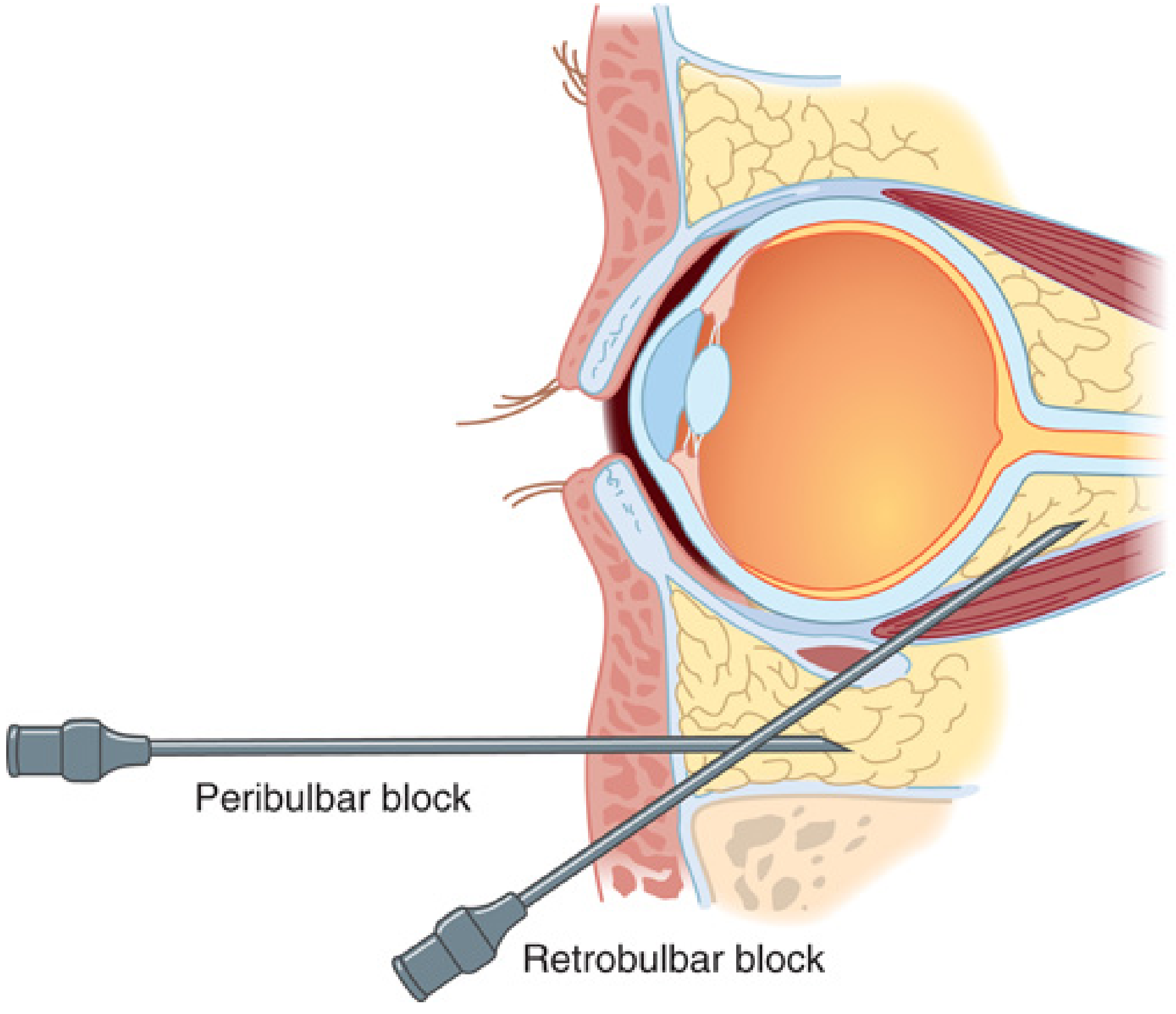
The four rectus muscles and their interconnecting fascial septa form a muscular cone extending from the optic foramen at the orbital apex to their insertions on the globe. In peribulbar block, the needle tip is placed extraconal - between the cone and the bony orbital wall. Cadaveric studies (Ripart et al.) have shown that extraconal injections diffuse into the intraconal space, explaining the eventual block of intraorbital structures despite extraconal placement.
Indications
- Cataract surgery (most common)
- Glaucoma surgery
- Corneal transplantation
- Vitreoretinal surgery
- Any intraocular surgery where regional anesthesia is preferred over general anesthesia
Technique
Patient position: Supine, looking straight ahead (primary gaze)
Needle: 23-25 gauge, 25 mm (max depth < 25 mm), short bevel needle
Classic Two-Injection Technique:
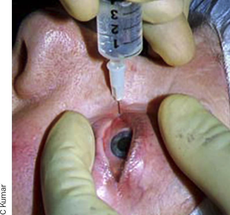
- Inferotemporal injection - At the junction of the medial and lateral thirds (some texts: middle and lateral thirds) of the lower lid, just above the inferior orbital rim. Needle directed vertically backwards, parallel to the orbital floor, no upward angulation. Advance to < 25 mm depth. Inject 5-10 mL of local anesthetic.
- Superonasal injection - At the superonasal quadrant, just below the orbital rim. Inject 2-5 mL.
Single Injection Technique:
A single inferotemporal injection of 5-10 mL suffices in many cases. After aspiration to confirm extravascular placement, slowly inject the local anesthetic.
Ocular compression (Honan balloon, 30 mmHg for 10 min, or gentle digital pressure) is applied after injection to spread the local anesthetic and reduce intraocular pressure.
Onset: Slower than retrobulbar (10-15 minutes vs. 3-5 minutes); may require supplemental injection if incomplete.
Local Anesthetic Agents
| Agent | Concentration | Onset | Duration |
|---|---|---|---|
| Lignocaine (Lidocaine) | 2% | Fast | Short (1-2 h) |
| Bupivacaine | 0.5-0.75% | Slower | Long (4-8 h) |
| Ropivacaine | 0.75-1% | Moderate | Long |
| Mixture | Lignocaine 2% + Bupivacaine 0.5% | Fast | Long |
Additives:
- Hyaluronidase 15-75 IU/mL: accelerates spread, shortens onset, reduces supplemental injection rates
- Epinephrine (1:200,000): prolongs duration, reduces bleeding
- Volume: Total 5-10 mL (larger than retrobulbar due to extraconal placement)
Effects of a Successful Block
- Analgesia - Block of nasociliary, lacrimal, and frontal branches of ophthalmic division of CN V
- Akinesia - Block of CN III, CN IV, CN VI (oculomotor, trochlear, abducens) - extraocular muscle paralysis
- Reduced IOP - Due to soft tissue pressure from volume injected + Honan balloon
- Chemosis (conjunctival edema) - Common due to extraconal injection, usually self-limiting
Note: Orbicularis oculi (CN VII) is OUTSIDE the cone and is NOT blocked - a separate facial nerve block (van Lint, Atkinson, or O'Brien technique) may be required to prevent lid squeezing.
Advantages over Retrobulbar Block
| Feature | Peribulbar | Retrobulbar |
|---|---|---|
| Needle depth | < 25 mm (extraconal) | 35 mm (intraconal) |
| Trajectory | Parallel to orbit floor | Toward orbital apex |
| Risk of retrobulbar hemorrhage | Lower | ~1% |
| Risk of optic nerve injury | Lower | Higher |
| Risk of globe perforation | Lower | Higher (esp. myopic eyes) |
| Onset | Slower (10-15 min) | Faster (3-5 min) |
| Chemosis | More common | Less common |
| Volume needed | More (5-10 mL) | Less (2-5 mL) |
Complications
Local:
- Chemosis (most common, benign)
- Subconjunctival hemorrhage
- Ecchymosis/hematoma
- Globe perforation (risk increased with axial length > 25 mm - high myopia)
Systemic (less common than retrobulbar):
- Brainstem anesthesia - spread via optic nerve sheath into CSF causing confusion, dysphagia, apnea, cardiac arrest (postretrobulbar apnea syndrome analog)
- Intravascular injection - seizures (ophthalmic artery retrograde flow to brain)
- Oculocardiac reflex - bradycardia, asystole (treat with atropine, stop surgery stimulus)
- Extraocular muscle damage
Contraindications
- Patient refusal
- Coagulopathy / anticoagulation (relative)
- Axial length > 26 mm (severe myopia) - higher risk of globe perforation
- Open globe injury
- Local infection / cellulitis
- Inability to cooperate or lie flat
- Allergy to local anesthetics
Key Exam Points to Remember
- First described 1986 (vs. retrobulbar which was standard before)
- Needle placed OUTSIDE the cone (extraconal) - this is the defining feature
- Needle depth < 25 mm and directed parallel to orbital floor (NO upward angulation toward apex)
- Larger volume (5-10 mL) needed vs. retrobulbar (2-5 mL)
- Hyaluronidase improves block quality
- Facial nerve NOT blocked - separate van Lint/Atkinson/O'Brien block needed for lid akinesia
- Main advantage: safer - reduces risk of retrobulbar hemorrhage and optic nerve injury
- Main disadvantage: slower onset, more volume, higher incidence of chemosis, may need supplemental injection
Sources: Miller's Anesthesia 10e, Ch. 65; Barash Clinical Anesthesia 9e, Ch. 49; Morgan & Mikhail's Clinical Anesthesiology 7e, Ch. 36
This is a shared conversation. Sign in to Orris to start your own chat.