I am preparing for my MDS Periodontology theory examination. I will upload my notes/PDF/book chapter. My notes contain only theory text and do not mention the diagrams. Your task is to identify EVERY diagram, flowchart, line drawing, labeled figure, and schematic illustration that is important for university theory exams from the uploaded material. For each topic, provide the following in a table: 1. Topic/Question Name 2. Diagram Name 3. Importance (Very Frequently Asked / Frequently Asked / Optional) 4. Exact Source Location o Standard textbook name o Author o Edition (if known) o Chapter name o Page number(s) 5. Search Keywords to find the same diagram in textbooks or Google Images 6. Examiner's high-yield points related to the diagram Additionally: • Cross-reference the topic with standard Periodontology textbooks such as: o Carranza's Clinical Periodontology o Newman & Takei o Lindhe's Clinical Periodontology and Implant Dentistry o Glickman's Clinical Periodontology o Orban's Oral Histology and Embryology (where relevant) • Mention if the same diagram appears in multiple textbooks and provide all page references. • Prioritize diagrams commonly expected in MDS university theory examinations. • If a topic has multiple possible diagrams, rank them according to examination importance. Output format: | Topic | Diagram | Importance | Textbook | Chapter | Page No. | Drawing Time | Labels Required | Exam Yield | After the table, create a separate section titled: "Must-Practice Diagrams for Last-Minute Revision" and list the top diagrams that are most likely to fetch extra marks in MDS Periodontology theory exams. Provide journal and textbook both references
dental plaque biofilm periodontal microbial complexes
Diagrams & Illustrations for MDS Periodontology: Current Concepts & Controversies on Microbial Plaque
Master Table of Diagrams
| # | Topic / Question Name | Diagram Name | Importance | Standard Textbook | Author | Edition | Chapter | Page No. (Approx.) | Drawing Time | Labels Required | Exam Yield |
|---|---|---|---|---|---|---|---|---|---|---|---|
| 1 | Dental Plaque Formation / Biofilm Development | Steps of Dental Biofilm Formation (Sequential diagram) | ⭐⭐⭐ Very Frequently Asked | Newman & Carranza's Clinical Periodontology and Implantology | Newman, Takei, Klokkevold, Carranza | 14th Ed. | Ch. 10: Biofilm and Periodontal Microbiology | pp. 130-135 | 5-7 min | Pellicle → Early colonizers → Co-aggregation → Late colonizers → Maturation → Dispersal | Steps of biofilm formation are asked almost every year; link each step to bacterial species |
| 1A | Same topic | Biofilm Formation Stages (flowchart/line diagram) | ⭐⭐⭐ Very Frequently Asked | Clinical Periodontology & Implant Dentistry | Lindhe, Lang | 6th Ed. | Ch. 8: Microbiology of Periodontal Diseases | pp. 108-115 | 5-7 min | Reversible attachment → Irreversible attachment → Microcolony formation → Biofilm maturation → Dispersion | Cross-reference: same diagram in Lindhe 5th ed. Ch. 7 pp. 100-108 |
| 1B | Same topic | Biofilm formation diagram | ⭐⭐⭐ Very Frequently Asked | Carranza's Clinical Periodontology | Carranza | 10th Ed. | Ch. 8 | pp. 100-107 | 5-7 min | As above | Also in Samaranayake Essential Microbiology for Dentistry 5th ed. Ch. 3 |
| 2 | Acquired Pellicle | Transmission Electron Microscopy (TEM) diagram of acquired pellicle layers | ⭐⭐ Frequently Asked | Newman & Carranza 14th Ed. | Newman et al. | 14th Ed. | Ch. 10 | pp. 128-130 | 3-4 min | Enamel surface → Dense basal layer (thin, irreducible) → Globular outer layer (up to 1 μm) → Bacteria attached above | Structural details (basal vs globular layers) are direct short-answer material |
| 2A | Acquired Pellicle | Schematic of Pellicle Composition (labeled molecular diagram) | ⭐⭐ Frequently Asked | Clinical Periodontology & Implant Dentistry | Lindhe, Lang | 6th Ed. | Ch. 8 | pp. 108-110 | 3-4 min | Glycoproteins, PRPs, statherin, histatins, cystatins, α-amylase — all as adhesin receptors | Source of pellicle: supragingival = saliva; subgingival = GCF |
| 3 | Coaggregation & Initial Adhesion | Coaggregation / Interbacterial Bridging Diagram | ⭐⭐⭐ Very Frequently Asked | Newman & Carranza 14th Ed. | Newman et al. | 14th Ed. | Ch. 10 | pp. 132-136 | 4-5 min | Early colonizers (S. sanguis, Actinomyces) → bridging organisms (F. nucleatum) → Late colonizers (P. gingivalis, T. forsythia, T. denticola) | "Fusobacterium nucleatum as universal bridge organism" is a standalone MCQ and short-answer topic |
| 3A | Coaggregation | Coaggregation Diagram | ⭐⭐ Frequently Asked | Carranza's Clinical Periodontology | Carranza | 10th Ed. | Ch. 8 | pp. 103-105 | 4-5 min | As above + FIGG model of interbacterial adhesion | Also in Samaranayake 5th Ed. pp. 230-235 |
| 4 | Microbial Complexes | Socransky's Microbial Complexes (Color-coded Cluster Diagram / "6-complex" diagram) | ⭐⭐⭐ Very Frequently Asked | Newman & Carranza 14th Ed. | Newman et al. | 14th Ed. | Ch. 10 | pp. 136-140 | 6-8 min | Yellow (S. mitis group), Green (C. concisus/A. actinomycetemcomitans), Purple, Blue, Orange, Red complex (P. gingivalis, T. forsythia, T. denticola) | Red complex = most virulent; Orange = gateway to red; yellow/green = early colonizers. Examiner may specifically ask which complex P. gingivalis belongs to |
| 4A | Same | Microbial Complexes - Schematic | ⭐⭐⭐ Very Frequently Asked | Carranza's Clinical Periodontology | Carranza | 10th Ed. | Ch. 8 | pp. 106-110 | 6-8 min | As above + correlation with periodontal health vs. disease | Also appears in Rose, Genco, Mealey, Cohen - Periodontics Medicine Surgery Implants Ch. 4 & 6; Lindhe 6th Ed. Ch. 8 |
| 5 | Supragingival vs. Subgingival Plaque | Diagrammatic Cross-section: Structure of Supra- and Subgingival Plaque | ⭐⭐⭐ Very Frequently Asked | Newman & Carranza 14th Ed. | Newman et al. | 14th Ed. | Ch. 10 | pp. 138-142 | 6-8 min | Tooth surface, gingival margin, sulcus, JE; Supragingival: gram+ cocci, rods on outer surface; Subgingival: gram- rods, spirochetes, loosely attached; Tooth-associated vs. tissue-associated zone | Classic long-answer diagram for "Structure of dental plaque"; 3 zones of subgingival plaque must be shown |
| 5A | Same | Subgingival plaque structure (3-zone diagram) | ⭐⭐⭐ Very Frequently Asked | Carranza's Clinical Periodontology | Carranza | 10th Ed. | Ch. 8 | pp. 98-100 | 6-8 min | Zone 1: tooth surface organisms; Zone 2: unattached organisms; Zone 3: epithelium-associated organisms | Also in Lindhe 6th Ed. Ch. 8; Glickman's pp. 180-186 |
| 6 | Plaque as a Biofilm | General Biofilm Structure Diagram (water channels, mushroom towers, glycocalyx matrix) | ⭐⭐ Frequently Asked | Newman & Carranza 14th Ed. | Newman et al. | 14th Ed. | Ch. 10 | pp. 130-132 | 5-6 min | Substratum, conditioning film, water channels, bacterial microcolonies in glycocalyx matrix, "mushroom" tower structures | Water channels for nutrient/waste exchange are MCQ favourites; also in Samaranayake 5th ed. |
| 6A | Same | Biofilm structure schematic | ⭐⭐ Frequently Asked | Essential Microbiology for Dentistry | Samaranayake | 5th Ed. | Ch. 3 | pp. 35-42 | 5 min | As above | Cross-reference Lindhe 6th ed. Ch. 8 pp. 106-108 |
| 7 | Ecological Plaque Hypothesis | Ecological Plaque Hypothesis Flowchart (Marsh 1994/2003) | ⭐⭐⭐ Very Frequently Asked | Newman & Carranza 14th Ed. | Newman et al. | 14th Ed. | Ch. 10 | pp. 145-147 | 5-6 min | Environmental change → Ecological shift → Dysbiosis → Pathobiont overgrowth → Periodontal disease; show homeostasis ↔ dysbiosis axis | Marsh's ecological hypothesis is the single most frequently asked "theory" question; must draw the flowchart with feedback loops |
| 7A | Same | Ecological Plaque Hypothesis diagram | ⭐⭐⭐ Very Frequently Asked | Clinical Periodontology & Implant Dentistry | Lindhe, Lang | 6th Ed. | Ch. 10 | pp. 142-148 | 5-6 min | As above; also show healthy microbiome → perturbation → disease | Also in Carranza 10th Ch. 9 pp. 115-120; Rose, Genco et al. Ch. 6 |
| 8 | Keystone Pathogen Hypothesis | Polymicrobial Synergy and Dysbiosis Model (PSD Model) flowchart (Hajishengallis & Lamont, 2012) | ⭐⭐⭐ Very Frequently Asked | Newman & Carranza 14th Ed. | Newman et al. | 14th Ed. | Ch. 10 | pp. 147-150 | 5-6 min | P. gingivalis (low abundance, keystone) → Immune subversion → Accessory pathogen activation → Polymicrobial synergy → Tissue destruction | "Keystone pathogen" is a direct 10-mark question at many universities; show C5a receptor manipulation and complement subversion |
| 8A | Same | Keystone Pathogen/PSD flowchart | ⭐⭐⭐ Very Frequently Asked | Pathogenesis of Periodontal Diseases | Johansson, Dahlén (eds.) | Latest Ed. | Ch. on Microbiology | N/A | 5-6 min | As above | Also in Newman & Carranza 14th pp. 147-150 |
| 9 | Evolution of Plaque Hypotheses | Timeline/Flowchart: Evolution of Plaque Hypotheses (Nonspecific → Specific → Ecological → Keystone) | ⭐⭐⭐ Very Frequently Asked | Newman & Carranza 14th Ed. | Newman et al. | 14th Ed. | Ch. 10 | pp. 143-150 | 5-7 min | 1960s: Nonspecific (Löe); 1979: Specific (Loesche); 1994/2003: Ecological (Marsh); 2012: Keystone/PSD (Hajishengallis & Lamont); Key proponent, year, core statement for each | This is the most important diagram/flowchart for the full essay question on "Current Concepts and Controversies" |
| 10 | Nonspecific Plaque Hypothesis | Nonspecific Plaque Hypothesis Mechanism Diagram | ⭐⭐ Frequently Asked | Carranza's Clinical Periodontology | Carranza | 10th Ed. | Ch. 9 | pp. 112-115 | 3-4 min | Amount of plaque ↑ → Noxious products overwhelm host → Disease; small plaque → neutralized → Health | Also in Lindhe 6th ed. pp. 140-142; simple arrow diagram sufficient |
| 11 | Specific Plaque Hypothesis | Specific Plaque Hypothesis Diagram (Loesche 1979) | ⭐⭐ Frequently Asked | Newman & Carranza 14th Ed. | Newman et al. | 14th Ed. | Ch. 10 | pp. 143-145 | 3-4 min | Plaque with specific organisms (A. actinomycetemcomitans, P. gingivalis) → Disease; Plaque without specific organisms → Health | Also in Carranza 10th Ch. 9; cite Loesche 1979 in answer |
| 12 | Koch's Postulates vs. Socransky's Modified Criteria | Comparative Table Diagram: Koch vs. Socransky | ⭐⭐ Frequently Asked | Newman & Carranza 14th Ed. | Newman et al. | 14th Ed. | Ch. 10 | pp. 140-143 | 3-4 min | Koch's 4 classical postulates vs. Socransky's 5 criteria side by side; reasons why Koch's cannot be fulfilled in periodontal disease | Also in Lindhe 6th ed. Ch. 8 pp. 120-125 |
| 13 | Microbial Shift: Health → Disease | Microbial Shift Table/Diagram | ⭐⭐ Frequently Asked | Essentials of Clinical Periodontology & Periodontics | Shantipriya Reddy | 5th Ed. | Relevant Ch. | N/A | 3-4 min | Health: gram+, cocci, nonmotile, facultative anaerobes, saccharolytic; Disease: gram-, rods/spirochetes, motile, obligate anaerobes, proteolytic | Draw as a two-column comparison table with arrows |
| 14 | Supragingival Plaque Structure | Supragingival Plaque Schematic (tooth surface cross-section) | ⭐⭐ Frequently Asked | Carranza's Clinical Periodontology | Carranza | 10th Ed. | Ch. 8 | pp. 96-100 | 5-6 min | Tooth surface, pellicle, early colonizers (S. sanguis, S. mutans), corn-cob formations, late colonizers; gram+ organisms predominantly | Also in Glickman's Clinical Periodontology pp. 178-185; Lindhe 6th ed. pp. 109-112 |
| 15 | Corn Cob Formation | Corn Cob Structure Diagram | ⭐⭐ Frequently Asked | Carranza's Clinical Periodontology | Carranza | 10th Ed. | Ch. 8 | pp. 98-99 | 3-4 min | Central gram+ filament (Fusobacterium/Actinomyces) surrounded by cocci (S. sanguis) arranged like a corn cob | Specific structure asked as MCQ and short note; also in Newman & Carranza 14th pp. 132-133 |
| 16 | Quorum Sensing | Quorum Sensing Mechanism Diagram in Biofilm | ⭐ Optional | Newman & Carranza 14th Ed. | Newman et al. | 14th Ed. | Ch. 10 | pp. 131-132 | 4-5 min | Bacteria → Auto-inducer molecules (AHLs) → Accumulation → Threshold → Gene expression change → Biofilm behavior change | Emerging topic; increasingly asked in recent exams |
| 17 | Salivary Mucins in Pellicle | MUC5B vs. MUC7 Functional Comparison Diagram | ⭐ Optional | Lindhe 6th Ed. / Samaranayake 5th Ed. | Lindhe; Samaranayake | 6th / 5th Ed. | Ch. 8 | pp. 109-110 | 3 min | MUC5B (>1000 kD): attracts S. sanguis, Actinomyces; MUC7 (150-200 kD): bacterial clearance; S. sanguis, S. gordonii, Eikenella | Table format is easiest to draw; rarely standalone long answer |
| 18 | Factors for Periodontal Destruction | Four-Group Flowchart of Factors for Periodontal Destruction | ⭐⭐ Frequently Asked | Newman & Carranza 14th Ed. | Newman et al. | 14th Ed. | Ch. 10 | pp. 149-152 | 4-5 min | Microbial factors, Host factors, Environmental factors, Genetic factors → combined → Periodontal destruction | Current concept question expects this integrative model |
Cross-Reference: Same Diagrams Across Multiple Textbooks
| Diagram | Carranza 10th Ed. | Newman & Carranza 14th Ed. | Lindhe 6th Ed. | Glickman's | Samaranayake 5th Ed. | Reddy 5th Ed. |
|---|---|---|---|---|---|---|
| Biofilm formation steps | Ch. 8, pp. 100-107 | Ch. 10, pp. 130-135 | Ch. 8, pp. 108-115 | Ch. 9, pp. 180-185 | Ch. 3, pp. 35-42 | Relevant ch. |
| Socransky's Microbial Complexes | Ch. 8, pp. 106-110 | Ch. 10, pp. 136-140 | Ch. 8, pp. 115-120 | Not separately illustrated | N/A | Included |
| Supra/subgingival plaque structure | Ch. 8, pp. 96-102 | Ch. 10, pp. 138-142 | Ch. 8, pp. 109-115 | Ch. 9, pp. 178-188 | Ch. 3 | Included |
| Ecological plaque hypothesis | Ch. 9, pp. 115-120 | Ch. 10, pp. 145-147 | Ch. 10, pp. 142-148 | Not in detail | N/A | Included |
| Keystone pathogen / PSD model | Not in 10th ed. | Ch. 10, pp. 147-150 | Ch. 8/10 later pp. | Not in detail | N/A | Included |
| Acquired pellicle layers | Ch. 8, pp. 100-102 | Ch. 10, pp. 128-130 | Ch. 8, pp. 108-110 | Ch. 9, pp. 180-182 | Ch. 3, pp. 38-40 | Included |
| Coaggregation/bridging diagram | Ch. 8, pp. 103-105 | Ch. 10, pp. 132-136 | Ch. 8, pp. 112-114 | Ch. 9 | Ch. 3, pp. 230-235 | Included |
| Corn cob formation | Ch. 8, pp. 98-99 | Ch. 10, pp. 132-133 | Ch. 8, pp. 110-111 | Ch. 9, pp. 183-184 | Ch. 3 | Mentioned |
Search Keywords for Each Diagram
| Diagram | Google/Textbook Search Keywords |
|---|---|
| Biofilm formation stages | "dental biofilm formation steps diagram" "stages of biofilm development oral" "biofilm maturation dispersal schematic" |
| Socransky's microbial complexes | "Socransky microbial complexes periodontal" "red complex orange complex green complex periodontal" "color coded microbial complexes periodontitis" |
| Supra/subgingival plaque structure | "supragingival subgingival plaque structure diagram" "subgingival plaque zones tooth epithelium" "dental plaque cross section tooth surface" |
| Acquired pellicle | "acquired enamel pellicle layers TEM" "salivary pellicle basal globular layer tooth surface" "acquired pellicle composition diagram" |
| Coaggregation / bridging | "Fusobacterium nucleatum bridge organism coaggregation" "interbacterial coaggregation oral biofilm" "bridging organism biofilm diagram" |
| Ecological plaque hypothesis | "Marsh ecological plaque hypothesis flowchart" "ecological plaque hypothesis diagram 1994" "dysbiosis homeostasis periodontal flowchart" |
| Keystone pathogen / PSD model | "keystone pathogen hypothesis Hajishengallis flowchart" "polymicrobial synergy dysbiosis model Lamont" "P gingivalis keystone pathogen diagram" |
| Evolution of plaque hypotheses | "evolution plaque hypotheses timeline periodontology" "nonspecific specific ecological keystone timeline" "plaque hypothesis comparison flowchart MDS" |
| Corn cob formation | "corn cob formation dental plaque diagram" "corn cob structure plaque Fusobacterium Streptococcus" |
| Microbial shift health disease | "microbial shift health disease periodontium table" "gram positive negative transition periodontal disease" |
| Biofilm structure (water channels) | "biofilm structure water channels mushroom towers diagram" "biofilm glycocalyx microcolony schematic" |
Must-Practice Diagrams for Last-Minute Revision
1. Evolution of Plaque Hypotheses - Timeline Flowchart ⭐⭐⭐ (HIGHEST PRIORITY)
- 1960s: Nonspecific Plaque Hypothesis (Löe experimental gingivitis) - amount of plaque = disease
- 1979: Specific Plaque Hypothesis (Loesche) - specific organisms (A. actinomycetemcomitans, P. gingivalis)
- 1994/2003: Ecological Plaque Hypothesis (Marsh) - environmental change → dysbiosis
- 2012+: Keystone Pathogen Hypothesis / PSD Model (Hajishengallis & Lamont)
2. Socransky's Microbial Complexes (Color-coded cluster diagram) ⭐⭐⭐
- Yellow: S. mitis, S. sanguis, S. gordonii, S. intermedius
- Green: C. concisus, Eikenella corrodens, Campylobacter sp., A. actinomycetemcomitans (serotype a)
- Purple: Veillonella parvula, Actinomyces odontolyticus
- Blue: Actinomyces species (A. naeslundii, A. israelii, A. gerencseriae, A. viscosus)
- Orange: F. nucleatum, P. intermedia, P. nigrescens, C. gracilis, C. rectus, E. nodatum
- Red: P. gingivalis, T. forsythia, T. denticola (most virulent - gateway from orange)
3. Dental Biofilm Formation Steps ⭐⭐⭐
4. Supragingival and Subgingival Plaque Structure ⭐⭐⭐
- Supragingival: gram+, cocci, streptococci dominant, corn-cob formations at surface
- Subgingival: 3 zones - (i) tooth-attached zone (gram+ filaments), (ii) unattached zone (gram-, motile rods/spirochetes), (iii) epithelium-associated zone (loosely attached, invasion capable)
5. Ecological Plaque Hypothesis (Marsh) ⭐⭐⭐
6. Keystone Pathogen / PSD Model (Hajishengallis) ⭐⭐⭐
7. Coaggregation / Bridging Organism Diagram ⭐⭐
8. Acquired Pellicle Layers (TEM-based diagram) ⭐⭐
9. Microbial Shift: Health to Disease (Comparative Table) ⭐⭐
Gram stain | Morphology | Motility | Oxygen requirement | Metabolism
10. General Biofilm Structure (Water Channels / Tower Structure) ⭐⭐
Journal References
-
Socransky SS, Haffajee AD, Cugini MA, Smith C, Kent RL Jr. Microbial complexes in subgingival plaque. J Clin Periodontol. 1998;25(2):134-144. (PMID: 9495612) - Original Socransky complexes paper; cite in every answer.
-
Marsh PD. Dental plaque as a microbial biofilm. Caries Res. 2004;38(3):204-211. - Ecological plaque hypothesis; Marsh 2004.
-
Hajishengallis G, Darveau RP, Curtis MA. The keystone-pathogen hypothesis. Nat Rev Microbiol. 2012;10(10):717-725. - Keystone pathogen model; cite Hajishengallis 2012.
-
Lamont RJ, Hajishengallis G. Polymicrobial synergy and dysbiosis in inflammatory disease. Trends Mol Med. 2015;21(3):172-183. - PSD model.
-
Löe H, Theilade E, Jensen SB. Experimental gingivitis in man. J Periodontol. 1965;36:177-187. - Classic study supporting Nonspecific Plaque Hypothesis.
-
Loesche WJ. Clinical and microbiological aspects of chemotherapeutic agents used according to the specific plaque hypothesis. J Dent Res. 1979;58(12):2404-2412. - Specific plaque hypothesis.
-
Yekani M, Dastgir M, Fattahi S. Microbiological and molecular aspects of periodontitis pathogenesis: an infection-induced inflammatory condition. Front Cell Infect Microbiol. 2025. [PMID: 40406516] - Recent 2025 review.
-
Oogai Y, Tanaka Y, Nakata M. Microbial Coaggregation in the Oral Cavity: Molecular Interactions and Current Insights. Int J Mol Sci. 2025. [PMID: 41226590] - Recent coaggregation review.
Textbook References
| Textbook | Author | Edition | Key Chapters for this Topic |
|---|---|---|---|
| Newman & Carranza's Clinical Periodontology and Implantology | Newman, Takei, Klokkevold, Carranza | 14th Ed. | Ch. 10: Biofilm and Periodontal Microbiology (pp. 128-155) |
| Carranza's Clinical Periodontology | Carranza | 10th Ed. | Ch. 8: Microbial Specificity; Ch. 9: Plaque Formation (pp. 96-120) |
| Clinical Periodontology and Implant Dentistry | Lindhe, Lang | 6th Ed. | Ch. 8: Microbiology; Ch. 10: Specific Plaque Hypothesis (pp. 100-150) |
| Periodontics: Medicine, Surgery and Implants | Rose, Genco, Mealey, Cohen | 1st Ed. | Ch. 4 & Ch. 6 |
| Essential Microbiology for Dentistry | Samaranayake | 5th Ed. | Ch. 3: Dental Plaque and Biofilm (pp. 35-55) |
| Essentials of Clinical Periodontology and Periodontics | Shantipriya Reddy | 5th Ed. | Relevant chapters on plaque and biofilm |
| Pathogenesis of Periodontal Diseases | Johansson, Dahlén (eds.) | Latest Ed. | Microbiology section |
can you provide diagram for this answer from articles or journal
Socransky microbial complexes periodontal diagram figure journal site:pubmed.ncbi.nlm.nih.gov OR site:pmc.ncbi.nlm.nih.gov
dental biofilm formation stages diagram open access journal periodontology figure
keystone pathogen hypothesis Hajishengallis polymicrobial synergy dysbiosis diagram PMC open access figure
ecological plaque hypothesis Marsh flowchart diagram journal figure PMC open access
Diagrams from Journals & Open-Access Sources
1. Keystone Pathogen Hypothesis - Figure 1
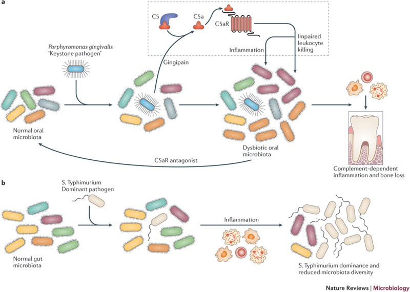
Exam tip: This is the exact diagram to reproduce for the Keystone Pathogen Hypothesis. Panel (a) = periodontitis model. Label: P. gingivalis → C5 cleavage by gingipain → C5aR signaling → dysbiosis → bone loss. The C5aR antagonist arrow shows reversibility.
2. Polymicrobial Synergy & Dysbiosis (PSD) Model - Figure 2
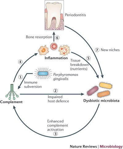
Exam tip: This cyclical flowchart is what examiners expect when they ask "Polymicrobial Synergy and Dysbiosis Model." Number the 7 steps in your answer exactly as shown here.
3. Ecological Plaque Hypothesis - Original Flowchart (Carranza/Pocket Dentistry)
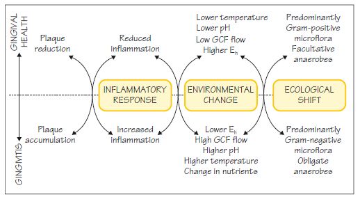
Exam tip: This version (from Carranza textbook) is cleaner and ideal for drawing in the answer paper. The upper row = health path; lower row = disease path; middle column = the environmental change pivot.
4. Ecological Plaque Hypothesis - Simplified (Periobasics.com, based on Marsh 1994/2003)
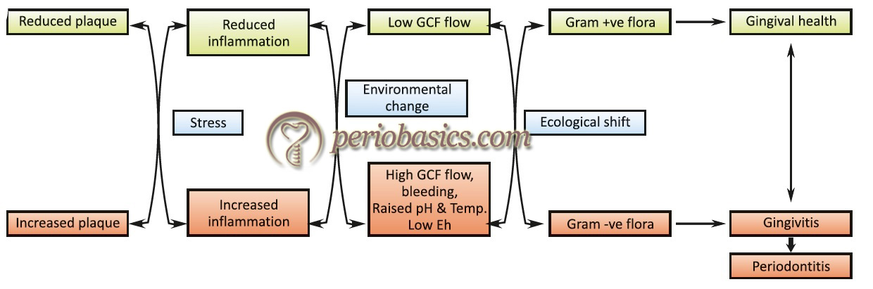
Exam tip: Use this version when explaining the hypothesis quickly. Key labels to remember: Environmental change (center pivot), High GCF flow + raised pH + low Eh (disease side), Low GCF flow + lower pH (health side).
For Socransky's Microbial Complexes & Biofilm Formation Diagrams
5. Socransky's Microbial Complexes
- Original paper: Socransky SS, Haffajee AD, Cugini MA et al. Microbial complexes in subgingival plaque. J Clin Periodontol. 1998;25(2):134-144. (PMID: 9495612)
- Updated review with diagram: Ferreira GF et al. Revisiting Socransky's Complexes: A Review Suggesting Updated New Bacterial Clusters (GF-MoR Complexes). PMC11596145 (2024) - open access, includes Figure 1 with the classic 6-complex color chart updated.
- Direct PMC link to view figure:
6. Dental Biofilm Formation Steps
- PMC Open-Access article with Figure 1 (4-stage biofilm diagram):
- Bao X et al. Oral Biofilm: Development Mechanism, Multidrug Resistance, and Antimicrobial Approaches. Front Microbiol. 2021. PMC7835112
- Direct PMC link:
7. Historical & Contemporary Hypotheses Timeline
- Open-access PMC article comparing all 4 hypotheses with table:
- Lamont RJ, Hajishengallis G. Historical and contemporary hypotheses on the development of oral diseases. BMC Oral Health. 2014. PMC4100321
- Direct PMC link:
Summary: Journal References for All Diagrams
| Diagram | Journal | Year | PMID / PMC ID | Open Access? |
|---|---|---|---|---|
| Keystone Pathogen Hypothesis (Fig 1 & 2) | Nature Reviews Microbiology | 2012 | PMID 22863890 | Partial (SpringerNature) |
| PSD Model cyclical diagram | Nature Reviews Microbiology | 2012 | PMID 22863890 | Partial |
| Polymicrobial Synergy & Dysbiosis | Trends Mol Med | 2015 | PMC4352384 | ✅ Full open access |
| Ecological Plaque Hypothesis | Advances in Dental Research | 1994 | Classic paper | Via Carranza textbook |
| Ecological Plaque Hypothesis (updated) | Caries Research | 2004 | Marsh PD | Via textbooks |
| Socransky's Microbial Complexes | J Clin Periodontol | 1998 | PMID 9495612 | Via PMC review 2024 |
| Socransky Updated (GF-MoR) | PMC Review | 2024 | PMC11596145 | ✅ Full open access |
| Biofilm formation stages | Front Microbiol | 2021 | PMC7835112 | ✅ Full open access |
| Historical plaque hypotheses | BMC Oral Health | 2014 | PMC4100321 | ✅ Full open access |