
Pathophysiology of Acute pulpal pain for 10 marks
pulpal pain pathophysiology dental pulp innervation diagram
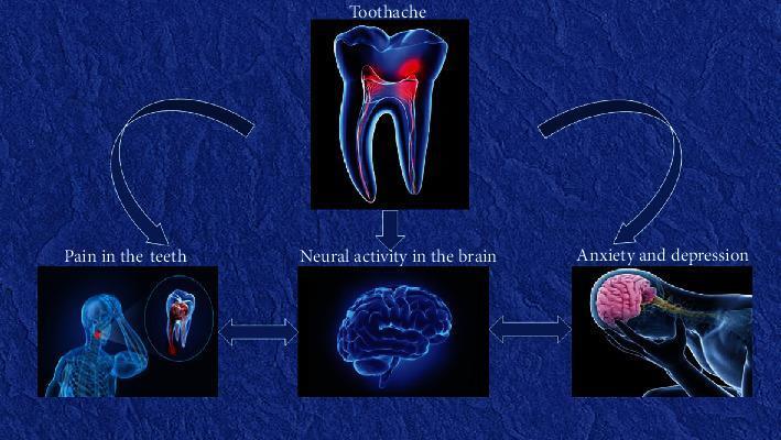
A pathophysiology diagram illustrating the multidirectional relationship between dental pain and neuropsychological states. The schematic follows a cyclical flow starting with 'Toothache', represented by a molar illustration with pulpal inflammation. A downward arrow connects this to 'Neural activity in the brain', depicted by a 3D model of the human brain. This central node is connected via double-headed horizontal arrows to two auxiliary states: 'Pain in the teeth' (visualized by a patient in distress with local dental inflammation) and 'Anxiety and depression' (visualized by a person holding their head with the brain highlighted). Curved arrows further link 'Toothache' to 'Pain in the teeth' and 'Anxiety and depression' back to 'Toothache', establishing a feedback loop. This educational visual demonstrates how chronic dental pain (odontalgia) triggers cerebral functional connectivity changes, which in turn correlate with mood disorders like anxiety and depression, potentially exacerbating the perception of pain.
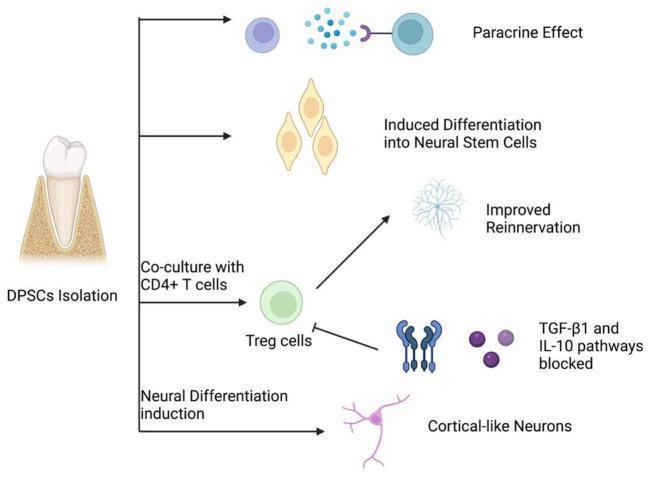
This medical pathophysiology diagram illustrates the regenerative mechanisms of Dental Pulp Stem Cells (DPSCs) in peripheral nerve repair. The flowchart begins on the left with 'DPSCs Isolation' from a human tooth, branching into four primary therapeutic pathways. 1) Paracrine Effect: Depicted by DPSCs secreting signaling molecules (blue spheres) that interact with target cell receptors. 2) Induced Differentiation into Neural Stem Cells: Shows DPSCs transforming into elongated, teardrop-shaped neural progenitor cells. 3) Immunomodulatory Pathway: Describes the co-culture of DPSCs with CD4+ T cells to produce Regulatory T cells (Treg cells), which promotes 'Improved Reinnervation' (visualized as a complex neuronal network) or blocks TGF-̢1 and IL-10 pathways to modulate the inflammatory environment. 4) Neural Differentiation Induction: Shows the direct maturation of DPSCs into 'Cortical-like Neurons' characterized by a distinct soma and branching processes. This diagram serves as a pedagogical summary of biotherapy and neuroregeneration concepts within regenerative medicine.
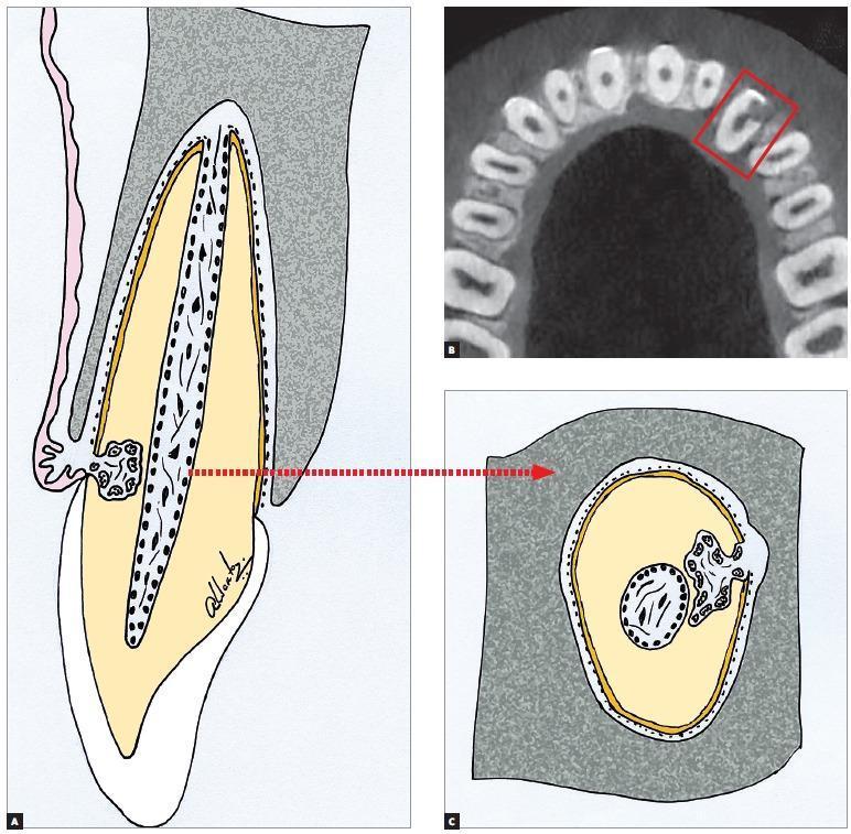
This composite educational resource illustrates external cervical resorption (ECR) through schematic diagrams and diagnostic imaging. Panel A shows a sagittal-view anatomical diagram of a tooth, highlighting a resorptive lesion originating at the cervical region and penetrating the dentine. Panel C provides a corresponding transverse-view diagram, emphasizing that the resorption circumvents the dental pulp without invading the pulp chamber, due to the protective layer of non-mineralized predentine. Panel B displays a Cone Beam Computed Tomography (CBCT) axial slice of the maxillary arch, with a red box identifying a radiolucent lesion on the cervical aspect of a tooth, correlating with the schematics. The visual demonstrates the key diagnostic feature of ECR: the preservation of the pulpal limits and a thin layer of mineralized dentine separating the lesion from the pulp space. This distinction is critical for differential diagnosis from internal resorption and informs treatment planning aimed at preserving pulp vitality. The material is intended for endodontic and orthodontic education, focusing on pathophysiology and radiographic interpretation.
acute pulpal pain pathophysiology inflammatory mediators neurogenic mechanism
Pathophysiology of Acute Pulpal Pain
Introduction
1. Anatomy Relevant to Pain Transmission
| Feature | A-delta Fibers | C Fibers |
|---|---|---|
| Myelination | Myelinated | Unmyelinated |
| Pain quality | Sharp, bright, well-localized | Dull, burning, poorly localized |
| Location | Extend 0.2-0.3 mm into dentinal tubules | Terminate in pulp chamber |
| Stimuli | Thermal, mechanical, osmotic | Thermal + inflammatory mediators (histamine, bradykinin) |
| Proportion | Minority | 50-75% of pulpal innervation |
| Clinical correlate | Early/reversible pulpitis | Irreversible pulpitis |
- Cummings Otolaryngology Head and Neck Surgery, Table 86.1
2. Initiating Events (Etiology)
-
Tooth fracture (direct pulp exposure)
-
Thermal or chemical trauma (deep restorations, erosion)
-
Retrograde infection through the apical foramen
-
Scott-Brown's Otorhinolaryngology Head & Neck Surgery
3. The Hydrodynamic Theory of Early Pulpal Pain (A-delta Mediated)
- Outward flow (e.g., cold stimulus causing fluid contraction or evaporation) or inward flow (e.g., heat causing expansion) mechanically deflects the A-delta fiber endings at the pulpo-dentinal junction.
- This produces a sharp, immediate pain that subsides when the stimulus is removed.
- Covering the tubules (e.g., with a smear layer or desensitizing agent) raises the threshold needed to provoke pain, directly confirming this mechanism.
- Cummings Otolaryngology Head and Neck Surgery
4. Inflammatory Cascade (Transition to Irreversible Pulpitis)
a. Classical Inflammation
- Mast cells and macrophages release histamine, bradykinin, and prostaglandins.
- These mediators directly sensitize C-fiber nociceptors - lowering their activation threshold and causing spontaneous firing even without an external stimulus.
- Bradykinin levels are significantly elevated in irreversible pulpitis, contributing to early-stage inflammation and pain.
- Prostaglandins (via COX pathway) further sensitize pulpal nerve endings.
b. Neurogenic Inflammation
- Substance P (SP) - measured directly in human pulp tissue via microdialysis; levels are significantly higher in irreversible pulpitis compared to non-infected teeth.
- Calcitonin Gene-Related Peptide (CGRP)
- Vasoactive Intestinal Polypeptide (VIP)
- Vasodilation - increasing blood flow and vascular permeability
- Plasma extravasation - raising tissue fluid pressure
- Further nociceptor sensitization - a positive feedback loop
- Cummings Otolaryngology Head and Neck Surgery, Frontiers in Dental Medicine 2024, PMC - Neurophysiology of Pulpal Pain
5. Peripheral Sensitization
- Previously high-threshold nociceptors lower their firing threshold.
- Silent/dormant nociceptors (previously unresponsive to stimuli) become activated and recruited.
- Activated neurons sprout additional terminal branches, widening the sensory field.
-
Hyperalgesia - exaggerated pain response to a normally painful stimulus (e.g., even mild warmth causes severe pain)
-
Allodynia - pain in response to normally innocuous stimuli
-
Pain becomes spontaneous, throbbing, persistent, and poorly localized
-
6. Central Sensitization and Pain Characteristics
-
With sustained nociceptor input, central neurons undergo NMDA receptor-mediated sensitization: neuropeptide substance P provides sufficient depolarization to remove the Mg²+ block from NMDA receptors, allowing glutamate from non-nociceptive A-beta fibers to activate pain neurons.
-
This is the mechanism of central sensitization and contributes to:
- Referred pain - lesions in mandibular teeth can refer to the maxilla, cheek, ear, or temple (convergence of afferents in the subnucleus caudalis)
- Spread of pain beyond the affected tooth
- Difficulty in pain localization, especially in irreversible pulpitis
-
Cummings Otolaryngology Head and Neck Surgery
7. Progression to Pulp Necrosis
-
Ischemic necrosis of the pulp ensues
-
Anaerobic bacteria colonize the necrotic root canal walls as a biofilm
-
Toxins and bacteria escape through the apical foramen into periapical tissues
-
This triggers acute periapical (apical) periodontitis - now characterized by severe, well-localized, throbbing pain that is exacerbated by percussion (biting), as the periodontal ligament and bone become inflamed
-
Scott-Brown's Otorhinolaryngology Head & Neck Surgery
8. Summary Flowchart
Bacterial invasion / dental caries
        ↓
Dentinal tubule exposure → Hydrodynamic fluid shifts → A-delta activation → SHARP PAIN (reversible)
        ↓
Bacterial products penetrate pulp
        ↓
Mast cell/macrophage activation → Histamine, Bradykinin, Prostaglandins
        ↓
C-fiber sensitization + activation → DULL, SPONTANEOUS PAIN
        ↓
Neuropeptide release (SP, CGRP) → Neurogenic inflammation → Vasodilation + ↑ tissue pressure
        ↓
Rigid pulp chamber → Cannot expand → Vascular occlusion → Ischemia
        ↓
Irreversible pulpitis: Spontaneous, throbbing, poorly localized pain
        ↓
Complete necrosis → Periapical spread → Acute apical periodontitis
Key Points for 10 Marks
- Dual fiber system: A-delta (sharp, early) vs C fibers (dull, later) - different stimuli and locations
- Hydrodynamic theory: fluid movement in dentinal tubules activates A-delta; explains dentinal hypersensitivity
- Rigid enclosure: the non-compliant pulp chamber amplifies inflammation and ischemia
- Inflammatory mediators: histamine, bradykinin, prostaglandins sensitize nociceptors
- Neurogenic inflammation: SP and CGRP from C fibers cause vasodilation and positive feedback
- Peripheral sensitization: silent nociceptors recruited; allodynia and hyperalgesia develop
- Central sensitization: NMDA-mediated changes in subnucleus caudalis explain referred pain
- Clinical progression: reversible pulpitis → irreversible pulpitis → necrosis → periapical disease
- Pain characteristics change: sharp/localized (early) → dull/throbbing/spontaneous/poorly localized (late)
- Trigeminal pathway: pulpal afferents → subnucleus caudalis → thalamus → somatosensory cortex