
Neuro muscler junction 6 marks
Reading File
Now let me search the library for neuromuscular junction content.
Finding Sources
Finding Sources
Good. Let me read the key physiology sources in parallel.
Reading File
Reading File
Reading File
Excellent content retrieved. Let me get a bit more on ACh synthesis, end plate potential, and the safety factor sections.
Reading File
Reading File
I now have comprehensive, detailed textbook content on the neuromuscular junction. Let me fetch the key diagram image and compile the full answer.
Loading Image
Loading Image
I now have everything needed for a thorough 6-mark answer. Here is the complete, exam-ready response:
Neuromuscular Junction (NMJ)
Definition
The neuromuscular junction (NMJ), also called the motor end plate, is the specialized chemical synapse between the terminal of a lower motor neuron (alpha motor neuron) and a skeletal muscle fibre. It converts an electrical nerve impulse into a chemical signal that triggers muscle contraction.
Structure
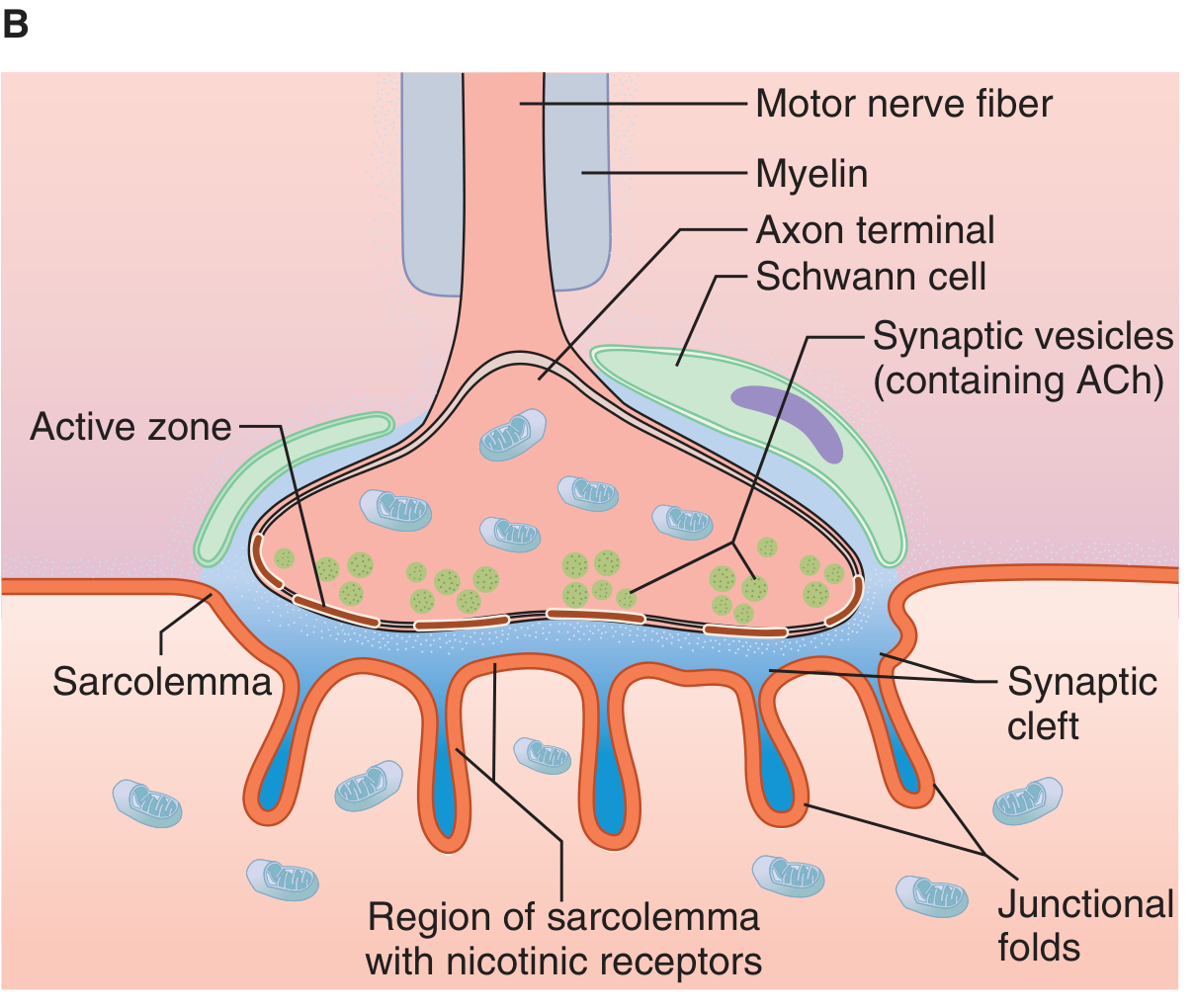
The NMJ has three main components:
1. Presynaptic (nerve) terminal
- The myelinated motor axon loses its myelin sheath near the muscle and divides into terminal boutons (knob-like endings)
- Each bouton contains ~300,000 synaptic vesicles, each packed with acetylcholine (ACh)
- Dense bars on the inner neural membrane flank voltage-gated Ca²⁺ channels - these are the active zones for vesicle docking and exocytosis
- Numerous mitochondria supply ATP for ACh synthesis
2. Synaptic cleft
- 20-30 nm wide space between nerve terminal and muscle membrane
- Contains the enzyme acetylcholinesterase (AChE), embedded in a collagen matrix, which rapidly degrades ACh
3. Postsynaptic (muscle) membrane - Motor End Plate
- The muscle membrane here (sarcolemma) is thickened and thrown into junctional (subneural) folds, greatly increasing surface area
- Nicotinic ACh receptors (nAChRs) are densely concentrated at the crests of these folds, directly opposite the active zones
- Voltage-gated Na⁺ channels are concentrated deeper in the folds
Each motor axon innervates several muscle fibres forming a motor unit; each muscle fibre receives input from only one nerve terminal.
Nicotinic ACh Receptor Structure
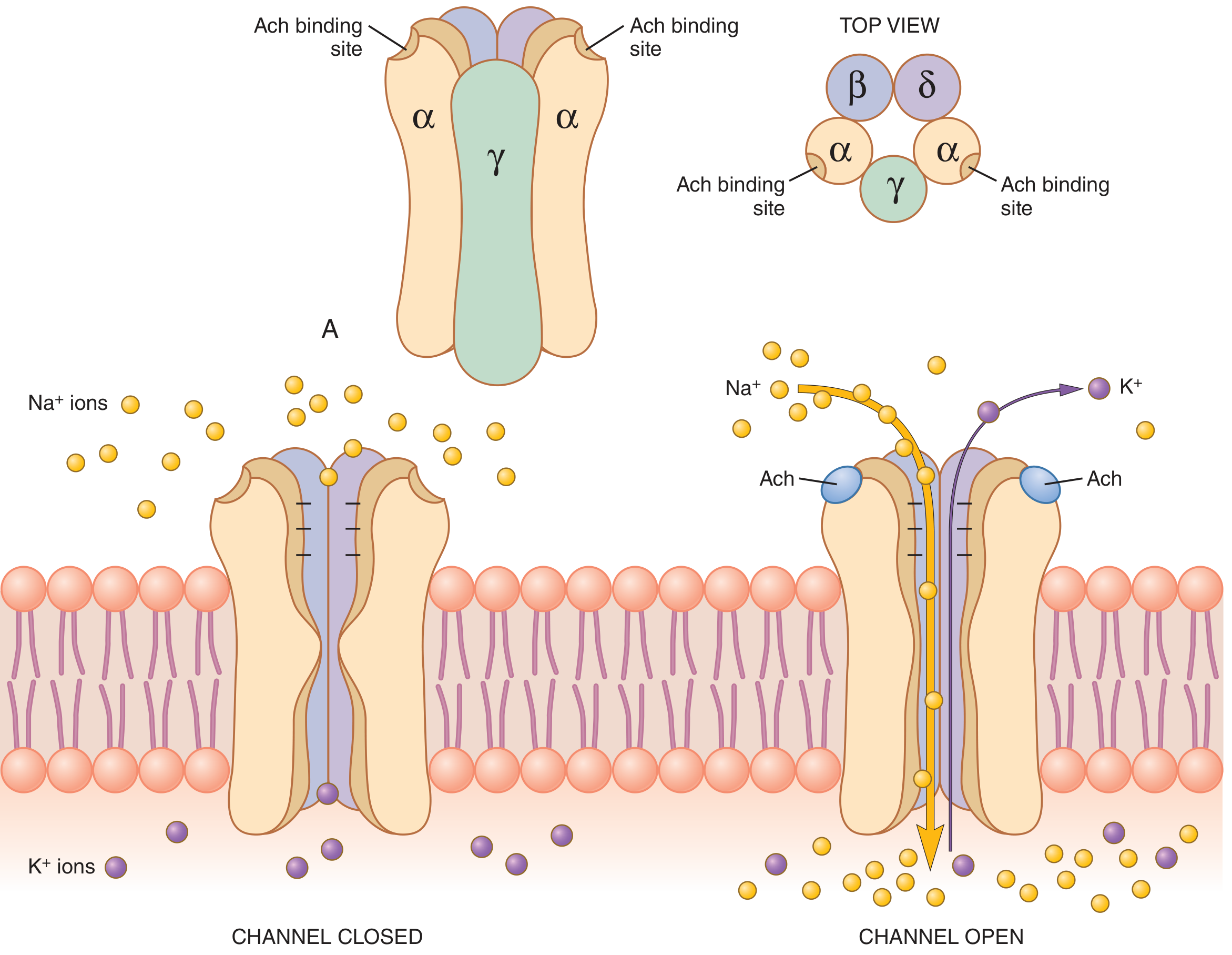
The nAChR is a pentameric ligand-gated ion channel with molecular weight ~275,000 Da:
- Subunits: 2α + 1β + 1δ + 1γ (fetal) or 1ε (adult) arranged around a central pore
- ACh binds simultaneously to both α subunits, causing a conformational change that opens the channel (~0.65 nm diameter)
- The open channel allows Na⁺, K⁺, and Ca²⁺ to pass; negatively charged channel mouth repels Cl⁻
- Net effect: large inward Na⁺ current (Na⁺ electrochemical driving force ~160 mV inward vs. K⁺ ~10 mV outward) → depolarisation
Sequence of Events at Transmission
| Step | Event |
|---|---|
| 1 | Action potential arrives at axon terminal |
| 2 | Voltage-gated Ca²⁺ channels open → Ca²⁺ influx |
| 3 | Ca²⁺ activates Ca²⁺-calmodulin kinase → phosphorylates synapsin proteins → vesicles freed from cytoskeleton |
| 4 | ~125 vesicles dock at active zones and undergo exocytosis → ACh released into synaptic cleft |
| 5 | ACh diffuses to nAChRs on junctional folds |
| 6 | Binding of 2 ACh molecules opens the ion channel → Na⁺ influx → End Plate Potential (EPP) of +50 to +75 mV |
| 7 | EPP depolarises adjacent muscle membrane beyond threshold → muscle action potential → muscle contraction |
| 8 | AChE rapidly hydrolyses ACh → choline is recycled back into the terminal for ACh re-synthesis |
End Plate Potential (EPP)
- A local graded depolarisation generated at the motor end plate
- Not propagated; it is always suprathreshold under normal conditions (the NMJ has a safety factor of ~3x - the EPP generated is approximately 3 times the minimum needed to fire an action potential)
- Subthreshold EPPs are produced by curare (competitive nAChR blocker) or botulinum toxin (blocks ACh release)
Safety Factor & Fatigue
- Under normal conditions, each nerve impulse produces ~3× the minimum EPP needed, ensuring reliable transmission
- At very high stimulation rates (>100/s for several minutes), ACh vesicle depletion can cause fatigue - impulses may fail to propagate to the muscle
- In normal activity, measurable NMJ fatigue is rare
Termination of ACh Action
AChE in the synaptic cleft hydrolyses ACh within a few milliseconds into choline + acetate:
- Prevents re-excitation of the muscle after each action potential
- Choline is actively re-taken up by the nerve terminal and used to re-synthesise ACh (via choline acetyltransferase + acetyl-CoA)
Clinical Relevance
| Condition | Mechanism |
|---|---|
| Myasthenia Gravis | Autoantibodies against postsynaptic nAChRs → reduced EPP → fatigable weakness |
| Lambert-Eaton Syndrome | Autoantibodies against presynaptic voltage-gated Ca²⁺ channels → reduced ACh release |
| Botulinum toxin | Cleaves SNARE proteins → blocks vesicle fusion → no ACh release → flaccid paralysis |
| Organophosphate poisoning | Inhibit AChE → ACh accumulation → sustained depolarisation → paralysis + cholinergic crisis |
| Curare / Non-depolarising NMBDs | Competitive nAChR blockade → no EPP |
| Succinylcholine (depolarising NMBD) | Persistent nAChR activation → phase I depolarising block |
Sources: Ganong's Review of Medical Physiology, 26th Ed. | Guyton & Hall Textbook of Medical Physiology | Boron & Boulpaep Medical Physiology
This is a shared conversation. Sign in to Orris to start your own chat.